Fig. 7.1
The patient lies in an articulated table and in the Lloyd-Davies position with the left arm abducted and 20° head-up tilt (reverse Trendelenburg position).
A vesical catheter and a nasogastric tube are applied after induction of anesthesia.
The surgeon works between the legs of the patient with the first assistant (camera assistant) on the right side of the patient. A further assistant is sometimes necessary and stands on the right side too. The scrub nurse and the instrument table are on the left side.
An electrocautery and a bipolar vessel sealing system are placed near the instrument table and main monitor with the laparoscopic instrumentation rack located above the patient’s right shoulder. 0,1,2,3 are future trocar positions
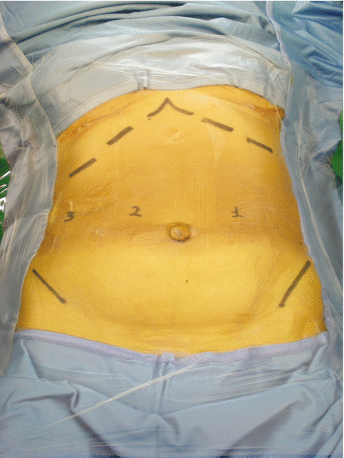
Fig. 7.2
The operative field is disinfected with povidone-iodine solution and protected with iodine-impregnated drapes. 0,1,2,3 are future trocar positions
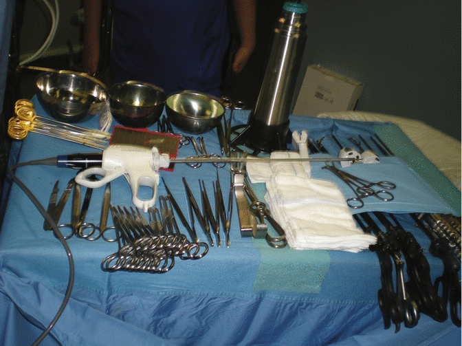
Fig. 7.3
The surgical procedure is carried up using a harmonic scalpel (Ultracision harmonic scalpel, Ethicon Endo-surgery Inc., Cincinnati, OH) for dissection and vessel sealing. Hemostasis is obtained with the utilization of bipolar forceps and application of titanium clips.
Anastomoses and duodenal and esophageal transection are achieved by the application of a 45 mm cartridge laparoscopic linear stapler (triple-staggered rows of staples).
Fenestrated bowel graspers (Johann and Croce-Olmi type) are used for manipulation and traction maneuvers. Curved and angled dissectors are useful for fine dissection, as curved scissors and fixed-tip electrode (J-hook type).
A five-finger fan retractor allows a good exposure of the hepatic hilum and of the esophagogastric junction.
A laparoscopic needle holder is utilized to perform intracorporeal sutures.
A laparoscopic irrigation-suction system and an endo-bag are used for maintaining clean the operative field and for taking out the specimen.
The video camera is connected to a 30° laparoscope and to a wide-screen high-definition monitor
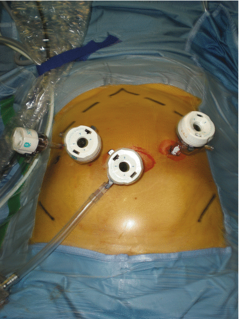
Fig. 7.4
Four trocars are used. One 12 mm trocar for laparoscope is inserted through the umbilicus (T0). Two 12 mm trocars are placed 2 cm above the umbilicus level in the left (T1) and right (T2) midclavicular line. The fourth 5 or 12 mm trocar (T3) is inserted two centimeters below the right costal margin in the hypochondrium
7.3 Laparoscopic Total Gastrectomy
7.3.1 Preliminary Maneuvers
The liver, diaphragm, serosal surfaces, omentum, bowel, mesentery, and pelvic organs are carefully inspected. Biopsy of suspicious lesions is done and documented pathologically by frozen section. Peritoneal lavage also is obtained and submitted for intraoperative cytology.
At this stage, intraoperative laparoscopic ultrasonography is carried up to scan the liver surface and assess the presence of deep liver metastases.
The identification of hepatic and peritoneal involvement could change the operating strategy.
7.3.2 Coloepiploic Detachment and Station 4sb Lymphadenectomy
Figs. 7.5 and 7.6
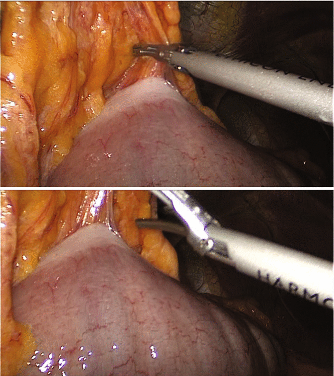
After exploration of the peritoneal cavity, the greater curvature is mobilized by dissection of the entire greater omentum from the transverse colon. Dissection is extended toward the lower pole of the spleen to ligate left gastroepiploic vessels and short gastric vessels and continued toward the right side to dissect the right gastroepiploic vessels at their roots
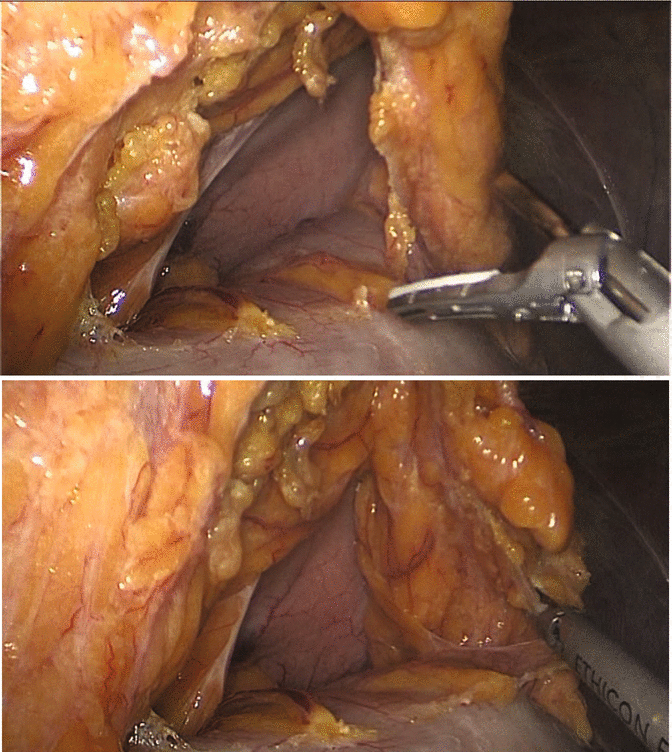
Figs. 7.7 and 7.8
The assistant inserts through T3 a fenestrated bowel grasper (Johann) in order to lift up the greater omentum and to expose the avascular plane for coloepiploic detachment. The surgeon, using harmonic scalpel and a bowel grasper via T1 and T2, begins the dissection at the middle third of the transverse colon and moves toward the origin of the left gastroepiploic vessels from the splenic artery and vein separating the gastrosplenic ligament and removing the lymph nodes of 4sb station
7.3.3 Station 4sa Lymphadenectomy
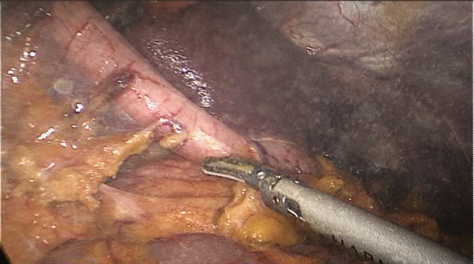
Fig. 7.9
Left gastroepiploic vessels are divided by a harmonic scalpel or sectioned between titanium clips. For a better exposure of the region, it is useful to dissect the adhesions between the posterior gastric wall and the anterior pancreatic surface opening the lesser peritoneal sac. The identification of the left gastroepiploic vessel roots is facilitated by a gentle mobilization of the pancreatic tail in order to expose the splenic vessels.
Using the same technique the surgeon proceeds cephalad to dissect short gastric vessels. The assistant, by a gentle traction, pulls the gastric fundus down while the surgeon, giving delicate traction on the anterior surface of the spleen, separates by harmonic scalpel the gastrosplenic ligament and the short gastric vessels as near as possible to the spleen to achieve a correct lymphadenectomy of group 4sa nodes
7.3.4 Station 2 Lymphadenectomy
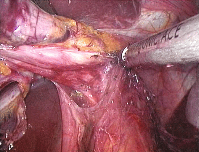
Fig. 7.10
Now the gastric fundus is free, and the left diaphragmatic crus is exposed. Pulling down the stomach and dissecting the peritoneum over the left diaphragmatic crus, the left paracardial nodes are removed (station n° 2)
< div class='tao-gold-member'>
Only gold members can continue reading. Log In or Register to continue
Related posts:


Stay updated, free articles. Join our Telegram channel

Full access? Get Clinical Tree


