Preoperative Evaluation and Staging of Patients with Pelvic Organ Prolapse

3 Preoperative Evaluation and Staging of Patients with Pelvic Organ Prolapse


image Video Clips online


The decision to perform a surgical intervention on a patient with pelvic organ prolapse (POP) should be based on the degree of interference that the prolapse creates in the patient’s daily life. This chapter discusses how to best take a history, perform a physical examination, and stage the prolapse appropriately. In addition, how to select the appropriate procedure to fit the patient’s condition and how to guide patients through the preoperative consent process are also presented.


Taking a Good History


Patients with POP may report symptoms directly related to the prolapse, such as vaginal bulge, pressure, and discomfort, as well as a plethora of functional derangements related to voiding, defecation, and sexual problems.


Patients with POP should be questioned about all aspects of pelvic floor dysfunction. In the author’s opinion, symptoms related to the lower urinary tract, the lower gastrointestinal tract, sexual function, and pelvic pain should be distinguished from those symptoms directly related to tissue protrusion or prolapse.


A variety of standardized questionaires (Burrows, 2004) can be used to assess fully the patient’s perception of her pelvic floor symptomatology. Preferably, the patient completes these questionnaires before beginning the interview process. Although not always possible, these questionnaires allow the health care provider to appreciate the daily impact of these various problems. A voiding diary, which a patient maintains for 1 to 3 days, may also be helpful; as many of these patients have significant lower urinary tract dysfunction.


The ultimate goal of the history is to appreciate—from the patient’s standpoint—which symptoms are the most bothersome and to determine whether the symptoms are related to the anatomic descent of her tissues. Ultimately, the surgeon needs to determine how anatomic correction of the prolapse will impact function.


Because the correlation between anatomic descent and functional derangement is somewhat unpredictable, patients need to be given realistic expectations regarding the impact of the surgical repair on their various symptoms. The role that these symptoms play in the continuum of the prolapse should be explained to the patient in a way that she can understand. Since many of these topics are extremely sensitive, questionnaires can significantly assist in the process of taking a history, especially when supplied to the patient before her visit. The answers to the various questions are used to guide the discussion and may permit the patient to share details that would otherwise be difficult to verbalize.


Appreciating any co-morbidities related to the patient’s ability to tolerate pelvic reconstructive surgery is also important. The history of significant cardiac disease, myocardial infarction, and pulmonary disease are examples of conditions that need to be evaluated before considering a surgical procedure. Pulmonary embolism is a major risk factor in all types of pelvic surgery, especially in those patients who require prolonged dorsal lithotomy positioning. Before any surgery, the surgeon should recognize risk factors for embolism and use proper interventions during and after the surgical procedure.


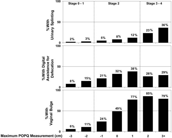
Vaginal prolapse in any compartment—anterior, apical, or posterior—can cause vaginal fullness, pain, and/or a protruding mass. In a recent study by Tan and colleagues (2005), the feeling of “a bulge or that something is falling outside the vagina” had a positive predictive value of 81% for POP, and the lack of this symptom had a negative predictive value of 76% for predicting prolapse at or past the hymen. Not surprisingly, an increased degree of prolapse, especially beyond the hymen, is associated with increased pelvic discomfort and the visualization of a protrusion. The association of Pelvic Organ Prolapse Quantification (POPQ) measurements during the examination with three commonly related symptoms—urinary splinting, digital assistance with defecation, and vaginal bulge—is shown in Figure 3-1.


Urinary incontinence and voiding difficulties can occur in association with anterior and apical vaginal prolapse. However, women with advanced degrees of prolapse may not have overt symptoms of stress incontinence because the prolapse may cause a mechanical obstruction of the urethra, thereby preventing urinary leakage. Instead, these women may perform manual replacement of the prolapse to accomplish voiding. Patients who require digital assistance to void, in general, have more advanced degrees of prolapse. In addition to the difficulty voiding, other urinary symptoms, such as urgency, frequency, and urge incontinence, are found in women with prolapse.


POP, especially in the apical and posterior compartments, can be associated with defecatory dysfunction, such as the need for manual assistance with defecation and anal incontinence of flatus, liquid, or solid stool. These patients often have outlet-type constipation, secondary to the trapping of stool within the rectocele, necessitating the need for splinting or for applying manual pressure in the vagina, rectum, or perineum to reduce the prolapse, which aids in defecation. Although defecatory dysfunction remains an area that is poorly understood in patients with prolapse, clinical and radiographic studies have shown that the severity of prolapse is not strongly correlated with increased symptoms.


Although the relationship between sexual function and POP is not clearly defined, questions regarding sexual dysfunction must be included in the evaluation of any patient with POP. Patients may report symptoms of dyspareunia and decreased libido and orgasms, as well as increased embarrassment because of an altered anatomy that affects body image. Some studies have reported that prolapse adversely affects sexual function, with subsequent improvement in sexual function after the repair of prolapse. (Barber et al, 2002; Rogers et al, 2001; Weber, Walters, Piedmonte, 2000) However, Burrows and associates (2004) showed little correlation between the extent of the prolapse and sexual dysfunction. Evaluating sexual function may be especially difficult in this patient population, secondary to the presence of factors other than prolapse, such as partner limitations and functional deficits.


Examination


When examining a patient with POP, a bimanual examination is performed to rule out co-existent gynecologic conditions.


The physical examination for prolapse should be conducted with the patient in the dorsal lithotomy position, as is used for a routine pelvic examination. If physical findings do not correspond to the reported symptoms or if the maximum extent of the prolapse cannot be confirmed, then the woman can be reexamined in the standing position. A rectal examination further identifies pelvic pathologic conditions and fecal impaction, the latter of which may be associated with voiding difficulties and incontinence in older women.


Initially, the external genitalia are inspected, and, if no displacement is apparent, the labia are gently spread to expose the vestibule and hymen. Vaginal discharge can mimic incontinence; therefore evidence of this problem should be sought and, if present, treated. Palpation of the anterior vaginal wall and urethra may elicit urethral discharge or tenderness that suggests a urethral diverticulum, carcinoma, or inflammatory condition of the urethra. The integrity of the perineal body is evaluated, and the extent of all prolapsed parts is assessed. A retractor, a Sims speculum, or the posterior blade of a bivalve speculum can be used to depress the posterior vagina to help visualize the anterior vagina and vice versa for the posterior vagina. The vaginal mucosa should be examined for atrophy and thinning, because both may affect the management of POP. Healthy, estrogenized tissue without significant evidence of prolapse will be well perfused, have rugation, and have physiologic moisture. Atrophic vaginal tissue consistent with hypoestrogenemia appears pale, thin, without rugation, and can be friable.


After the resting vaginal examination, the patient is instructed to perform a Valsalva maneuver or to cough vigorously. During this maneuver, the order and extent of the descent of the pelvic organs is noted, as is the relationship of the pelvic organs to each other at the peak of increased intraabdominal pressure. The presence and severity of anterior vaginal relaxation, including cystocele and proximal urethral detachment and mobility or anterior vaginal scarring, are estimated. Associated pelvic support abnormalities, such as rectocele, enterocele, and uterovaginal prolapse, are also noted. The amount or severity of prolapse in each vaginal segment should be measured and recorded using a standardized reproducible system for staging (see pages 34-37). A rectovaginal examination is required to evaluate fully the prolapse of the posterior vaginal wall and perineal body. Digital assessment of the bowel contents in the rectovaginal septum during the straining examination can help diagnose an enterocele. Inspection should also be made of the anal sphincter; as fecal incontinence can be associated with posterior vaginal support defects. Women with a torn external sphincter may have scarring or what has been termed a dovetail sign. (See Video 3-1, “Live Demonstrations of a Variety of Women with Advanced Prolapse” image)


Anterior vaginal wall descent usually represents bladder descent with or without concomitant urethral hypermobility. The anterior vaginal wall prolapse is believed to be the result of a midline defect, a paravaginal defect, or, less commonly, a transverse defect. These defects may co-exist or occur in isolation. Although clinicians have attempted to describe techniques to differentiate these various defects during the physical examination, researchers have shown that the preoperative predictability is not particularly reliable or accurate. In a study by Barber and associates (1999) of 117 women with prolapse, the sensitivity of the clinical examination to detect paravaginal defects (92%) was good, yet the specificity (52%) was poor. Despite a high prevalence of paravaginal defects, the positive predictive value was only 61%. Less than two thirds of women believed to have a paravaginal defect on physical examination were confirmed to possess the same at surgery. Another study by Whiteside and colleagues (2004) demonstrated poor predictability and reproducibility of the clinical examination to differentiate various anterior vaginal wall defects. Thus the clinical value of attempting to determine preoperatively the defect that is responsible for the anterior wall prolapse is low.


Clinical examinations do not always accurately differentiate rectoceles from enteroceles in posterior prolapse. Thus some investigators have advocated performing imaging studies to delineate further the exact nature of the posterior wall prolapse. Traditionally, most clinicians believe that they are able to detect the presence or absence of these defects without anatomically localizing them. However, little is known regarding the accuracy or use of clinical examinations in the evaluation of the anatomic locations of prolapsed small or large bowel or of specific defects in the rectovaginal space. Burrows and associates (2003) found that clinical examinations often did not accurately predict the specific location of defects in the rectovaginal septum that was subsequently found intraoperatively. Clinical findings corresponded with intraoperative observations in 59% of patients and differed in 41%; sensitivities and positive predictive values of clinical examinations were less than 40% for all posterior defects. However, the clinical consequence of not preoperatively detecting defects remains unclear.
< div class='tao-gold-member'>

Only gold members can continue reading. Log In or Register to continue

Stay updated, free articles. Join our Telegram channel

Jul 24, 2016 | Posted by in UROLOGY | Comments Off on Preoperative Evaluation and Staging of Patients with Pelvic Organ Prolapse

Full access? Get Clinical Tree

Get Clinical Tree app for offline access