Fig. 2.1
Narrow band imaging filter technology. Light source and video processor are used to deliver high-definition light through the cystoscope (Courtesy of Olympus)
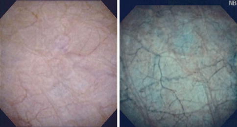
Fig. 2.2
Normal bladder. White-light imaging (WLI) cystoscopy (left) and narrow band imaging (right). Mucosa is bland on WLI cystoscopy; NBI shows prominent green submucosal vessels and brown superficial capillaries, but no evidence of concentrated enhancement (neovascularity) (From Herr [27], with permission)
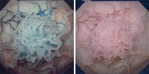
Fig. 2.3
Papillary tumor shown by NBI cystoscopy (left) and WLI cystoscopy (right) (From Herr [27], with permission)
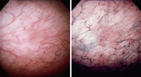
Fig. 2.4
Small papillary tumor missed on WLI cystoscopy (left) is clearly seen on NBI cystoscopy (right) (From Herr [27], with permission)
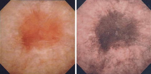
Fig. 2.5
Carcinoma in situ of bladder visualized on WLI cystoscopy (left) and NBI cystoscopy (right) (From Herr [27], with permission)
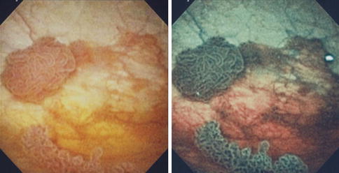
Fig. 2.6
Papillary tumors on WLI cystoscopy (left) appear dark green on NBI cystoscopy (right), but surrounding brown lesions indicate associated carcinoma in situ (From Herr [27], with permission)
NBI Detection of Bladder Tumors
Bryan et al. [4] were the first to describe the results of flexible NBI cystoscopy in 29 patients with recurrent bladder tumors. They found 15 additional tumors in 12 patients compared with WLI cystoscopy. Table 2.1 lists results from nine series, all showing enhanced detection of tumors using NBI diagnostic cystoscopy [4–12]. Although the series by Zhu et al. [11] included only 12 patients, they were evaluated by NBI for positive urine cytology and negative WLI cystoscopy. NBI found evidence of carcinoma in situ in 5 (42 %) patients. Collectively, the studies show superior sensitivity and negative predictive value (>90 %) for NBI over WLI cystoscopy, making it useful for identifying abnormal lesions and for excluding a diagnosis of bladder tumor. Although more negative biopsies of false-positive lesions (lower specificity) occurred with NBI, this is not believed to be significant or detrimental, since many patients have denuded mucosa and positive urine cytology, indicating carcinoma in situ (CIS), and additional biopsies have not led to increased toxicity. Negative tumor margins of suspicious lesions indicated by NBI also confirm a complete resection. Table 2.2 shows NBI enhances detection of biopsy-proved carcinoma in situ.
Table 2.1
Detection of bladder tumors by white-light imaging (WLI) and narrow band imaging (NBI) cystoscopy
Series | No. of cases | Cystoscopy | Sensitivity (%) | Specificity (%) | PPV (%) | NPV (%) |
|---|---|---|---|---|---|---|
Bryan et al. [4] | 29 | WLI | 82 | 79 | 70 | 81 |
NBI | 96 | 85 | 61 | 92 | ||
Herr and Donat [5] | 427 | WLI | 87 | 85 | 66 | 96 |
NBI | 100 | 82 | 63 | 100 | ||
Cauberg et al. [6] | 95 | WLI | 79 | 75 | – | – |
NBI | 95 | 69 | – | – | ||
Tatsugami et al. [7] | 104 | WLI | 57 | 86 | 69 | 79 |
NBI | 93 | 71 | 63 | 95 | ||
Shen et al. [8] | 78 | WLI | 77 | 82 | – | 79 |
NBI | 92 | 73 | – | 87 | ||
Xiaodong et al. [9] | 64 | WLI | 79 | 76 | – | – |
NBI | 97 | 68 | ||||
Geavlete et al. [10] | 95 | WLI | 84 | – | – | – |
NBI | 95 | – | – | – | ||
Zhu et al. [11] | 12 | WLI | 50 | 91 | – | – |
NBI | 78 | 80 | – | – | ||
Chen et al. [12] | 179 | WLI | 97 | 81 | – | – |
NBI | 79 | 79 | – | – |
Table 2.2
Detection of carcinoma in situ by WLI and NBI cystoscopic biopsy
Series | No. of cases | Cystoscopy | Sensitivity (%) | Specificity (%) | PPV | NPV |
|---|---|---|---|---|---|---|
Herr and Donat [5] | 67 | WLI/NBI | 83; 100 | 72; 76 | 36;36 | 97;100 |
Tatsugami et al. [7] | 30 | WLI/NBI | 50;90 < div class='tao-gold-member'>
Only gold members can continue reading. Log In or Register to continue
Stay updated, free articles. Join our Telegram channel
Full access? Get Clinical Tree
 Get Clinical Tree app for offline access

|


