and Hubert Lepidi1
(1)
UER Médecine, Aix-Marseille Université, Marseille, France
Abstract
The tissue of the corpora cavernosa clitoridis, or cavernous tissue, is erectile tissue, meaning its histological structure enables these bodies to fill with blood, increase in size and harden. This tissue is made up of a complex network of venous sinuses that appear as intercommunicating anfractuous cavities (cavernous labyrinth) and of a thick fibrous envelope, the albuginea, which was long believed, erroneously, to be inextensible. The sinusoid cavities, or sinuses of the corpora cavernosa, most often have wide-open lumina (though sometimes collapsed) that are very irregular. Their dimensions vary greatly. Their shapes are more often polyhedral or narrow and elongated rather than circular or oval. These sinuses present numerous recesses. The inter-sinus communications are clearly visible and the main characteristic of the sinuses is that they are richly anastomotic (Fig. 6.1). The wall of the sinuses is indeed a vessel wall, comprising an endothelial layer and a conjunctive axis made of collagenous bundles in which smooth muscle fibres may be seen. Specific stains (orcein) demonstrate the existence of a rich network of elastic fibres (Fig. 6.2). It should be noted that the walls of the sinuses are thin, especially if compared to the size of the vessel lumina. The endothelial cells, made apparent by the immunolabelling with an anti-factor VIII antibody, have junctions of varying tightness and rest on a basal lamina. Extensions originate from the sinus walls and float in the vessel lumina. Their role is probably to direct blood flow during filling. Most of these extensions appear as fingers that are curved to varying degrees (like a bent index finger). They may also take the shape of small mounts, pyramids, bull horns, points, hooks or clubs. There are also complete or incomplete bridges between two opposite sinus banks, the sinus septa, which contribute to making the sinus labyrinth even more complex. Numerous small vessels (arterioles or venules) are also housed in the sinus walls. These small vessels are very sinuous and their walls are often thick. The arterioles and their branches are called “helicine” due to their often helicoidal arrangement (see Chap. 10). The larger vessels are located at intersections in the inter-sinus connective tissue. Otherwise, a fair number of nerve endings are found in the sinus walls themselves and in the inter-sinus connective tissue (Fig. 7.2).
6.1 Cavernous and Spongy Tissue
6.1.1 Cavernous Tissue
The tissue of the corpora cavernosa clitoridis, or cavernous tissue, is erectile tissue, meaning its histological structure enables these bodies to fill with blood, increase in size and harden. This tissue is made up of a complex network of venous sinuses that appear as intercommunicating anfractuous cavities (cavernous labyrinth) and of a thick fibrous envelope, the albuginea, which was long believed, erroneously, to be inextensible. The sinusoid cavities, or sinuses of the corpora cavernosa, most often have wide-open lumina (though sometimes collapsed) that are very irregular. Their dimensions vary greatly. Their shapes are more often polyhedral or narrow and elongated rather than circular or oval. These sinuses present numerous recesses. The inter-sinus communications are clearly visible and the main characteristic of the sinuses is that they are richly anastomotic (Fig. 6.1). The wall of the sinuses is indeed a vessel wall, comprising an endothelial layer and a conjunctive axis made of collagenous bundles in which smooth muscle fibres may be seen. Specific stains (orcein) demonstrate the existence of a rich network of elastic fibres (Fig. 6.2). It should be noted that the walls of the sinuses are thin, especially if compared to the size of the vessel lumina. The endothelial cells, made apparent by the immunolabelling with an anti-factor VIII antibody, have junctions of varying tightness and rest on a basal lamina. Extensions originate from the sinus walls and float in the vessel lumina. Their role is probably to direct blood flow during filling. Most of these extensions appear as fingers that are curved to varying degrees (like a bent index finger). They may also take the shape of small mounts, pyramids, bull horns, points, hooks or clubs. There are also complete or incomplete bridges between two opposite sinus banks, the sinus septa, which contribute to making the sinus labyrinth even more complex. Numerous small vessels (arterioles or venules) are also housed in the sinus walls. These small vessels are very sinuous and their walls are often thick. The arterioles and their branches are called “helicine” due to their often helicoidal arrangement (see Chap. 10). The larger vessels are located at intersections in the inter-sinus connective tissue. Otherwise, a fair number of nerve endings are found in the sinus walls themselves and in the inter-sinus connective tissue (Fig. 7.1).
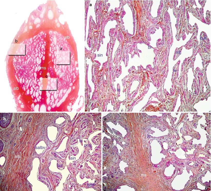
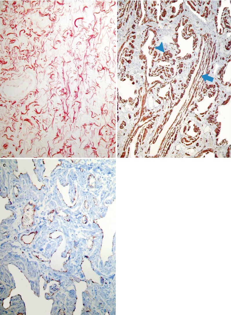
Fig. 6.1
Details of a frontal section through the descending part of the clitoral body showing the organisation of the corpora cavernosa. Part (a) of the section: the cavernous labyrinth; part (b) of the section: tunica albuginea and corpus cavernosum; part (c) of the section: the median septum and the corpora cavernosa. ad advance, alb tunica albuginea, brid bridge, bul bulge, cc corpus cavernosum, fe finger-like extension, ms median septum, proj projection, sin sinus, sl suspensory ligament, salbv sub-albugineous vein
Fig. 6.2
Microarchitecture of the clitoral corpora cavernosa (histochemical study by specific staining of frontal sections of the clitoral body). (a) Staining by orceine: highlighting the elastic fibres. (b) Staining by smooth actin: highlighting the bundles of smooth muscle fibres (wine coloured) oriented in all directions, transversely (blue arrow) or longitudinally (blue arrowhead). (c) Staining by factor VIII: highlighting the endothelial cells: brown colour of the endothelia of the cavernosus maze and of the vascular endothelia. These endothelial cells secrete NO!
It is now proven that the work on corpora cavernosa of the penis can be transposed to the clitoral corpora cavernosa (A. Toesca et al.). Without negating the previous description, this work, notably that of A.M.B. Goldstein et al., demonstrates that instead of viewing the cavernous tissue as a labyrinth of sinuses with walls made of collagenous fibres and smooth muscle fibres, it should be considered as a fibrous collagenous tissue, rich in smooth muscle fibres, in which a network of intercommunicating vascular spaces are housed.
The inter-sinus connective tissue is made up of collagenous fibrous bundles which have the morphological characteristic of being wavy when in a state of rest (Fig. 6.3). According to E. Wespes, who for the most part, studied the penis corpora cavernosa, it is the presence of elastic fibres arranged in a bridge in contact with the collagenous bundles that enables the collagenous fibres to maintain this pleated appearance when in the flaccid phase and, most of all, to return to it as sexual arousal wanes.
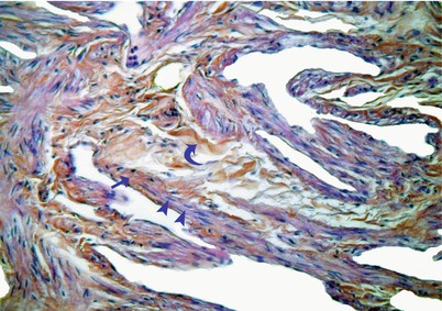
Fig. 6.3
Microarchitecture of the clitoral corpora cavernosa (Details of the cavernous network on a section of the clitoral body). The collagen fibres and the fibroblasts have an orange colour. The smooth muscular cells have a mauve colour. Note: At rest, the collagen fibres have an undulating shape (curved blue arrow). The smooth muscular cells, with flat and elongated nuclei (blue arrowheads), settle on the bundles of collagen fibres (right blue arrow)
The smooth muscle fibres of the clitoral corpora cavernosa were clearly identified using immunohistochemistry methods as the anti-desmin antibody affixes to them specifically. Alpha-smooth actin staining may also be used (Fig. 6.2). These muscle fibres have a longitudinal or transversal orientation within the inter-sinus connective tissue (A. Toesca). They are arranged as bundles or packets and end on collagenous fibres (Fig. 6.3) from which they will eliminate the folds to enable clitoral tumescence. These smooth muscle fibres are also present at the large arteriole and venule adventitia and then disappear on the small vessels. All of these smooth muscle fibres are interconnected. They therefore play a manifest contractile role in the regulation of sinus blood flow, as indicated by the author cited above. Their spontaneous myogenic activity corresponds to the flaccid state and their release only occurs during sexual arousal when the NO neurotransmitter is produced (see Chap. 7). Studied by electron microscopy (E. Wespes), these smooth muscle fibres demonstrate all characteristics of myogenic cells: uniform nucleus, fine basement membrane and above all, in the cytoplasm, mitochondria, contractile filaments and glycogen granules (energy nutrient).
Related posts:


Stay updated, free articles. Join our Telegram channel

Full access? Get Clinical Tree


