Fig. 14.1
The central liver includes the right anterior lobe and the left medial lobe
14.2 Definition of Mesohepatectomy
Mesohepatectomy is the resection of the right anterior and left medial lobes of the liver (ΙV, V, VIII±I in Couinaud’s scheme). The indications for this procedure are tumors that lie between the right anterior and left medial lobes, as well as tumors in the right anterior and left medial lobes that invade the middle hepatic vein which requires en bloc resection.
14.3 Evolution of Mesohepatectomy
In 1965, Wu et al. first reported the use of a mesohepatectomy to treat central hepatic tumor [2].
In 1972 McBride was the first to define mesohepatectomy as resection of Couinaud’s segments IV, V, and VIII, which laid the anatomical foundation for this procedure.
In the 1980s, due to the technical difficulty of the procedure and the high incidence of complications, most central hepatic mass lesions were still treated by extended hemihepatectomy or trisegmentectomy [3–5].
In the 1990s, understanding of liver anatomy and relevant surgical techniques evolved, and mesohepatectomy was gradually accepted as a treatment for central hepatic mass lesions. During this period, further investigations were performed regarding the protection of hepatic vascular structures, neoadjuvant chemotherapy, and portal vein embolization [6–9].
In 2002, Chinese scholars suggested that central hepatocellular carcinoma should be defined as a tumor lying within 1 cm of the bifurcation of portal vein, the confluence of the three main hepatic veins to the IVC, or the retrohepatic IVC trunk.
In the early 2000s, with the increasing use of laparoscopic techniques, Machodo et al. were the first to report laparoscopic mesohepatectomy using an intrahepatic Glissonian approach [10].
In 2013, Zeng Yong of West China Hospital was the first to report an anatomical classification of central hepatic mass lesions [11].
14.4 Indications for Mesohepatectomy
Tumors of the central liver, including primary hepatocellular carcinoma, liver metastasis, hepatic hemangioma, gallbladder carcinoma, and hilar cholangiocarcinoma, as well as other processes such as central liver hepatolithiasis, damage of the central liver which cannot be fixed, chronic liver abscess and hepatic echinococcosis, etc., are all suitable for mesohepatectomy, especially for patients with poor liver function or cirrhosis.
14.5 Conventional Classification of Mesohepatectomy and Its Key Technical Points
14.5.1 In Classical Regular Mesohepatectomy
Glisson’s pedicle is first dissected; then, the afferent and efferent canals of the central liver are dissected and divided, and the liver parenchyma is transected.
Advantage:
Precision
Disadvantage:
Technically demanding
Key technical points:
First dissect the left Glisson’s pedicle. Dissect the first porta hepatis, and expose the confluence of the left and right hepatic ducts and the bifurcation of the proper hepatic artery. Dissect along the left hepatic artery until its left medial branch is exposed, and suture and divide this branch. Be careful to protect the left hepatic duct, as it is accompanied by the trunk of the left hepatic artery in the inferior aspect of the ligamentum teres hepatis. Beneath the divided left medial branch of the left hepatic artery is the left medial branch of the left hepatic duct, which should be divided and sutured; alternatively, this branch can be divided during the transection of the parenchyma. The left hepatic artery and the left hepatic duct are gently lifted to expose the left portal branch which lies beneath theses structures, and dissection should be continued anteriorly to expose the left medial branch of the portal vein, which should be carefully clamped and sutured. This completes the dissection process for the left Glisson’s pedicle; a similar process should be performed on the right Glisson’s pedicle. At the bifurcation of the proper hepatic artery, the common hepatic duct should be pulled to the left to provide a better exposure as the right hepatic artery is commonly found the behind common hepatic duct. The right hepatic artery is dissected along the right portal vein fissure to expose the right anterior and posterior branches of the right hepatic artery, and its right anterior branch points to the gallbladder bed while the right posterior branch points to the bottom right almost vertically [20]. The right anterior branch of the hepatic artery is sutured and divided, and then the right anterior branch of the right hepatic duct, which is commonly found next to the hepatic artery, should be divided and sutured carefully. The division of the right bile duct can also be carried out during transection of the parenchyma. In the end, the liver parenchyma is transected, along with the relevant canals. Now that the central liver is basically devascularized, the dividing line between the central liver and the left lateral lobe, as well as the dividing line between the central liver and the right posterior lobe, can be seen on the surface. The transection line is marked using an electronic knife, and the liver parenchyma is transected using CUSA, a water jet dissector, a LigaSure, or a hemostat. During this process, relatively large relevant canals should be divided and sutured, ligated, or clipped, using titanium clips.
14.5.2 Mesohepatectomy with Transection of the Glisson’s Pedicle
Advantage:
There is no need to dissect the Glisson’s pedicle when dissecting the hepatic pedicle.
Disadvantage:
A lack of precision creates a certain risk of subsidiary injury, especially for individuals with complicated anatomical variation of canals entering the liver.
Key technical points:
First dissect the gallbladder, and then find the hepatic pedicle of the right anterior lobe at the intersection of the longitudinal axis of the gallbladder and the lower limb of the liver parenchyma. Bluntly dissect the lateral parenchyma to within 0.5 cm of this intersection, and then use straight hemostatic forceps to take a suture from the back of the hepatic pedicle of the right anterior lobe and ligate the pedicle. The dividing line between the right anterior and posterior lobes can be seen on the parenchyma surface. After ligating and dividing the Glisson’s pedicles from the right to the round ligament one by one, the left dividing line can be exposed. The key technical point is to avoid opening the Glisson’s pedicle; rather, bluntly dissect between the pedicle and the parenchyma. Now several portal branches of the left medial lobe are ligated and divided, while the arteries are not involved. The left ischemic line can be seen after all portal branches of the left medial lobe are ligated. Finally, transect the liver parenchyma [11].
14.5.3 Irregular Mesohepatectomy
Compared with the two procedures described above, irregular mesohepatectomy is less demanding and less complicated. In this procedure, the transection line is directly marked on the liver surface without dissecting the porta hepatis. The right transection line extends from the notch of the right liver to the inner side of the confluence of the right hepatic vein and the IVC. The left transection line is the right margin of the falciform ligament. The actual transection line is placed 0.5 cm medially to the aforementioned lines, in order to avoid the right and left hepatic veins. This procedure is not precise but still provides satisfactory results. The key point is the use of patience and care during the operation in order to avoid hemorrhage [5, 12].
14.5.4 Laparoscopic Mesohepatectomy
Disadvantage:
There is a high incidence of complications such as uncontrolled hemorrhage and bile duct damage because of the special anatomy of the central liver and anatomical variants of the intrahepatic vessels.
Advantage:
Anatomical structures are magnified with the laparoscope; thus, regional structures are seen more clearly than in an open procedure. The dissections of the hepatic artery, the portal vein, and the hepatic vein are performed one by one and can be managed by careful dissection using instruments.
Key technical points:
Set up pneumoperitoneum in a routine fashion, insert the instruments, and tilt the operating table l5–30° to the left as required. The surgeon stands between the patient’s legs, assistants stand by either side of the surgeon, and the instrument nurse stands by the patient’s right foot. Five or six ports are created (Fig. 14.2). Normally, the observing port is located 1 cm beneath the umbilicus. The main port is located 2–4 cm beneath the xiphoid when resecting the left aspect of the central liver and dissecting the left lobe’s Glisson’s pedicle, 4–6 cm beneath the xiphoid when resecting the right aspect of the central liver and dissecting the right lobe’s Glisson’s pedicle. Two or three ancillary ports are located at the intersections of the right costal margin with the midclavicular line and anterior axillary line. Pretreatment blocking is set up routinely at the porta hepatis, and the transection area is marked according to preoperative imaging, intraoperative exploration, and anatomical markers of the liver. First dissect the porta hepatis. Dissect the hepatoduodenal ligament, and mobilize the common bile duct, the left and right hepatic ducts, the proper hepatic artery and its two branches, and the portal trunk and its two branches, respectively. Clip and divide the left hepatic artery, the left branch of portal vein, and the left medial branch of the left hepatic duct, respectively. Suture and divide the right hepatic artery, the right branch of the portal vein, and the right anterior branch of the right hepatic duct, respectively, in the same way. Then transect the liver parenchyma, which is commonly performed using a laparoscopic ultrasonic scalpel. Finally, handle the transection and relevant canals using the same method as in an open procedure.
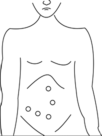
Fig. 14.2
Laparoscopic mesohepatectomy; five or six ports are created
14.6 West China Classification of Central Hepatic Mass Lesions [11]
14.6.1 Background
Because of the shortage of the literature and guidelines regarding mesohepatectomy, as well as this procedure’s difficulty and its high incidence of complications, many surgeons prefer to perform extended left or right hemihepatectomy instead. However, extended hemihepatectomy resects 60–70 % of the liver, leading to a high risk of postoperative liver failure and even death [5, 12, 13]. Although hepatectomy has progressed in precision due to techniques regarding donor liver resection, complications such as intraoperative hemorrhage, damage of afferent and efferent hepatic vessels, and postoperative bile leakage still pose great challenges for surgeons. Investigators at West China Hospital of Sichuan University collected clinical data from 356 patients diagnosed with central hepatic mass lesions between Jan 2005 and Dec 2011 and created a classification of mesohepatectomy and key points for each type of procedure, in an attempt to characterize and simplify each type of mesohepatectomy, prevent postoperative complications, and ensure safety [11].
14.6.2 Foundations of the Classification
① Established location of the lesion in the central liver, ② relationship of the lesion to the bile ducts and the portal vein branches of the porta hepatic, ③ relationship of the lesion to the hepatic veins of the second hepatic hilum, and ④ relationship of lesion to IVC.
Related posts:


Stay updated, free articles. Join our Telegram channel

Full access? Get Clinical Tree


