Magnetic Resonance Imaging

Chapter 31 Magnetic Resonance Imaging


INTRODUCTION


Magnetic resonance imaging (MRI) is increasingly used by gynecologists and especially reproductive surgeons to assess the genital tract in a variety of clinical situations that require precise detail of the reproductive organs. Sufficient detail is provided in most circumstances by transvaginal ultrasonography (TVUS) or saline infusion sonohysterogram. In some instances, such as in determining the size and location of fibroids or discerning adenomyosis, neither TVUS nor saline infusion sonohysterogram is definitive. Furthermore, accurate determination of a müllerian anomaly may allow more precise preoperative assessment and even potentially eliminate a surgical step, such as a diagnostic laparoscopy for resection of a uterine septum.


Magnetic resonance imaging is gaining widespread acceptance and, in many instances, is a cost-effective tool in the evaluation of abnormal uterine bleeding. It is noninvasive, differentiates uterine anatomy in response to exogenous hormones or the normal menstrual cycle, and reliably localizes pelvic pathology and size of lesions. When uterine conservation is desired in women with fibroids, and TVUS or saline infusion sonohysterogram is indeterminate in localizing the depth of myometrial involvement of a fibroid, MRI should be considered as a part of the clinical algorithm. The precision of MRI localization of submucosal fibroids can obviate the need for hysterectomy and permit a skilled surgeon to hysteroscopically resect the fibroids. If the clinical examination is suspicious for adenomyosis and the TVUS is nondiagnostic, the clinician should strongly consider MRI. When the results of the imaging study would influence surgical route and planning, MRI should be considered in the preoperative evaluation.


This chapter gives an overview of the physics of MRI and gives a visual overview of the potential clinical applications of this imaging modality.


PRINCIPLES OF MAGNETIC RESONANCE IMAGING


Clinical MRI takes advantage of the physical properties of the protons in the body to produce anatomic images. All nuclei—in this case, hydrogen nuclei—have the physical properties of charge and spin. Depending on the charge and spin, certain atoms, including hydrogen nuclei (protons), behave as tiny magnets and have both polarity and an associated magnetic field with the properties of magnitude and direction. When placed in a magnetic field, they tend to align with the field in a lower energy state or against the field in a higher energy state.


When the system is at rest, in equilibrium, there are a relatively tiny number of excess protons in the lower energy state. These are the protons that are available for use in MRI. Because of the physical properties of the system, these magnetic fields do not remain perfectly aligned with the main magnetic field but rather they rotate or precess about the central axis of the main magnetic field, much as a spinning top precesses in the presence of gravity. This precession is very important in MRI because it is essentially the only parameter in the system that we can either change or measure.


The Larmor equation tells us that the precessional frequency is equal to the strength of the magnetic field in which a particular proton of interest resides times a constant called the gyromagnetic ratio. The gyromagnetic ratio is fixed for a given nucleus. At the magnetic field strengths used in MRI, the precessional frequencies are in the radiofrequency range. If we know the strength of the magnetic field in a particular location, we can predict the precessional frequency of the protons in that location. Analogously, if we change the magnetic field in a predefined manner and if we can measure the precessional frequencies of various protons, we can calculate their positions as long as we know the magnetic field variations.


Precessing protons will only absorb energy applied at their precessional frequency. When we introduce radiofrequency energy at the appropriate frequencies, the protons precessing at these frequencies absorb the energy and move to a higher energy state. As these protons relax back to equilibrium, radiofrequency signals can be detected at their particular precessional frequencies.


In MRI, we use these principles to create images. During imaging, appropriate radiofrequency energy is put into the system while supplementary magnetic fields, called gradients, are added to or subtracted from the main magnetic field in a predefined linear fashion. Because the scanner can vary the gradients in all directions, the process of spacial localization, identifying the location of a particular signal in three-dimensional space, can be achieved. Multiple iterations of the imaging examination are performed and the scanner collects and reconstructs the obtained data, assigning the signal from the protons to the appropriate location on the image until the entire image is formed.


Varying certain acquisition parameters, especially timing, changes the images so that their contrast varies depending more on their interactions with each other (T1-weighted imaging) or on their interactions with their local environment (T2-weighted imaging). Many other techniques and strategies are employed to vary the contrast, sensitivity, and efficiency of MRI and much more complete descriptions are available to the interested reader.1


MRI VERSUS OTHER IMAGING MODALITIES


MRI has many advantages over ultrasound and computed tomography (CT) for the evaluation of pelvic anatomy, congenital anomalies, and pathologic disorders. Ultrasound, the workhorse of pelvic imaging, is considered convenient, efficient, and cost-effective. Ultrasound is easily accessed by gynecologists for evaluation of their patients and is an ideal screening examination. Among its many uses are the identification and follow-up of cysts, endometrial abnormalities, and fibroids. However, even in the best of hands, ultrasound remains operator-dependent and patient limited.


Pelvic CT, performed in the axial plane, is limited by surrounding osseous structures and by the soft tissue contrast produced by differential absorption of the X-ray beam. The patient is exposed to ionizing radiation and likely to an intravenous contrast medium to exploit variations in tissue blood supply. MRI is also useful in the identification and differentiation of simple, proteinaceous, or hemorrhagic fluid as well as in the identification of fluid septations and loculations.


The strengths of MRI include sensitivity to soft-tissue contrast unparalleled in other imaging modalities, multiplanar capability, less limitation by uterine and in most cases patient size (especially relative to ultrasound), and the lack of ionizing radiation. With recent advances in sequence and hardware technology, the classic MRI limitation of sensitivity to motion, whether it derives from patient respiration or peristalsis, is decreasing.


The following sections include illustrative cases of normal anatomy and benign pathology frequently encountered by reproductive surgeons. The focus is on congenital and acquired pathology, but we do not discuss differentiation of a benign or malignant cyst. The assessment of malignant versus benign cysts is most commonly determined by ultrasonography (see Chapter 30).


NORMAL ANATOMY


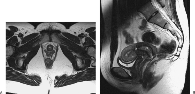
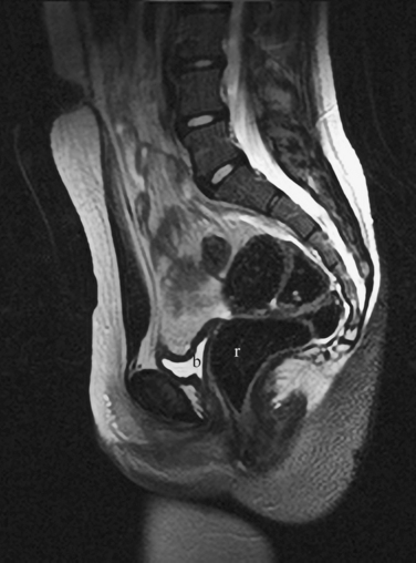
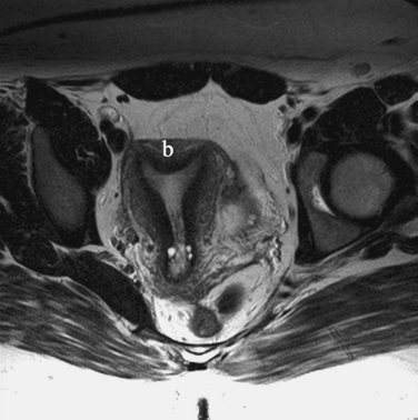
The normal uterus, cervix, vagina, endometrium, and ovaries can be readily identified on T2-weighted images in a variety of planes. Also the plicae palmitae, the anterior and posterior fornix, the cul-de-sac, rectum, urinary bladder, and urethra are easily identified (Fig. 31-1). The uterine MRI anatomy can be subdivided. The signal intensity and thickness of these layers can vary based on the hormonal milieu. The myometrium is seen externally and tends to be intermediate in signal intensity. Centrally is the endometrium, which tends to be of increased signal on T2-weighted sequences. Between the two layers is a much darker, uniform layer, called the junctional zone. Although histologically similar to the adjacent myometrium, the difference in signal intensity is believed to result from a lower water content relative to the adjacent myometrium3 and the compact longitudinally oriented smooth muscle bundles oriented parallel to the endometrium4 (see Fig. 31-1). The radiologist, when assessing the uterus for pathology, evaluates the thickness, signal characteristics, and distinctness of each of these zones.


CONGENITAL ANOMALIES OF THE REPRODUCTIVE TRACT


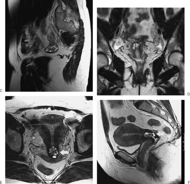
The MRI appearance of müllerian anomalies directly corresponds to the findings on both hysterosalpingography and gross examination. Several potential anatomic observations can be made. In cases of uterine agenesis, no vaginal or uterine tissue is present (Fig. 31-2).


The arcuate uterus is a frequent müllerian anomaly that usually has little clinical significance and is identified as a contour or “bulge” of the fundus, with no changes associated with the endometrial canal (Fig. 31-3).


Several anomalies are easily determined by MRI, such as a unicornuate uterus where a single horn can be identified (Fig. 31-4). However, others are more difficult, especially when the decision of whether to proceed to surgery relies on the MRI diagnosis.


Stay updated, free articles. Join our Telegram channel

Aug 27, 2016 | Posted by in UROLOGY | Comments Off on Magnetic Resonance Imaging

Full access? Get Clinical Tree

Get Clinical Tree app for offline access