■ Both arms are tucked to the patient’s side with the thumbs facing up. This allows the surgeon, assistant, and camera driver plenty of room to maneuver during the case.
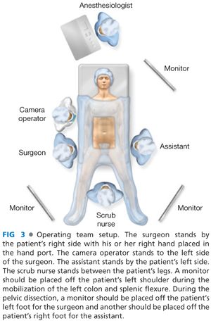
■ A monitor should be placed off the patient’s left shoulder during the mobilization of the left colon and splenic flexure. During the pelvic dissection, a monitor should be placed off the patient’s left foot for the surgeon and another should be placed off the patient’s right foot for the assistant.
TECHNIQUES
PORT PLACEMENT AND OPERATIVE TEAM SETUP
■ There are several options for the position of the hand port:
■ The hand port can be placed through either a Pfannenstiel or a midline incision. The suprapubic position allows for direct visualization into the pelvis. The hand port can then be used to facilitate the rectal dissection, for division of the distal rectum, for the performance of the anastomosis, and to address any pelvic complications such as bleeding or anastomotic failure.
■ The periumbilical position allows the surgeon to put his or her nondominant hand through the hand port.
■ A left lower quadrant (LLQ) position uses a muscle-splitting incision and allows for the right hand to be placed into the abdomen to facilitate the lateral and splenic flexure mobilizations of the left colon.
■ For the purposes of this chapter, the suprapubic hand port position is discussed (FIG 2).
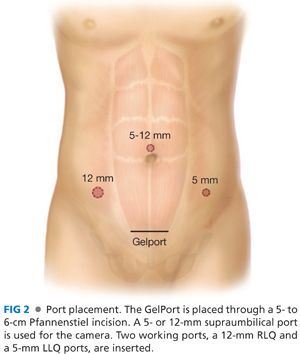
■ The 5-mm or 12-mm camera port is placed in the supraumbilical position. The camera needs to be above the umbilicus, as the wound protector portion of the hand port extends several centimeters beyond the edges of the incision.
■ The primary laparoscopic working port (12-mm port) is placed in the right lower quadrant (RLQ), at an equal distance between the hand port and the camera port and lateral to the rectus muscle.
■ A 5-mm working port for the first assistant is placed in the LLQ. This will allow the assistant to help with the lateral and splenic flexure mobilization and the pelvic dissection. The lower the port is placed, the less time the assistant works in a reverse motion to the camera during the lateral and splenic flexure mobilization.
■ The surgeon stands by the patient’s right side with his or her right hand placed in the hand port. The camera operator stands to the left side of the surgeon. The assistant stands by the patient’s left side (FIG 3).
TRANSECTION OF THE INFERIOR MESENTERIC ARTERY
■ The patient is placed in a steep Trendelenburg position and in airplane position with the left side up to use gravity to place the small bowel in the right upper quadrant (RUQ) and the omentum in the upper abdomen to expose the transverse colon and splenic flexure. This helps to expose the inferior mesenteric artery (IMA) at its origin off the aorta and the inferior mesenteric vein (IMV) at the level of the ligament of Treitz.
■ The surgeon’s right hand is placed through the hand port and an energy source is placed through the RLQ working port.
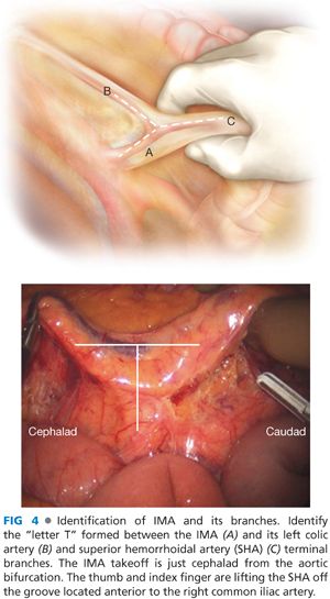
■ The retroperitoneum is accessed at the level of the sacral promontory. The superior rectal artery is grasped and elevated (FIG 4). A wide incision is made in the peritoneum dorsal to this artery; the wider the incision, the more the artery can be more elevated to obtain better exposure. Because of the curve of the pelvis at this point, the sigmoid mesentery curves up and away from the visual field. Therefore, the retroperitoneal plane is higher than expected, so the more mobile the arterial pedicle is, the easier it is to visualize the correct plane.
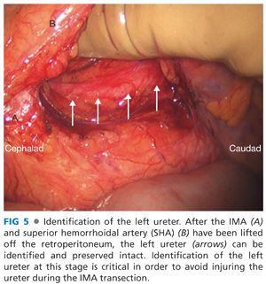
■ Identification of the left ureter is necessary before the IMA can be ligated (FIG 5). The following text is a four-step algorithm to identify the left ureter.
■ Mobilization of the superior rectal artery is as described earlier and the ureter is identified.
■ At the level of the IMV: The IMV is grasped and elevated. The peritoneum is incised dorsal to the IMV and the retroperitoneum is accessed. The retroperitoneum is flat in this area and is often more easily accessed. Once in the correct plane, the dissection is carried in a caudad fashion to meet up with the initial plane under the superior rectal artery.
■ If the ureter is still not identified, the sigmoid and left colon is mobilized in a lateral to medial fashion.
■ Finally, the top of the hand port can be removed and the left ureter can be located via an open fashion.
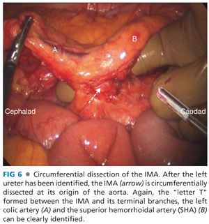
■ After the left ureter is identified and swept into the retroperitoneum, the IMA can be isolated at its origin (FIG 6). The index finger elevates the superior rectal artery and the middle finger is used to sweep down the retroperitoneum along the course of the IMA. This motion continues until the bare area is exposed cephalad to the IMA and medial to the IMV.
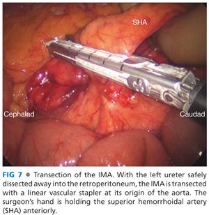
■ It is important to sweep down the retroperitoneal tissue in this area to help preserve the sympathetic plexus around the IMA. Once the IMA is safely isolated and the left ureter is clearly out of harm’s way, the vascular pedicle can be ligated at its origin from the aorta with the surgeon’s energy source of choice or with a linear stapler with a vascular cartridge (FIG 7).
TRANSECTION OF THE INFERIOR MESENTERIC VEIN
■ The IMV courses parallel to the left colic artery. The previous IMA dissection plane is carried cephalad with Endo Shears and 5-mm energy device (sweeping the retroperitoneal tissues dorsally) until the left colic artery separates from the IMV as it courses toward the splenic flexure at the level of the ligament of Treitz.
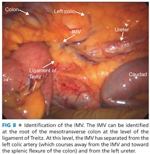
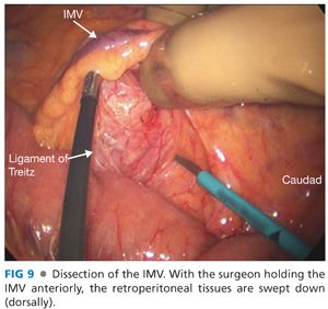
■ Now that the IMV is elevated off the retroperitoneum, it is isolated at the inferior border of the pancreas and near the ligament of Treitz (FIG 8). It can be isolated with the same technique used for the IMA: The index finger and thumb elevate and create tension on the IMV and the middle finger and/or dissecting instrument sweeps the retroperitoneum dorsally along the course of the vein (FIG 9).
■ A bare area is then created near the inferior border of the pancreas that allows the IMV to be safely isolated.
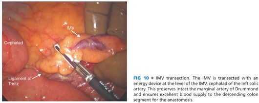
■ Once isolated, the IMV can be safely transected with an energy device (FIG 10). The IMV should be transected cephalad to the left colic artery in order to preserve the marginal artery blood supply to the descending colon intact.
MOBILIZATION OF THE LEFT COLON
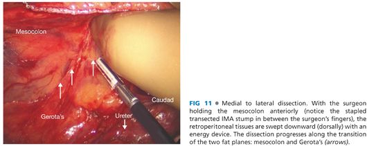
■ The left colon mesentery is now dissected off the retroperitoneum using a medial to lateral dissection approach (FIG 11) all the way out to the lateral abdominal wall.
■
Related posts:
 End and Diverting Loop Ileostomies: Creation and Reversal
 Parastomal Hernia
 Surgical Management of Complicated Diverticulitis: Perforation and Colovesical Fistula
 Low Anterior Resection and Total Mesorectal Excision/Coloanal Anastomosis: Open Technique
 Hand-Assisted Laparoscopic Abdominoperineal Resection
 Total Abdominal Colectomy: Laparoscopic Technique
Stay updated, free articles. Join our Telegram channel

Full access? Get Clinical Tree


