Fig. 16.1
Transverse sonogram of the liver, the arrows indicate a hypoechoic nodule due to sarcoidosis (Reprinted with permission from Kessler et al. [6])
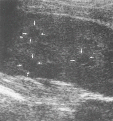
Fig. 16.2
Splenic US, multiple hypoechoic focal lesion, each less than 1 cm in diameter are shown by the arrows (Reprinted with permission from Kessler et al. [6])
Confluent granulomas forming tumorlike nodules (“sarcoidoma”) appear as space-occupying lesions and must be further differentiated by biopsy. Enlarged abdominal lymph nodes occur in up to 30 % of cases with abdominal sarcoidosis [6, 7].
MRI appears to demonstrate hepatic nonspecific abnormalities in sarcoid patients more sensitively than ultrasound [6].
CT scan can show diffuse hepatosplenomegaly and has detected focal hepatic lesions in 38 % of patients with proved hepatic involvement [6, 8].
In uncomplicated hepatic sarcoidosis, CT and MRI do not add significant diagnostic information to ultrasound findings.
Liver biopsy is the method of choice to demonstrate hepatic granulomas. If granulomas are present on the liver surface, they appear laparoscopically as small (≤2 mm) and well-delineated whitish nodules. Even if no granulomas are seen laparoscopically, liver biopsy usually will yield positive results. Although hepatic nodules are not specific for sarcoidosis, it should be considered among the possible etiologies in appropriate patients.
Histology: Sarcoid granulomas are epithelioid and noncaseating. Occasionally some granulomas may show a central fibrinoid necrosis which must not be mistaken for caseation. Granulomas are spread diffusely throughout the liver and preferentially located in the portal/periportal areas. Usually all granulomas are approximately the same developmental stage. Early acinar granulomas are composed of relatively few loosely assembled epithelioid cells, while mature cells, while mature lesions are sharply rounded and may also coalesce to form granulomatous aggregates.
Large confluent granulomas may form pseudotumors. Involvement of central veins or portal venous branches may cause granulomatous phlebitis. Nodular regenerative hyperplasia, probably due to the disturbance of microcirculation, develops in 6 % of cases. Healing of large granulomas by fibrosis may lead to nodular transformation of the liver with consequent portal hypertension [9–11].
16.1.5 Therapy
The presence alone of granulomas in an organ in sarcoidosis does not dictate treatment.
The decision to treat should be based on symptoms and severity of disease.
Although hepatic involvement usually is asymptomatic, a minority of patients progress to chronic cholestatic disease, portal hypertension, and cirrhosis that may require liver transplantation. Treatment of hepatic sarcoidosis should be reserved for patients who manifest this spectrum of disease.
Glucocorticoid treatment is the first-line therapy for hepatic sarcoidosis, improving symptoms and abnormal laboratory values but generally having no effect on the progression of disease.
In addition to glucocorticoids, immunosuppressive agents such as azathioprine, methotrexate, hydroxychloroquine, and infliximab have been used with some positive effects on symptoms, liver enzyme abnormalities, and hepatomegaly, but none has been shown to prevent progression of disease (Table 16.1).
Table 16.1
Drugs used in the treatment of severe hepatic sarcoidosis
Prednisone | 40 mg/day |
Methotrexate | 15 mg qw |
Azathioprine | 20–200 mg/day |
Chloroquine phosphate | 200–400 mg/day |
Ursodeoxycholic acid | 10–15 mg/kg body weight/day |
Ultimately, in case of overt liver failure, liver transplantation is the definitive treatment [4].
16.2 Hepatic Endometrioma
Endometriosis is a condition characterized by the presence and proliferation of endometrial tissue outside the uterus. Ectopic endometrium has been described in almost every location of the female and even in the male body. The only organ in the abdominal cavity that is apparently refractory to the disease is the spleen [12, 13].
Hepatic endometriosis has an extremely rare occurrence characterized by the presence of ectopic endometrium in the liver. At present only 15 cases of hepatic endometriosis have been previously reported in literature [14].
16.2.1 Clinical Presentation
Abdominal pain is the main symptom, even not related to menses. The physical exam can reveal no palpable mass.
The gross and microscopic pathological appearances of endometriosis vary widely depending on the location, extent, age, and endocrine response of lesion. The appearance of endometriotic tissue depends on the degree of its response to the normal hormonal fluctuation of the menstrual cycle. Endometriotic foci may enlarge to produce nodules, cysts, or both.
Because of a wide range of morphological features of endometriomas, there are no characteristic findings with which to distinguish either pelvic or extrapelvic endometriosis from other processes; therefore, clinical history is important for proper diagnosis [15].
Endometriomas typically have a fibrotic wall of variable thickness and are more commonly covered by dense fibrous adhesions that may result in fixation to adjacent structures. The wall structure has been described as smooth, membranous, ragged, or undulating at imaging or morphological examination; the content may be cystic (mostly) or heterogeneous, frequently including septation and/or punctate calcification of the wall. The location of the hepatic lesion has been described as completely intrahepatic and intraparenchymal as well as peripheral and not completely intraparenchymal at surgery [16].
Computed tomography scan can show a well-limited hypovascularized cystic mass with hemorrhagic contents in the lumen, with or without septation [14] (Fig. 16.3).
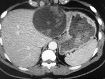
Fig. 16.3
Hepatic CT scan showing a cystic endometriotic mass in the left lobe of the liver (Reprinted with permission from Rivkine et al. [14])
At MRI, endometrial implants usually demonstrate signal intensity similar to that of normal endometrium on T1- ant T2-weighted images. However, because endometrial implants can exhibit various degrees of hemorrhage due to hormonal stimulation, implants may demonstrate a spectrum of appearances depending on the age of hemorrhage [17].
Hepatic endometriosis can be added to the differential diagnosis list of peripherally located cystic or heterogeneous liver mass with septations and wall structure in women, with or without pelvic endometriosis. The radiologist must be aware that the appearance of endometriotic tissue can vary depending on hormonal response, especially on MR.
A diagnosis of hepatic endometriosis is established after surgery. The diagnosis is made according to a histological examination of the whole surgical sample.
16.2.2 Therapy
Some authors propose the systematic use of intraoperative frozen sections to avoid radical hepatectomy in order to decrease morbidity and mortality.
A diagnosis of endometriosis must be considered in the differential diagnosis of cystic liver masses, particularly in patients with known endometriosis.
16.3 Cystic Lymphangioma of the Liver
Lymphangiomas are congenital malformations of the lymphatic system.
Related posts:


Stay updated, free articles. Join our Telegram channel

Full access? Get Clinical Tree


