(1)
Department of Gastrointestinal Surgery, Kameda Medical Center, Kamogawa, Japan
Keywords
Laparoscopic right colectomyFascial compositionFusion fasciaSubperitoneal fascia6.1 Introduction
In the laparoscopic right colectomy (LapRC) surgical technique (including ileocaecal resection), it is easy to understand the fascial composition based on the medial-retroperitoneal approach, and thus the surgical technique becomes evident. Our laparoscopic right colectomy surgeries have a general rule that the procedures involving division of the mesentery, ligation of vessels, and anastomosis are to be performed under small auxiliary laparotomy of the epigastric region due to its “rapidity” and “safety”.
6.2 Resection Range and Degree of Lymph Node Dissection
As has already been described in the Chap. 1, since the concept of the surgical trunk by Gillot [1] (Fig. 6.1) was first introduced in Japan in the 1970s, two main views are prevalent with regards to lymph node dissection for right colon cancer. Namely, lymph node dissection of the main lymph node based on the dominant artery root and lymph node dissection of the surgical trunk based on the lymph flow of Gillot [2]. No definitive conclusions have been reached as to which is more correct; however, the former calls for the dissection of portions where the lymph flow is missing, while in the latter the theory of lymph flow is only emphasised and whether the dissection of all this part is actually clinically meaningful has not been determined.
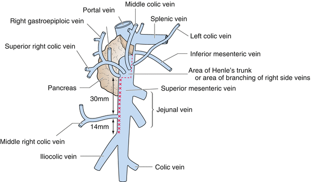

Fig. 6.1
The concept of surgical trunk by Gillot. The surgical trunk refers to an approximately 44-mm section from the cranial side of the ileocolic vein to the most caudal side of Henle’s trunk. Lymph nodes are present in the ventral and lateral side of the superior mesenteric vein
6.3 Fascial Composition of the Right-Side Colon
As for the configuration of the right colon, it can be considered as divided into a simple fusion fascia between the colon and retroperitoneum at the caudal side (Fig. 6.2) and a relatively confused fusion fascia at the cranial pancreatoduodenal portion (Fig. 6.3). In the former, the right colon and retroperitoneum are fused to become the right fusion fascia of Toldt and the ascending colon is buried in the retroperitoneum (Fig. 6.2a, b). In the latter, it is easy to understand the fascial composition if the embryologic processes are divided into two stages (Fig. 6.3a, b). That is, the second portion of the duodenum that was accompanied by a dorsal mesentery falls to the right, forming the posterior pancreatic fascia of Treitz between the parietal peritoneum and the pancreatoduodenal region (Fig. 6.3c). Then, the ascending colon covers the head of the pancreatoduodenal region when the intestinal rotation is completed, and it forms the right fusion fascial of Toldt and the anterior pancreatic fascia (Fig. 6.3e). In other words, the right fusion fascia of Toldt is divided into the posterior pancreatic fascia of Treitz dorsally and the anterior pancreatic fascia ventrally at the margin of the second portion of the duodenum (Fig. 6.3e). Additionally, the hepatic flexure is located at the transition of the transverse colon with the mesocolon and the ascending colon, in which the mesocolon is fused with the posterior abdominal wall.
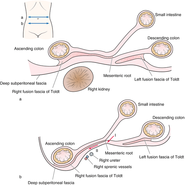
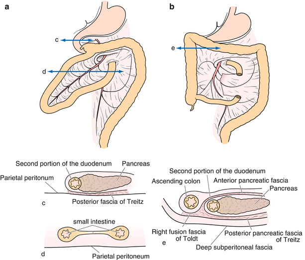

Fig. 6.2
Fusion and its cross-section with the ascending mesocolon (a, b). The right colon and retroperitoneums are fused to become the right fusion fascia of Toldt and the ascending colon is buried in the retroperitoneum (b). Arrow I indicates the first incisional position of the retroperitoneal approach. Arrow II indicates a dissecting layer between the fusion fascia of Toldt and the deep subperitoneal fascia (b)

Fig. 6.3
Intestinal rotation and the relationship of each mesenterium. Originally, two sheets of the dorsal mesentery are rotated around the superior mesenteric artery. It is easy to understand the fascial composition of the pancreatoduodenal portion if the embryological process is divided into two stages (a, b). That is, the second portion of the duodenum that was accompanied by a dorsal mesentery falls to the right, forming the posterior pancreatic fascia of Treitz between the parietal peritoneum and the pancreatoduodenal region (c). Then, the ascending colon covers the head of the pancreatoduodenal region when the intestinal rotation is completed, and it forms the right fusion fascia of Toldt and the anterior pancreatic fascia (e)
In addition, with regards to the relation between the transverse colon and the second portion of the duodenum, three fusion patterns can be considered in three degrees of fusion. That is, there are fusions between the ventral leaf of the transverse colon and about half of the second portion of the duodenum (Fig. 6.4a), between the ventral leaf of the transverse colon and the entire second portion of the duodenum (Fig. 6.4b), and almost no fusion (Fig. 6.4c)
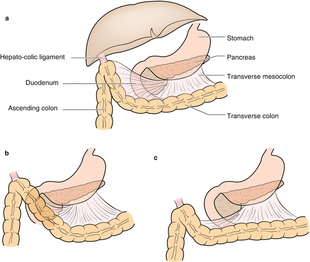

Fig. 6.4
Fusion between the transverse colon and the duodenum. Three fusions can be considered in three degrees of the fusion. That is, cases can be divided where there is a fusion between the ventral leaf of the transverse colon and about half of the second portion of the duodenum (a), between the ventral leaf of the transverse colon and the half second portion of the duodenum (b), and almost no fusion (c)
In order to dissect the right colon from the pancreaticoduodenum, the relationship between the anterior pancreatic fascia, the posterior pancreatic fascia of Treitz, and the right fusion fascia of Toldt, and between the transverse mesocolon and the anterior pancreatic fascia must be considered. In right colon cancer, in order to dissect the colon from the retroperitoneum, it is necessary to dissect between the right fusion fascia of Toldt and the deep subperitoneal fascia. However, since the right colon is depressed on its dorsal side compared with the left colon, the lateral approach of the right colon tends to be a difficult and uncertain procedure (Fig. 6.2b). The right fusion fascia of Toldt is close to the deep subperitoneal fascia in the paracolic gutter of the lateral of the ascending colon (Fig. 6.2b). Thus, if one attempts to dissect the colon from this side, there is a tendency to enter the dorsal side of the deep subperitoneal fascia, and place the urinary tract and the spermatic vessels on the discharge side, and be in danger of lifting the right kidney. In contrast, it is easier to dissect between the right fusion fascia of Toldt and the deep subperitoneal fascia along the mesenteric root because this region is rich in fat and connective tissue (Fig. 6.2b I, II). In addition, since the deep subperitoneal fascia in the pancreaticoduodenal section continues onto the dorsal side of the posterior pancreatic fascia of Treitz behind the pancreas head, dissection of the right colon from the dorsal side is easy provided that the ventral side of the deep subperitoneal fascia is maintained (Fig. 6.3e). As the dissection is continued using this approach, the right colon can be dissected freely without any damage to the other organs because the urinary tract and the spermatic vessels are now located on the dorsal side of the deep subperitoneal fascia. From the above discussion, it can be deduced that laparoscopic right colectomy is recommended to be performed using the medial-retroperitoneal approach.
6.4 Operative Procedure (in Men)
The medial-retroperitoneal approach that precedes the dissection between the right fusion fascia of Toldt and the deep subperitoneal fascia is a basic procedure. This surgical procedure takes advantage of laparoscopic surgery as it may be easier to dissect the colon from the retroperitoneum and this may mobilise the colon earlier.
6.4.1 Incision of the Peritoneum in the Mesenteric Root
Scope: umbilicus
The operator’s right hand: an electrosurgical knife with spatula-type blade
The operator’s left hand: provides a surgical plane where the dissection is to be performed and divides in countertraction together with the assistant. Usage of bowel forceps
The assistant’s right hand: grips the root of the appendix or the caecum with bowel forceps.
The assistant’s left hand: grasps the terminal ileum or the mesentery of the small intestine with bowel forceps to orient the surface towards the operator in countertraction with the right hand.
The patient lies in the head-down position on the operating table. The scope is inserted through the umbilical port. The assistant lifts the mesentery of the small intestine to the ventral side with two forceps to establish a surgical plane for the mesentery and to prevent the small intestine from entering the operative field (Fig. 6.5). The operation begins with the incision of the peritoneum at the base of the mesentery of the small intestine near the second portion of the duodenum moving from the cranial-medial side to the caudal-lateral side with a spatula-type electrosurgical knife (Fig. 6.5, arrow I, II). Along this course, as the deep subperitoneal fascia with the retroperitoneum is lifted ventrally, the incision should be performed for several centimetres along the ventral side (Fig. 6.6a) and the deep subperitoneal fascia should be dissected towards the dorsal side from the fusion fascia of Toldt (Fig. 6.6b).
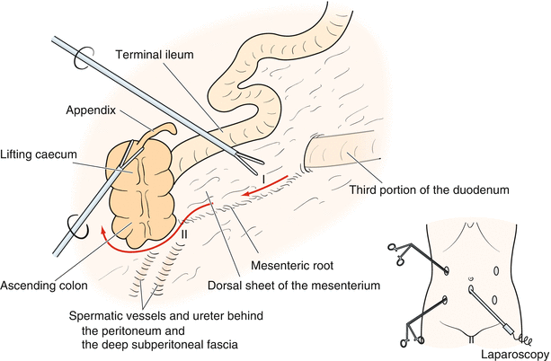
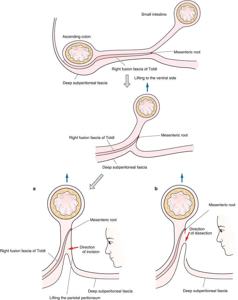

Fig. 6.5
Cutting and dissection at the base of the mesentery of the small intestine. The assistant lifts the mesentery of the small intestine to the ventral side using two forceps. The operation can prevent the small intestine from entering into the surgical field, as it is possible to secure a good field of vision. The operation begins with the incision of the peritoneum at the base of the mesentery of the small intestine near the second portion of the duodenum moving from the cranial-medial side to the caudal-lateral side with an electrosurgical knife with spatula-type blade

Fig. 6.6
Appearance and interpretation of the incision and dissection line. The deep subperitoneal fascia with the retroperitoneum is lifted to the ventral side and the incision is performed along several centimetres along the ventral side (a). The deep subperitoneal fascia should be dissected from the fusion fascia of Toldt towards the dorsal side (b)
If the duodenum cannot be seen through the fascia from the retroperitoneum, it may be easier to direct the mobilisation of the cecum to the cranial side. In this case, however, it is necessary to be careful not to proceed further into deeper layers. Therefore, it is important to dissect the fascia dorsally while correcting the fascia composition of the caecal wall that is being lifted ventrally. Lifting the caecum and the terminal ileum, the dissection is performed just dorsal of the right fusion fascia of Toldt and slightly enough cranially (Figs. 6.2b II and 6.5 II).
Stay updated, free articles. Join our Telegram channel

Full access? Get Clinical Tree


