(1)
Department of Gastrointestinal Surgery, Kameda Medical Center, Kamogawa, Japan
Keywords
Colorectal cancerFascial compositionFusion fasciaVascular anatomy of the colon1.1 Introduction
Laparoscopic surgery is generally performed for digestive diseases. Laparoscopic procedures are requested particularly for patients with colorectal cancer. Given the fine vision currently available to surgeons with the development of advanced laparoscopic surgery systems, an improved understanding of the fascial composition of the abdomen is necessary. Modern surgical procedures of the colon and the rectum have thus required surgeons to revise their understanding of the surgical anatomy of this region. Complications can be reduced by careful dissection of the correct tissue plane in the abdominal cavity. However, descriptions of the anatomy of the fascial composition have also involved observations that are unrelated to better-known fundamental embryological concepts, causing confusion in the explanation of operative procedures since various terms are often used without a specific definition. A better understanding of the fascial composition is useful not only for the laparoscopic approach but also for open surgery procedures.
1.2 Definition of the Terminology—Dissection, Cutting, Fusion, and Adhesion
In laparoscopic surgery, visual, tactile, and motor skills are limited compared to open surgery or laparotomy. The basic techniques of digestive surgery include dissecting and cutting. To overcome these limits to dissecting or cutting away, the selection of the body posture and the position of the port are important in laparoscopy. The use of a 30° view angle laparoscopy or laparoscopy with a flexible tip enables a multidirectional observation of the surgical field as in laparotomy. With a close view of the operative field, dissection and disconnection also become more precise procedures. Conversely, the close operative field narrows the operative view, and reduces dissections to one small stroke, inevitably increasing the time required for an operation. For these reasons, dissection of the adhesion can be performed using an electrosurgical knife, but an ultrasonically activated device (USAD) is required for dissection with haemostasis.
1.2.1 The Concept of Dissection and Cutting
The aim of surgical treatment is to distinguish between the parts to be preserved and those to be resected, and to leave the former and remove the latter. It follows that the understanding of the fascial composition is of utmost importance. Especially in radical surgical treatment for malignant disease, it is necessary to distinguish the boundaries of the tissue to be removed from those to be retained. Resecting along layers that are reasonably and strictly defined is called “dissection”. In particular, the dissection of lymphatic vessels and lymph nodes is referred to as “lymphatic dissection”.
The boundaries that are strictly defined are represented as single thin lines. If a line consists of a thick width, the resection procedure along the boundary must be defined as “cutting” or “dividing” rather than “dissection” [1].
1.2.2 The Difference Between Adhesion and Fusion
Adhesion and fusion are common terms in digestive surgery, and the difference between them in clinical anatomy must be clearly understood. Resection in the gastrointestinal tract is possible if we dissect the dorsal side of the fusion fascia in accordance with the embryonic fascial anatomy. Therefore, if the concept of fusion fascia is not understood, it will be difficult to understand the clinical anatomy and the surgical procedure will not proceed smoothly. Of course, this does not lead to better surgical education.
Here, we must reconfirm the definition of the word “fascia”. This is because there are many frequent errors in the usage of the terms fascial configuration and fascial anatomy. First, fascia is a term applied to masses of connective tissue large enough to be visible to the unaided eye. Such structures are highly variable, but, in general, collagen fibres in the fascia tend to be interwoven and seldom show the compact, parallel orientation seen in tendons and aponeuroses [2, 3]. Furthermore, “fascia is not necessarily intended to cover the muscles. It is those that wrap the surface of other organs (such as the glands) and those that form the membrane as a partition of the loose connective tissue. In addition, the sheath that may be seen around the thick blood vessels also can be described as a kind of fascia” [4].
Frequently used as a reference textbook of clinical anatomy is the French, Cahiers D’Anatomie Abdomen (I) (L. Perlemuter and J. Waligora) [5]. In many of the figures in this book, there are arrows indicating the planes of fusion fascia. For example, two arrows in Fig. 1.1 show the left fusion fascia of Toldt, but these arrows have given rise to misunderstandings regarding the dissecting plane. By definition “In fusion fascia, the mobility of the adjacent serosa is lost and these eventually fuse into a single sheet of connective tissue” [4, 7, 8], the interior of the fusion fascia cannot be dissected. In addition, it is important to understand the difference between fusion and adhesion. Although sometimes the term “physiological adhesion” is used, it is ambiguous whether the word means fusion or pathological adhesion. Thus, the term will not be used herein.
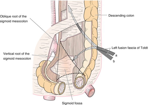
Fig. 1.1
Fascial composition and fusion fascia of the sigmoid colon. Although these arrows do not show signs indicating dissection, they indicate a plane where the left fusion fascia has been formed [6, with permission]
The fascia structure present along the body wall is embryologically represented by the term superficial or deep, defined in relation to the fascia of the skin. The terms anterior and posterior are not used because of their arbitrariness. Thus, the terms ventral, dorsal, left, right, cranial, and caudal are used to define direction and location. These can be expressed definitively irrespective of the direction of the patient. In addition, as a note about the use of directional terms, if there is an anatomical term defined as “ventral”, there must also be a “dorsal” side. In addition, with respect to the term “dissection”, only two possibilities exist: either dissection between the two fasciae or dissection of the ventral or dorsal side of one fascia. Lymphatic dissection refers to cutting the border of the lymphatic vessels and lymph nodes that lie sandwiched between two or more fasciae, which have been dissected.
1.2.3 The Concept of Dissection in Endoscopic Surgery
The structure of the original gastrointestinal tract is three-dimensional, but laparoscopic surgery is two-dimensional, and dissection is carried out in a one-dimensional plane. To proceed with dissection along a one-dimensional line, it is necessary to obtain a suitable dissecting surface by creating tension from the coordinated movement of the operator’s left hand and the assistant’s hands. In laparoscopic surgery, as the lines can be clearly visible, it is possible to perform an accurate dissecting procedure. Therefore, the most important thing in performing dissecting manoeuvres in laparoscopic surgery is to visually identify the dissecting layer. Thus, by finding the correct dissecting layer and the dissecting line, it is possible to enter the correct layers using any device. It is possible to find the boundary between the fusion fascia and another fascia. Dissecting the correct layer should not require any haemostasis. In this context, the understanding of the fascia configuration is of fundamental importance.
1.3 Peritoneal Configuration, Body Wall, and Intestinal Rotation in Foetal Life
The basic structure of the peritoneal configuration and body walls in foetal life is shown in Fig. 1.2. A basic understanding of the fascial composition of the body circumference and its interpretations has been provided by Tobin et al. [9] and Sato [10]. According to these interpretations, the structure of the body below the diaphragm can be simplified as a straight intestine within a cylindrical body. Basically, the body composition can then be divided into the composition of the peritoneal cavity (consisting of the cylinder) and the composition of the body wall (consisting of the cylinder wall). In the former, the cranial abdomen includes the dorsal and ventral mesentery, and the caudal abdomen includes only the dorsal mesentery involving the intestine. The latter has a ringed composition, and the body walls are symmetrical in relation to the central position of the muscle layer (Fig. 1.2). The trunk has typically been regarded as an onion-like multi-layered structure [11].
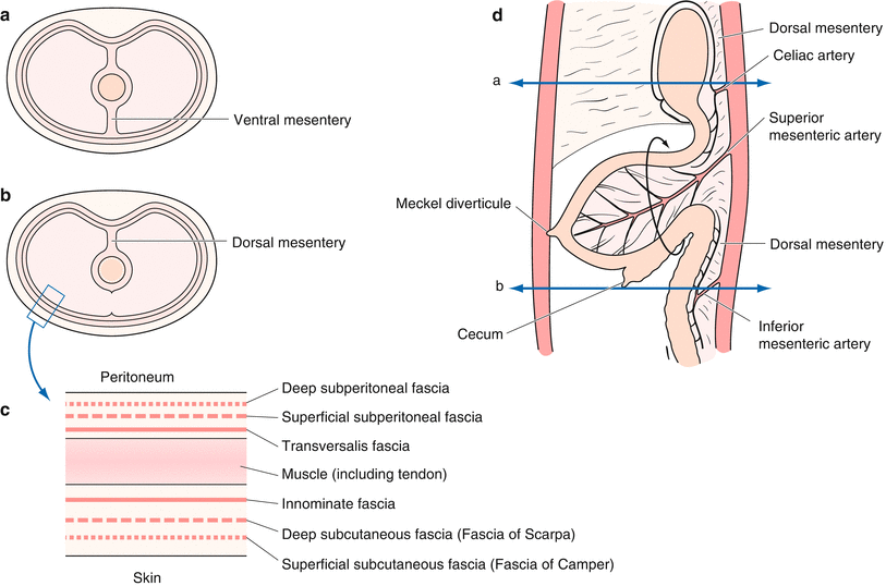
Fig. 1.2
Peritoneal configuration, body wall, and intestinal rotation in foetal life. According to the interpretations of Tobin et al. and Sato, the internal structure of the body can be simplified as a straight intestine within a cylindrical body (a, b). The basis of body composition can then be divided into the composition of the peritoneal cavity and the composition of the body wall. The body walls are symmetrical relative to the central position of the muscle layer (c) [6, with permission]
The fascial structure present in the body wall is embryologically represented by the term superficial or deep, defined in relation to the superficial skin surface. The terms anterior and posterior are not used because of their arbitrariness. Analogously to the subcutaneous superficial fascia and subcutaneous deep fascia, the deep subperitoneal fascia and superficial subperitoneal fascia exist circumferentially around the abdominal wall (Fig. 1.2). It is important that the superficial subperitoneal fascia and the deep subperitoneal fascia are independent of each other (Fig. 1.2c) and the two fasciae sandwich the aorta from ventral and dorsal sides directly toward the pelvic space.
Unless the continuity of the two subperitoneal fasciae to the pelvic space is guaranteed, this basic configuration (Fig. 1.2) also becomes imperfect. Therefore, to examine the fascial composition in the pelvic space, the terms deep subperitoneal fascia and superficial subperitoneal fascia will be used herein to refer to the pelvic space in consideration of its continuity from the peritoneal cavity. Unless the continuity of the two subperitoneal fasciae is guaranteed, the textbook of surgical procedures also becomes imperfect.
1.4 Intestinal Rotation and Peritoneum
With regards to the relationship between intestinal rotation and each mesentery, the dorsal mesentery (Fig. 1.2) rotates around the superior mesenteric artery (SMA) (Fig. 1.3) [13]. The concept of fusion fascia is thus indispensable when considering the relationship between the peritoneum and the mesentery at the terminal end of rotation of the intestine. After fusion of the bilateral mesentery with the peritoneum, the fixation of the colon to the retroperitoneum is complete.
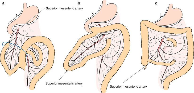
Fig. 1.3
Intestinal rotation and fusion. The dorsal mesentery is rotated around the superior mesenteric artery (SMA) (a, b). The fusion is formed at the end of the intestinal rotation and most of the colon is fixed to the retroperitoneum (c). The SMA is thus defined as the artery that heads to the ileum, 50–100 cm from the terminal ileum. The blue solid line indicates the direction of the intestinal rotation [12, with permission]
When we dissect the plane between the fusion fascia of the colon and the deep subperitoneal fascia, it is often possible to see another fascia, for example in the ascending colon or in sigmoid colon surgery. The mesentery of the colon may not necessarily be constituted by two layers. The peritoneum is the widest among the serosa of the body and is divided into the parietal peritoneum and visceral peritoneum. The parietal peritoneum covers the abdominal cavity, the pelvic cavity, and the diaphragm surface. The visceral peritoneum covers the internal organs of the abdomen and pelvis, and is generally considered to also include the mesentery. Originally, the peritoneum completely covers the entire intestinal tract. The mesentery, including the parietal peritoneum, is made of a fibrous layer (the tunica subserosa) and a surface layer of mesothelium (tunica serosa) [14]. The deep subperitoneal fascia is extended to the colon wall through the dorsal mesentery (Fig. 1.4) [10]. Thus, it may be natural that the mesentery is composed of four fasciae. These features are important in the clinical anatomy of the dissection at the hepatic flexure of the right hemicolectomy and for the dissection of the dorsal side at the Denonvilliers’ fascia. The fascia seen on dissection should not be unconditionally believed to be artifacts resulting from surgical interventions. However, what is important is not the number of fasciae, but that the fasciae are used to indicate the correct plane to be dissected.
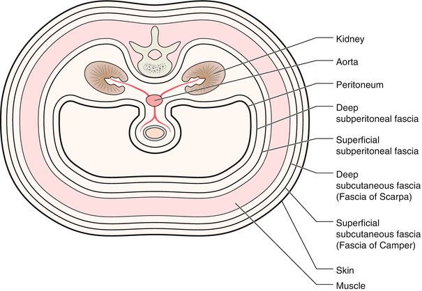
Fig. 1.4
Sato’s concept. According to this concept, the deep subperitoneal fascia covers the entire circumference through the dorsal mesentery and the vessel corridor is located between this fascia. However, if this concept were to refer to all the fields within the abdominal cavity, this concept would be unable to respect the operative views in the clinical setting
1.5 Relationship Between the Stomach and Transverse Colon—Particular Relationship at the Centre of the Transverse Colon
It is not possible to ignore the relationship between the stomach and the colon in order to understand the overall image of the colon. With regards to the description of this area, there are many errors in many existing books. To understand the boundaries for dissecting and cutting, you must understand whether the fascia belongs to the stomach or to the transverse colon in every stomach or pancreatic operation.
By applying the intestinal rotation and fusion fascia to this part, it is possible to understand its fascial composition. In other words, what is referred to as the gastro-colic ligament is the ligament stretching between the stomach and the transverse colon, which is none other than the omentum. Originally, two sheets of dorsal mesentery became four sheets and then formed the omentum that extends between the stomach and transverse colon. The third sheet of the dorsal mesentery is the posterior wall of the omental bursa, and the fourth sheet of the dorsal mesentery fused with the ventral side of the transverse mesocolon in foetal life (Fig. 1.5). The omentum is tissue belonging to the stomach, which was formed by the original dorsal mesentery (also known as the posterior gastric mesentery) that is stretched caudally. The transverse mesocolon is composed of a ventral and dorsal leaf (generally an anterior and a posterior leaf); it has no relationship at all with the stomach. The root of the transverse colon lies in the caudal edge of the pancreatic body and tail and the transverse mesocolon extends from there as a characteristic fan-shaped mesentery (Fig. 1.3). In the final process of bowel rotation, the fourth sheet of the dorsal mesentery (omentum) and the ventral leaf of the transverse mesocolon are fused (Fig. 1.5). To better understand these relationships, it helps to consider the connections between the omentum and the transverse mesocolon as if they are divided into two stages during the course of intestinal rotation. In the sagittal sectional view at the centre of the transverse colon, the omentum and mesocolon are irrelevant (Fig. 1.5a), however, the fusion fascia is formed (Fig. 1.5c).
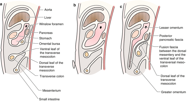
Fig. 1.5


Relationship between the stomach and the transverse colon by the upper abdominal sagittal sectional view. The omentum is tissue belonging to the stomach, which is formed by the original dorsal mesentery stretched caudally. The transverse mesocolon has ventral and dorsal leafs, which are unrelated to the stomach (a, b). However, the fourth sheet of the dorsal mesentery (omentum) and the ventral leaf of the transverse mesocolon are fused in the final stroke of the intestinal rotation (c)
Related posts:


Stay updated, free articles. Join our Telegram channel
Full access? Get Clinical Tree
