The indication for use of laparoscopy, in the pediatric population, was initially for diagnostic purposes. As confidence with the technology and utility grew, it began to be applied for therapeutic indications. With equivalent surgical outcomes and decreased morbidity, the usefulness of a laparoscopic approach became more apparent, and today minimally invasive surgery is an indispensable tool in the management of many pediatric urologic conditions. The management of renal pathologies using laparoscopy is now currently the approach of choice for most pediatric renal maladies.
Key points
- •
Laparoscopy has become a mainstay approach in the management of pediatric renal maladies.
- •
Transperitoneal and retroperitoneal laparoscopic approaches are safe and effective.
- •
Advancing technologies, such as single-site surgery and robotics, are becoming increasingly common in the management of pediatric patients.
Introduction
The indications for simple laparoscopic nephrectomy range from mitigation of renin-mediated hypertension and severe proteinuria, to removal of infected, poorly functioning renal units, or severely hydronephrotic kidneys. The use of minimally invasive surgery (MIS) further extended to malignant pathologies, hence radical laparoscopic nephrectomy is frequently offered for pediatric renal malignancies, such as Wilms tumor or renal cell carcinoma. On the other hand, although partial nephrectomy is traditionally used for management of renal malignancies in adults, its role in the pediatric population is more commonly limited to duplex kidneys with or without ureteroceles.
Historically, laparoscopic nephrectomies in pediatric and infant patients were initially described by Ehrlich and colleagues (pediatric) and Koyle and colleagues (infant) in the early 1990s. In both cases they removed a multicystic dysplastic kidney. Since that time, the general principles of transperitoneal laparoscopic nephrectomy have largely remained the same.
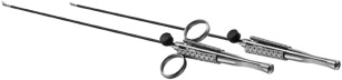
Although pediatric laparoscopy was hindered at first by the size of the access ports and the length of the instrumentation, advances in technology and development of dedicated pediatric instruments have led to fast adoption and dissemination of this surgical approach. Today, there are 3-mm to 5-mm instruments with respective optics widely available, and technical advances by some companies (eg, GIMMI [GIMMI GmbH, Tuttlingen, Germany]; Fig. 1 ) have allowed even further miniaturization to 2.7-mm instruments and optics.
Laparoscopy in children is a unique procedure when compared with adults, and the nuances of this approach in the pediatric population are not only related to size of the patients or instruments, but also pertain to the physiology. For instance, the relative distance between the access point and any intra-abdominal structures has the potential to cause significant damage as instruments are advanced into the abdominal or retroperitoneal cavity. In addition, there can be more significant physiologic effects of pneumoperitoneum on the respiratory, cardiovascular, renal, and gastrointestinal tracts, particularly at higher pressures. Surgeons embarking on incorporating laparoscopy in their practice are advised to have a clear and deep understanding of the evolving technology and the physiology of children.
Introduction
The indications for simple laparoscopic nephrectomy range from mitigation of renin-mediated hypertension and severe proteinuria, to removal of infected, poorly functioning renal units, or severely hydronephrotic kidneys. The use of minimally invasive surgery (MIS) further extended to malignant pathologies, hence radical laparoscopic nephrectomy is frequently offered for pediatric renal malignancies, such as Wilms tumor or renal cell carcinoma. On the other hand, although partial nephrectomy is traditionally used for management of renal malignancies in adults, its role in the pediatric population is more commonly limited to duplex kidneys with or without ureteroceles.
Historically, laparoscopic nephrectomies in pediatric and infant patients were initially described by Ehrlich and colleagues (pediatric) and Koyle and colleagues (infant) in the early 1990s. In both cases they removed a multicystic dysplastic kidney. Since that time, the general principles of transperitoneal laparoscopic nephrectomy have largely remained the same.
Although pediatric laparoscopy was hindered at first by the size of the access ports and the length of the instrumentation, advances in technology and development of dedicated pediatric instruments have led to fast adoption and dissemination of this surgical approach. Today, there are 3-mm to 5-mm instruments with respective optics widely available, and technical advances by some companies (eg, GIMMI [GIMMI GmbH, Tuttlingen, Germany]; Fig. 1 ) have allowed even further miniaturization to 2.7-mm instruments and optics.

Laparoscopy in children is a unique procedure when compared with adults, and the nuances of this approach in the pediatric population are not only related to size of the patients or instruments, but also pertain to the physiology. For instance, the relative distance between the access point and any intra-abdominal structures has the potential to cause significant damage as instruments are advanced into the abdominal or retroperitoneal cavity. In addition, there can be more significant physiologic effects of pneumoperitoneum on the respiratory, cardiovascular, renal, and gastrointestinal tracts, particularly at higher pressures. Surgeons embarking on incorporating laparoscopy in their practice are advised to have a clear and deep understanding of the evolving technology and the physiology of children.
Nephrectomy
Equipment
The array of laparoscopic equipment available in the market today is immense, although still far less so in the pediatric market as compared with the adult market. In general, we prefer to have a standard set up for all laparoscopic renal cases ( Table 1 ), with additional specialized equipment available on standby to be used as needed. Standardization of equipment placement and organization of the various cables and tubes is essential for safety and efficiency during the case. For instance, we recommend using a dedicated Mayo stand to keep the basic equipment in a predictable location and easily within reach.
| Case | General Equipment | Special Equipment |
|---|---|---|
| Nephrectomy or partial nephrectomy | 30° scope (3, 5, or 10 mm depending on the procedure to be performed); scissor with cautery, Maryland, bowel, and right-angle graspers; hook; suction irrigator; clip applier; specimen-retrieval bag | Laparoscopic stapler, specialized coagulation device (eg, Harmonic, Enseal, LigaSure, Thunderbeat) |
| Retroperitoneal nephrectomy or partial nephrectomy | In addition to above, narrow S-retractor or right-angle retractor |
General Positioning
Like any surgical procedure, one of the first, and most critical, steps in a laparoscopic nephrectomy is patient positioning, padding, and retention to the table. We recommend that positioning be done as a joint effort among the surgeon, anesthesiologist, and nursing staff to ensure that all aspects and potential implications of the patient position are accounted for. When performing simple nephrectomy, positioning can vary from supine to partial flank to full flank, depending on multiple factors, such as body habitus, operative side, kidney size, planned port placement, and anticipated exposure needs. In any of the positions, the break point in the bed and/or kidney rest may be used to help widen the angle between the lower ribs and pelvic brim, effectively increasing the operative space. In general, however, the benefit of the kidney rest and/or bed break is fairly limited in pediatric patients because of their small size. When a partial or full-flank position is used, the positioning of the arms and legs are critical to ensure there is no strain on the respective joints. Once positioning is established, the focus shifts toward appropriately padding pressure points. Once positioned and padded, the patient needs to be secured to the bed with sturdy tape (2–3-inch cloth tape) to provide complete security and allow the bed to be maximally deflected in any direction without risking significant shifting of the patient on the bed. Furthermore, key points that need to be secured with tape include upper and lower legs, hips, chest and shoulders, arms, and head. If simple bed movement is planned, such as mild Trendelenberg, then it is often necessary to tape only the legs and chest/shoulders. Any degree of side-to-side bed adjustment should mandate taping around the hips, arms, and head as well.
Transperitoneal simple nephrectomy
Access
When performing a transabdominal approach, we recommend open access to the peritoneal cavity via the umbilical region, with the point of entry at the fusion of the fascial layers to form the linea alba. We find this anatomic landmark to be the most predictable point of entry into the abdomen.
Once the umbilicus is everted, the lateral aspects are grasped with Allis clamps and skin is incised in a vertical transumbilical fashion using a #11 blade ( Fig. 2 ). The open umbilicus is carefully separated until the point in which the peritoneum joins the skin is seen. Once the peritoneal cavity is entered, the skin is then separated from the peritoneum as described in the Bailez technique ( [CR] ). A 2-0 absorbable suture u-stitch is placed around the peritoneal opening to facilitate traction during port placement, limit gas leakage around the port, and for use in fascial closure at the conclusion of the case. With the previously placed 2-0 Vicryl in place and before complete tightening of the suture, traction is applied, and the 5-mm or 10-mm laparoscopic trocar is placed. A 30° scope is then inserted under direct vision, and after confirming that the trocar is properly placed intraperitoneally by visualizing, bowels insufflation is started (pressure of 12 mm Hg or less dependent on the age and weight of patient with incremental flow of 1–3 L/min).
Two accessory additional ports are then placed under direct vision. Ideally the 2 working ports are placed along the ipsilateral rectus border. In case splenic or liver retraction is needed, a third accessory port can be placed in the midline just below the xiphoid. Because the abdominal cavity is small and finger pressure may completely obscure and mislead trocar site insertion, we traditionally use the following method. For each port, the target entry site is first identified using a 22-gauge needle while visualized from the intraperitoneal cavity. Once the correct site is adequately identified, local analgesia is injected followed by incision using an 11 blade through the skin, abdominal wall, and peritoneum. Using the same trajectory, a straight hemostat is then used to gently spread the fascia and thereafter the laparoscopic trocar ( [CR] ). We find that this trocar placement method limits gas leak and decreases the chance of dislodging the trocar, especially in long procedures.
Technical Details
- •
Mobilization the colon overlying the kidney while maintaining the lateral renal attachments ( [CR] ).
- •
Colon is dissected along the white line of Toldt from the splenic flexure on the left or hepatic flexure on the right.
- •
Once the ureter is identified, as it courses over the psoas muscle, it is followed cephalad to the hilum and can be used for traction.
- •
The hilum should be carefully dissected using gentle scissor or blunt dissection.
- ○
The suction irrigator also can be useful as a relatively atraumatic dissection device.
- ○
- •
Efforts should be made to isolate the artery first and vein second.
- •
Once isolated, the vessels can be ligated separately with clips ( Fig. 3 ) and hilum is secured.
Fig. 3
Prone positioning. Note the ability of the abdomen to expand without compression against the surgical table.
- •
The remaining superior and lateral attachments are divided along with the ureter.
- •
In certain circumstances and especially when the kidney is hydronephrotic, the hilum may be effaced and is composed of many small vessels that need precise dissection and clipping. Hence, caution should be exercised when dissecting the kidney superiorly or posteriorly to avoid aberrant vessels.
Specimen Removal
With small kidneys being removed for nononcologic indications, the specimen can often be removed via the umbilical incision. The 10-mm lens is exchanged for a 5-mm lens to allow visualization via one of the accessory ports. A 10-mm heavy grasper can be placed through the umbilical port and the specimen removed along with the port. If necessary, the fascial and/or skin incision can be extended to accommodate specimen retrieval. If the specimen is very large or needs to remain intact, such as with a renal tumor, it should be placed in a retrieval bag and extracted via extension of the incision of one of the ports or via an alternate incision, such as a Pfannenstiel incision, in the lower abdomen.
The resection bed should be inspected for hemostasis. Routine placement of a drain is not necessary. Closure of the umbilicus is accomplished at the fascial level by tying down the 2-0 absorbable suture placed during initial access. We do not close the 5-mm port sites and we approximate skin with absorbable suture and skin glue. We consider that the type of trocar (dilating tip) and method we use for trocar insertion to be minimally disruptive to the fascia or overlying muscles hence the risk of hernia is low. In our practice, we have not yet seen any patient presenting with a hernia from the trocar site. Nevertheless, we recommend that surgeons use their own judgment for these minor surgical details.
The key steps of the transperitoneal nephrectomy are shown in [CR] .
Comments
Although transperitoneal laparoscopic nephrectomy is popular, the retroperitoneal approach is considered a neat direct approach to tackle a benign pathology in the pediatric population. Children have minimal perinephric fat and anatomically are very suitable for a retroperitoneal laparoscopic approach.
Retroperitoneal flank approach
Retroperitoneal laparoscopic nephrectomy in children was first described by Diamond and colleagues in 1995. In this series, patients were positioned in a lateral decubitus position with access established via an incision in the costovertebral angle. Prone positioning also has been used, with access achieved in a similar fashion. Traditionally, enlargement of the retroperitoneal potential space is accomplished using balloon dilators, Foley catheters, condoms, and glove fingers, but we find a soaked wet sponge to be sufficient to create the space in children.
Positioning
When performing a retroperitoneal laparoscopic nephrectomy in the flank position, the patient’s back should be flush with the table edge, hence allowing full and free anteroposterior planes for instruments to prevent instrument collision with the surface of the bed when dissecting the renal anterior attachments.
Access
A 1-cm incision is made approximately 1 cm off the tip of the 12th rib. The muscle layers are sequentially split using sharp dissection and advancing the tips the of scissors all the way to the retroperitoneum. Once the tips of the scissors are in the retroperitoneum, narrow right-angle or “S” retractors are used to identify and confirm that the peritoneum is not violated (bowels should not be seen) and retroperitoneal space is reached (presence of perinephric fat, albeit minimal, is seen). A damp sponge is then fed through the incision and into the retroperitoneal space to begin developing the cavity. In children older than 1 year, the entire sponge can generally be inserted. In children younger than 1 year, typically only half of the sponge is needed. With the sponge in place, a 2-0 absorbable suture is placed as a u-stitch on the external fascia to provide traction and minimize gas leak. With traction on the untightened 2-0 Vicryl, a 10-mm trocar is placed into the retroperitoneum, and once perinephric tissues and fat are seen (ie, scope is not in muscle layers), then insufflation initiated. Pressure should be set at 12 mm Hg or less, with flow started at 1 L/min and gradually increased to 3 L/min.
Technical Details
- •
Dissection and creation of the retroperitoneal space is created with both blunt dissection and the pressure of direct insufflation, using the 30° laparoscope.
- •
The retroperitoneal space is further developed by moving the camera and trocar as a unit posteriorly along the psoas muscle toward the anterior abdominal wall and then medially to mobilize the peritoneum away from the surgical space.
- ○
This maneuver needs to be delicate and under vision to avoid violating the peritoneum.
- ○
- •
Once the retroperitoneal space is adequate and edge of peritoneum is medially positioned, the 2 additional 5-mm ports are placed under direct vision.
- ○
The first port is placed anterior to the paraspinous muscles and traditionally is used to further develop the retroperitoneal space by further mobilizing the peritoneum medially.
- ○
The second trocar is inserted 10 to 15 mm superior to the anterior superior iliac spine in the axillary line.
- ○
It is imperative that this anterior port is placed after sufficiently displacing the peritoneum anterior and medially. Failure to do so risks transperitoneal port insertion.
- ○
- •
Once all trocars are in place, the kidney is approached posteriorly so as to identify the renal pedicle.
- ○
A key anatomic landmark is the psoas muscle, which we use to be fully oriented to the retroperitoneal cavity.
- ○
- •
An instrument is typically needed to help lift the kidney away from the psoas muscle to allow visualization and dissection of the renal hilum.
- •
With the kidney still attached to the peritoneum anteriorly, it is customary to identify the ureter, which makes it quite easy to follow cephalad toward the hilum.
- •
Ligation of the renal vessels and ureter can be performed.
- ○
The retroperitoneal approach makes it easy to first identify the artery and ligate before visualizing the vein.
- ○
Nevertheless, it is common to again have the vessels effaced by a large hydronephrotic kidney or even small and flattened by a small shrunken renal unit, hence making it easy to miss small vessels, which may cause bleeding if not identified and adequately ligated.
- ○
Specimen Removal
As with the transperitoneal approach, the specimen either can be extracted via the larger 10-mm port site (using an alternative port with a 5-mm camera), which often has to be extended, or, if the specimen is larger, removing the kidney in pieces may be achieved. With the intracorporeal space being very small, it is usually difficult to place a retrieval bag.
The 10-mm port site can be closed at the fascial level, with the 2-0 absorbable suture placed during the retroperitoneal access. The remaining port sites are closed at the skin level only with an absorbable suture and skin glue.
Comments
Although the retroperitoneal flank approach provides a direct access to the kidney and hilum, it needs lots of dexterity and may be difficult to teach. For instance, the mere fact that dissection and hilar control may need to be done with one instrument while the other is retracting the kidney anteriorly makes it cumbersome, especially because the space is very limited and may be difficult to accommodate an extra trocar for retraction. One approach to overcome this surgical limitation is to dissect the whole kidney and then tackle the hilum with the kidney floating in the retroperitoneal space. Although this technique is a useful alternative, it may be difficult to use on all cases, especially when the kidney has aberrant multiple vessels. Thus, we recently incorporated the prone retroperitoneal approach into our surgical armamentarium. This approach is quite permissive, not only because peritoneal cavity violation does not pose any limitation on surgical exposure, but rather because it allows the use of both accessory trocars for dissection and manipulation.
Related posts:


Stay updated, free articles. Join our Telegram channel

Full access? Get Clinical Tree


