(1)
Department of Gastrointestinal Surgery, Kameda Medical Center, Kamogawa, Japan
Keywords
Laparoscopic abdominoperineal resection of the rectumCircumferential resection marginWaistingShafix’s conceptKraske operation5.1 Introduction
In the treatment of rectal cancer, the spotlight has been placed on how to preserve the anus. In fact, it has become possible to perform anal sparing surgery in many cases using intersphincteric resection of the rectum (ISR) for rectal cancer. However, the indication for laparoscopic abdominoperineal resection (LapAPR) of the rectum for rectal cancer has not diminished. The surgical technique involved requires the laparoscopic resection of the low anterior rectum. However, procedures beyond the rectosacral ligament must maintain sufficient distance from the rectum and/or the anal canal, and is defined as the circumferential resection margin.
5.2 Stoma Site Marking and Preoperative Treatment
Stoma site marking is performed together with a certified wound-ostomy-continence nurse.
5.3 Operative Procedures (in Men)
5.3.1 Treatment of the Anus
The anus is closed using two purse-string sutures with a 0-monofilament suture. The handling of the needle is necessary to fully raise the tissue using fine sutures in order to prevent leakage of stool during the operation.
5.3.2 Intraoperative Positions
The patient is placed in the lithotomy position with LevitatorTM stirrups with both thighs strapped to linearize to the trunk as much as possible. In order to improve the field of view in the anal operation, the knee is bent further, which is a modification of the Lloyd-Davies position [1]. As a result, the anus is widely accessible, and an optimal field of view of the ventral and lateral sides of the anal canal is ensured (Fig. 5.1).
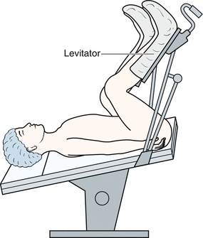
Fig. 5.1
Modified Lloyd-Davies position. The knee position is bent further, which is a modification of the Lloyd-Davies position, the anus is widely accessible, and ensures an optimal field of view of the ventral and lateral sides of the anal canal
5.3.3 Operative Procedures
See the relevant laparoscopic sigmoidectomy (LapS; Chap. 3) and laparoscopic low anterior resection (LapLAR; Chap. 4) sections for the dissection and mobilization of the sigmoid colon and the rectum.
5.3.3.1 Additional Dissection in the Lesser Pelvis (Especially in the Retrorectal Space)
Scope: umbilicus
The operator’s right hand: an electrosurgical knife with spatula-type blade
The operator’s left hand: maintains traction of the fascia propria of the rectum to the ventral-caudal side
The assistant’s right hand: assists in the fine manoeuvres of the operator
The assistant’s left hand: lifts the fascia propria of the rectum to the ventral side as required
The procedure to reach the pelvic floor after dissection of the recto-sacral ligament is similar to that of LapLAR; however, after reaching the pelvic floor, the operative procedure differs.
At this point, the caudal edge of the S4 pelvic splanchnic nerve that rises from the sacrum can be confirmed. Here, the rectum is dissected bilaterally to the caudal-ventral side, the supralevator space resembles a comma-like space, but the procedure must not extend to the nearby the anorectal region (Fig. 5.2).

Fig. 5.2
(a, b) Disconnection of the anococcygeal raphe. This dissection of the left and right sides of the mesorectum is performed minimally and distally to the hiatal ligament. The anococcygeal raphe is divided near the coccyx and the levator ani muscle is divided into a U-shape using an ultrasonically activated device (USAD)
In the retrorectal space, when dissection is performed toward the caudal side, the anococcygeal raphe is visually recognized as directed from the coccyx to the rectum. If the dissection is performed slightly distant from the midline, the anococcygeal raphe is clearly exposed (Fig. 5.2). A minimal dissection of the bilateral mesorectum is performed far from the hiatal ligament. The anococcygeal raphe is divided near the coccyx and the levator ani muscle is divided into a U-shape using an ultrasonically activated device (USAD) (Fig. 5.2). Distancing from the rectum as far as possible, the iliococcygeus muscle and the pubococcygeus muscle (the levator ani muscle) are divided to the ventral side into a U-shape by the USAD, to expose the granular fatty tissue of the ischiorectal fossa (Fig. 5.3).
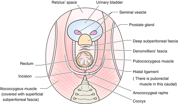
Fig. 5.3
Division of the levator ani muscles. Distancing from the rectum as much as possible, the iliococcygeus muscle and the pubococcyeus muscle (the levator ani muscle) are divided to the ventral side in a U-shape by USAD, to expose the granular fatty tissue of the ischiorectal fossa
When bleeding occurs before reaching the pelvic floor, the field of view is no longer visible for the operation. Thus, sufficient haemostatic control is mandatory.
5.3.3.2 Further Dissection of the Ventral Side of the Rectum
Scope: umbilicus
The operator’s right hand: a spatula-type electrosurgical knife and an USAD
The operator’s left hand: controls the ventral side of the rectum
The assistant’s right hand: controls the prostate to the ventral side
The assistant’s left hand: controls the prostate to the ventral side
It is important to dissect sufficiently as far as possible between the prostate and Denonvilliers’ fascia. Denonvilliers’ fascia is separated from the prostate at the terminal end by an USAD. In addition, at the lateral portions of the rectum, the rectal branches from the pelvic plexus are further divided using the USAD.
5.3.3.3 Division of the Sigmoid Colon
Scope: umbilicus
The operator’s right hand: an electrosurgical knife with spatula-type blade and Endo-GIATM
The operator’s left hand: grasps the sigmoid mesocolon with the forceps
The assistant’s right hand: grasps the sigmoid mesocolon with the forceps
The assistant’s left hand: maintains traction of the sigmoid colon to the cranial side
After determining where the sigmoid colon is to be divided, the sigmoid mesocolon is divided from the cranial stump obtained from the lymph node dissection. In cooperation with the assistant, a view of the mesocolon is obtained and the mesocolon is divided with the USAD and the sigmoid colon is then divided using a flexible Endo GIATM (Fig. 5.4).
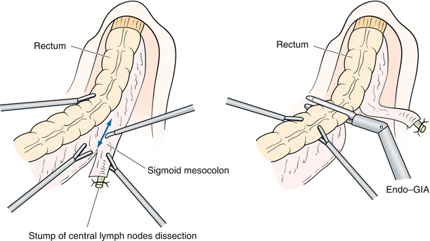
Fig. 5.4
Division of the sigmoid colon. The sigmoid mesocolon is divided from the cranial stump of the central lymph node dissection. In cooperation with the assistant, a view of the mesocolon is obtained and the mesocolon is then divided using a USAD. The sigmoid colon is divided using the flexible Endo GIATM
5.3.3.4 Operation from the Perinaeum
The anus is sutured and closed with purse-string sutures. The patient is in the modified Lloyd-Davies position with further bending, and the perinaeum is fully visible. Following a second thorough disinfection of the perineum, an incision is made several centimetres distant from the anal verge (Fig. 5.5). The anatomical landmarks are the coccyx on the dorsal side, bilateral ischial tuberosity on the lateral side, and the perineal body on the ventral side in men. Although the skin guideline acts as the midpoint between the above and the closed anus, a broad skin excision does not lead to a better dissection. A Loan-Star wound retractorTM is used to raise the edge of the skin incision (Fig. 5.5). The subcutaneous fat is excised. If the lesion is a far advanced cancer, the incision is made using the medial edge of the gluteus maximus muscle as an index.
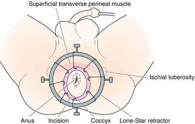
Fig. 5.5
Perineal procedures. The anus is sutured and closed with purse-string sutures. The patient is in the modified Lloyd-Davies position and bent further, the perinaeum is fully visible. Following thorough disinfection, an incision is made several centimetres distant from the anal verge and a Loan-Star wound retractorTM is used to raise the edge of the skin incision
Without exposing the gluteus maximus muscle, which is located dorsally and bilaterally, the dissection proceeds towards the coccyx tip as the anatomical landmark, there the granular fatty tissue of the ischiorectal fossa becomes visible. It is possible to reach the peritoneal cavity easily on the ventral side of the coccyx. Since the levator ani muscles have been divided into a U-shape, the passage between the abdominal cavity and the perineum may be incised into a U-shape at this point (Fig. 5.3)
Related posts:


Stay updated, free articles. Join our Telegram channel

Full access? Get Clinical Tree


