Set-up
Notes
Patient’s positioning
Patient in the supine position, arms tucked at the sides, head down at an angle of 20–30°
Narrow laparoscopic muscle hook (KS Hook)
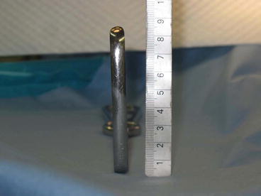
8 × 80-mm muscle hook developed by our department to gently dissect the rectus abdominis from the posterior rectus sheath.
Camera
Olympus, 5-mm, 30° laparoscope.
Forceps
We use forceps designed for standard laparoscopic surgery. This is because roticulator forceps are difficult to maneuver in the restricted working space within the preperitoneum.
Insufflator
We use an insufflation pressure of 8 mmHg
Laparoscopic coagulating shears or electric scalpel
Standard type
Mesh
We use a 12 × 10-cm or larger mesh
Mesh delivery system
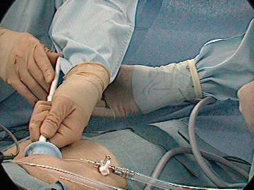
Mesh is rolled into a plastic cylinder and inserted into the preperitoneal space
Absorbable clips
These are used to secure the mesh
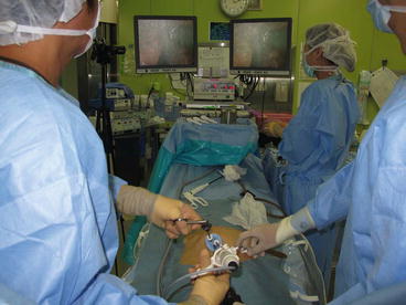
31.3.1 Patient Positioning (Fig. 31.1)

Fig. 31.1
Intraoperative scene of actual set-up
The patient is placed in a supine position, arms tucked at the sides, head down at an angle of 20–30° (Trendelenburg position). For Single-TEP, the surgeon stands closer to the patient’s head than for standard TEP, so the surgical procedures are facilitated if the patient’s arms are kept close to the sides.
31.3.2 Instruments and Mesh

Fig. 31.2
Narrow laparoscopic muscle hook (KS Hook (Takasago))
The instruments used for this operation include a 5-mm, 30-degree oblique view laparoscope (Olympus Medical, Tokyo, Japan), standard straight forceps and laparoscopic coagulating shears or electro-cautery. It is difficult to maneuver roticulator forceps in the restricted working space in the pre-peritoneum. An insufflation pressure of 8 mmHg is maintained, the same as that used for standard laparoscopic surgery. Careful monitoring is required to avoid an insufflation pressure above 15 mmHg, which can cause subcutaneous emphysema. An 8 × 80 mm retractor, developed in our department is used to gently dissect the rectus abdominis from the posterior rectal sheath. (KS Hook, Takasago Medical Industry Co., Ltd., Japan (Fig. 31.2). A 12 × 10-cm or larger mesh is routinely used [2] (Microval Mesh JG, MicroVal SA, France). The mesh is rolled into a plastic cylinder and inserted into the pre-peritoneal space (Mesh Delivery System) (Fig. 31.3). The aim of this maneuver is to prevent damage to the inferior epigastric vessels and the transverse fascia during mesh insertion. Absorbable clips are applied to secure the mesh.


Fig. 31.3
Mesh delivery system
31.4 Indications
This technique can be used on any type of hernia, with the exception of huge, nonreducible, scrotal hernias. Preferred indications are recurrent hernias, mainly after conventional repairs, with the advantage of avoiding anterior scar tissue, bilateral hernia where both sides can be approached through the same access, and hernias with massive destruction of the posterior abdominal wall (a defect diameter greater than 3 cm or pantaloon hernias). Contraindicated included the patients under treatment with anticoagulants.
Two methods of approach are available:
1.
Multi-trocar approach. Insertion of three trocars through a single incision.
2.
Multi-channel approach. Use of a commercially available multi-channel access port.
In our department, standard practice is to use the multi-channel approach because it facilitates trocar insertion into the pre-peritoneum and insufflation.
Stay updated, free articles. Join our Telegram channel

Full access? Get Clinical Tree


