Fig. 2.1
Algorithm for endoscopic submucosal dissection of gastrointestinal neoplasms
Absolute and Expanded Indications of ESD for EGC
Currently accepted indications for endoscopic resection of EGC include the resection of small intramucosal EGCs of intestinal histology type. The rationale for this recommendation is based on the knowledge that larger lesions or diffuse histology lesions are more likely to extend into the submucosal layer and thus have a higher risk of lymph node metastasis. In addition, en bloc resection of large lesions was not technically feasible until the ESD procedure was developed. Therefore, at present, the accepted indications for EMR according to the gastric cancer treatment guidelines published in 2001 by the Japanese Gastric Cancer Association are: (1) well-differentiated elevated cancers less than 2 cm in diameter and (2) small (<1 cm) depressed lesions without ulceration. Also, these lesions must be moderately or well-differentiated cancers that are confined to the mucosa and have no lymphatic or vascular involvement [5, 6]. However, it has been observed clinically that the accepted indications for EMR may be too strict, which leads to unnecessary surgery (Table 2.1) [7].
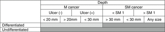
 Guideline criteria for EMR,
  Surgery
Table 2.1
Indications for extension of EMR
Guideline criteria for EMR, SurgeryFurther studies by Gotoda et al. have defined new criteria for expanding the indications for endoscopic treatment of gastric cancer. Endoscopic submucosal dissection was developed to dissect directly along the submucosal layer using specialized devices. Preliminary studies have been published on the advantages of ESD over conventional EMR for en bloc removal of larger or ulcerated EGC lesions. Thus, ESD allows for precise histological assessment of the resected specimens, possibly preventing residual disease and local recurrence. Gotoda et al. analyzed more than 5,000 EGC patients who underwent gastrectomy with meticulous D2 level node dissection; they provided important information on the risks of lymph node metastasis, wherein differentiated gastric cancers with no lymphovascular involvement, correlating with a nominal risk of lymph node metastasis, were defined [8]. Thus, they proposed the expanded criteria for endoscopic resection: (1) mucosal cancer without ulceration, irrespective of tumor size; (2) mucosal cancer with an ulcer <3 cm in diameter; and (3) minute (<500 μm from the muscularis mucosa) submucosal invasive cancer <3 cm in size (Table 2.2). However, extending the indications for ESD remains controversial because the long-term outcomes of these procedures have not been fully documented.
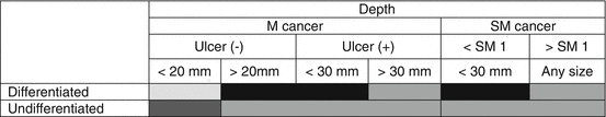
 Guideline for ESD:
  Expanded indication,
  Consider surgery,
  Surgery
Table 2.2
Indications for extension of ESD
Guideline for ESD: Expanded indication, Consider surgery, SurgeryRecent Evidence for Expanded Indications of ESD for EGC
Although the absolute indication is applicable only to mucosal cancer, there have been some studies about ESD for submucosal cancer. Of the 145 well-differentiated tumors that had invaded less than 500 μm into the submucosa and were smaller than 30 mm in diameter, none showed evidence of lymph node metastasis, provided that there was no lymphatic or venous invasion. Based on these findings, it was suggested that the criteria for EMR and ESD as local treatment for EGC should be extended [9–13].
According to a recent study of patients who had surgery for EGC at Seoul National University Bundang Hospital [14], of 132 patients with mucosal cancers, 129 met the extended indications for EMR or ESD while three (2.3 %) had lymph node metastasis. Of the 52 submucosal cancer cases that met the extended indications for EMR or ESD, two (4 %) had lymph node metastasis. Differentiated mucosal cancers without ulcer formation did not have lymph node metastasis, irrespective of size. These data suggest that a well-differentiated mucosal cancer of any size without ulceration may be considered as an extended indication for EMR or ESD. However, data from this study showed that 2.8 % of tumors meeting the extended criteria for EMR or ESD had positive lymph nodes, and the authors suggest that if EMR or ESD had been performed in these patients, it would not have been curative.
Regarding the expansion of indications to EGC with undifferentiated histology, the supporting data are continuously being reported. Ye and colleagues reported that EGC with undifferentiated histology has no lymph node involvement, provided that the cancer is smaller than 25 mm and is confined to the mucosa or upper third of the submucosa and has no lymphatic involvement [15]. A similar study for signet ring cell carcinoma was reported by Park et al. [16]; EGC with signet ring cell histology has a high risk for nodal and organ metastases, while smaller cancers of less than 25 mm that are confined to the SM2 layer and have no lymphovascular involvement demonstrated no lymph node involvement.
In another Korean study on the lymph node metastasis of poorly differentiated adenocarcinomas, a retrospective analysis was performed on 234 patients with poorly differentiated EGC who underwent radical gastrectomy with D2 lymph node dissection [17]. Of the 234 lesions with poorly differentiated EGC, half (n = 116) showed submucosal invasion in the resection specimen, and 25.9 % (30/116) of those were limited to the upper third (SM1). Of the lesions confined to the mucosa, lymph node metastasis was found in 3.4 % (4/118). With minor submucosal infiltration (SM1), the lymph node metastasis rate was non-existent (0/30). However, with SM2/3 invasion, the lymph node metastasis rate increased sharply to about 30 %. Therefore, poorly differentiated EGC confined to the mucosa or with minimal submucosal infiltration could be considered for curative ESD due to the low risk of lymph node metastasis. Another Korean study [18] focusing on endoscopic resection for undifferentiated-type cancer such as poorly differentiated adenocarcinoma and signet ring cell carcinoma showed interesting results. In this study, a total of 58 lesions with undifferentiated EGC (17 poorly differentiated; 41 signet ring cell) were treated by endoscopic resection. The en bloc and complete resection rates in poorly differentiated cases were 82.4 % and 58.8 %, whereas those in signet ring cell were 85.4 % and 70.7 %, respectively. Interestingly, all of the histologically incomplete resections in poorly differentiated cases were vertical cut end-positive, whereas 83.3 % of these resections in signet ring cell were lateral cut end-positive. The recurrence rate was 5.1 % in complete resection during the follow-up period. Therefore, the authors suggested that endoscopic resection may be a feasible local treatment for undifferentiated EGC if complete resection can be achieved. However, the indication for poorly differentiated cancers is still controversial. Further follow up periods and accumulation of a larger number of cases are still required to clarify this issue.
Yamaguchi et al. reported clinical outcomes of ESD according to indication criteria. A total of 589 EGC lesions were divided into the guideline group and the expanded group [6]. En bloc, complete, and curative resections were achieved in 98.6 % and 93.0 %, 95.1 % and 88.5 %, and 97.1 % and 91.1 % of the guideline and expanded criteria lesions, respectively; the differences between the two groups were significant for all types of resection. The expanded criteria lesions were at significantly higher risk for ESD-associated bleeding and perforation. Overall survival was adequate, irrespective of indication, and the disease-specific survival rates were 100 % in all groups.
Limitations of ESD
However, more aggressive cases have been encountered. Walter et al. reported one case [19] with early gastric cancer that was initially treated by ESD. Esophagogastroduodenoscopy showed a slightly elevated, centrally depressed lesion about 15 mm in diameter with a very small ulceration in the center (type IIa + IIc) and biopsies showed only focal high-grade intraepithelial neoplasia. The resected specimen showed a submucosal infiltration depth of greater than 500 μm. Therefore, the patient underwent gastrectomy. The postoperative stage was pT1 (sm3), pN0 (0/58), cM0, L0, V0, G2 (UICC stage Ia). Three months later, an ultrasound revealed a new mass in the liver, and biopsy showed a rapidly growing metastasis of the gastric adenocarcinoma. This case highlights the risk of affected lymph nodes in early gastric cancer and the consequent risk of metastasis, which increases with greater depth of infiltration into the submucosa.
Another obstacle in EMR and ESD is the presence of micrometastasis [20–24]. Even after curative surgical resection for EGC, the recurrence rate is about 1.7–3.4 %, which could be the result of micrometastasis. According to Cai and colleagues, tumor size, macroscopic type, accompanying ulcers, and depth of invasion are strongly associated with micrometastasis in lymph nodes. Therefore, tumors with suspected submucosal invasion, large size, accompanying ulcers, and undifferentiated histology may have a risk of recurrence owing to micrometastasis, which may indicate the inappropriateness of EMR or ESD.
One more problem should be solved as follows: (1) We cannot be aware of the presence of ulcers before ESD and, as a result, it is very difficult to resect the lesion. We also have to pay attention to the fact that there are some differences in the definition of ulceration among physicians. (2) The way lesion size is measured is also somewhat different among institutions. Therefore, a uniform, standard way for measuring lesion size may become necessary in the future.
Long-Term Follow-Up Data
Long-term follow-up data are needed for the clinical application of the expanded indication. One Japanese study [25] from the National Cancer Center Hospital, involving a total of 1,955 EGC patients enrolled from January 1999 to December 2005, showed that there were no significant differences in the overall 5-year survival rates of the curative resection group, as defined by the expanded criteria, and the non-curative resection group after additional surgery. These data suggest that ESD using the expanded criteria can show excellent long-term outcomes.
Related posts:
 Management of Gastrointestinal EMR and ESD Perforation: From Lab to Practice
 Submucosal Fluid Cushion Injection Fluid: Western Perspective
 Submucosal Endoscopy: From ESD to POEM and POET
 Electrocautery for ESD
 Advanced Endoscopic Imaging in the Upper Gastrointestinal Tract
 Endoscopic Submucosal Dissection for Superficial Esophageal Cancer
Stay updated, free articles. Join our Telegram channel

Full access? Get Clinical Tree


