Fig. 7.1
The ViOptix (tissue oximetry) silicone surround sensor
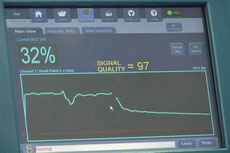
Fig. 7.2
Digital display of the ViOptix sensor
The ViOptix Tissue Oximeter (T.Ox™) [(ViOptix Inc., Fremont, CA)] utilizes near-infrared spectroscopy to measure local tissue oxygen saturation (StO2). Near-infrared spectroscopy has been assessed as a noninvasive technique for tissue investigation in various animal models [13–15]. Furthermore, various investigators have analyzed the clinical applications of near-infrared tissue oximetry [8, 16–20]. Keller was able to evaluate the role of StO2 as an indicator of tissue hypoxia by measuring the StO2 drop rate (change in StO2/change time) [16]. Oximetry technology has also been utilized in breast cancer during free-flap reconstruction [15, 16] and applied in post-occlusive reactive hyperemia to evaluate extremities of patients with peripheral vascular disease [17]. It has also been employed in men with erectile dysfunction to quantify penile oxygen saturation [18]. Furthermore, tissue oximetry has been applied in measuring renal ischemia during hilar clamping in a porcine model [19].
Partial nephrectomy for renal carcinoma involves temporarily occluding the renal artery, rendering the kidney ischemic during tumor resection. Warm ischemia time is the strongest modifiable surgical risk factor for postoperative chronic kidney disease [8]. Prior studies evaluating renal parenchymal oxygen saturation levels in response to hilar occlusion during partial nephrectomy are scarce. The first study evaluating hyperspectral imaging as a noninvasive method to assess renal oxyhemoglobin saturation during renal hilar occlusion was by Holzer et al. [21]. In a porcine model, he showed that renal oxygenation decreases rapidly after clamping, and nadir oxyhemoglobin is attained within 10 min of hilar occlusion and returns to baseline 30 min after reperfusion [21].
Cadeddu et al. [22] described the initial intraoperative hyperspectral imaging experience during open partial nephrectomy in 2011 on a porcine model. This was executed by illuminating the kidney with light arrays specific for hemoglobin (520–645 nm). Reflectance images were captured at each pixel and digitally processed to determine the percent of oxyhemoglobin [22].
Hyperspectral imaging has also been used to characterize renal oxygenation during laparoscopic partial nephrectomies [23–25]. In 2011, Cadeddu and colleagues [23] used this real-time technology to monitor renal perfusion/ oxygenation during hilar occlusion and kidney recovery. They found a baseline percent hyperspectral oxyhemoglobin in the kidney to be 74.6 %. After 10 min of hilar occlusion, there was a median 20.0 % decrease from pre-occlusion baseline, where it remained for the duration of kidney ischemia. Upon reperfusion, the percent of oxyhemoglobin returned to baseline at a median of 5 min [24].
In 2011, Olweny, et al. [25] investigated the association between tissue oxygen saturation %HbO(2) and estimated glomerular filtration rate (eGFR). They customized the hyperspectral digital light imaging (DLP(®)-HSI) for use during laparoscopic partial nephrectomy by combining a hyperspectral illumination source with a conventional laparoscopic light and merging it with a digital data device [25]. They found the median %HbO(2) dropped after hilar clamping and recovered to baseline levels shortly after unclamping. Furthermore, baseline %HbO(2) was inversely associated with preoperative eGFR (τ = − 0.38; P = 0.036) and eGFR at postoperative follow-up (τ = − 0.38; P = 0.036). Baseline and ischemic tissue oxygen saturation levels exhibited no correlation with hypertension, diabetes, and age and/or tobacco history. Overall, the laparoscopic hyperspectral imaging device successfully characterized dynamic changes in renal oxygenation during partial nephrectomy and may potentially to predict postoperative individual kidney function [25].
A study by Lee et al. [19] compared tissue oximeter to a digital imaging software which was programmed to track ischemia by detecting color change in renal parenchyma during renal hilar clamping. The digital imaging detected “red” when the computer was directed at renal tissue in which oxygen was bound to hemoglobin. And during warm ischemia, loss of bound oxygen to hemoglobin resulted in the development of a cyanotic or “blue” kidney. Tissue oximetry levels decreased during hilar clamping then returned to baseline after unclamping. Digital image software produced a histogram which demonstrated red and blue differences that correlated with nonischemic and ischemic levels in the respective tissues. Tissue oximetry and digital image analysis were both able to calculate a drop in tissue oxygen saturation during periods of acute renal ischemia.
Using tissue oximetry, Lee et al. [26] also demonstrated clamping artery alone resulted in less ischemic damage compared to clamping artery and vein together. Yorkshire swines were subjected to hilar clamping (either artery versus artery and vein together), and near-infrared renal oximetry measurements were obtained at baseline, during warm ischemia (15- and 30-min trials), and after unclamping. Tissue oximetry analysis during renal arterial clamping alone demonstrated larger drops in tissue oxygen saturation and faster recovery of tissue ischemia compared to combined renal artery/vein clamping together, indicating reduced ischemic changes when clamping artery alone. These findings suggest that clamping artery alone may protect against renal reperfusion injury during renal hilar clamping [26].
In summary, hyperspectral imaging is a real-time noninvasive method to assess renal oxyhemoglobin saturation intraoperatively throughout the kidney. In addition, tissue oximetry has been used to evaluate kidney perfusion during ischemia caused by renal hilar clamping during partial nephrectomy. This technology may allow us to assess future surgical or pharmacological interventions ability to minimize ischemic injury during renal hilar clamping.
References
1.
Pagtalunan ME, Olson JL, Tilney NL, Meyer TW. Late consequences of acute ischemic injury to a solitary kidney. J Am Soc Nephrol. 1999;10:366–73.PubMed
< div class='tao-gold-member'>
Only gold members can continue reading. Log In or Register to continue
Related posts:
 Endoscopic Fluorescence Imaging of Bladder Cancer: Photodynamic Diagnosis and Confocal Laser Endomicroscopy
 Optical Coherence Tomography in Bladder Cancer
 Multiphoton Microscopy in Urologic Surgery
 Endoluminal Ultrasonography
 Multiparametric Magnetic Resonance Imaging for Prostate Cancer
 Ultrasound-Guided Prostate Cryotherapy
Stay updated, free articles. Join our Telegram channel

Full access? Get Clinical Tree


