Hormones and the Kidney


Hormones and the Kidney


Sola Aoun Bahous

Maya Khairallah

Joumana T. Chaiban

Kamal F. Badr


HORMONAL MODULATION OF NEPHRON FUNCTION: AN OVERVIEW


Modulation of Glomerular Filtration Rate

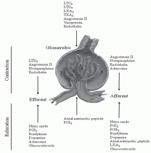
The major physiologic determinants of single nephron glomerular filtration rate (GFR) are glomerular plasma flow (QA), glomerular transcapillary hydraulic pressure (PGC), and the ultrafiltration coefficient (Kf).1 These variables are determined, in part, by the contractile state of the afferent arteriole, efferent arteriole, and mesangial cells. Kf also varies with alterations in the hydraulic permeability of the capillary filtration barrier, which consists of endothelial cells, visceral epithelial cells, and the glomerular basement membrane. By binding to specific receptors on cellular and structural components of the glomerulus, circulating and locally produced hormones influence one or more of the physiologic determinants of GFR. Figure 8.1 summarizes the effects of different hormones on pregomerular, glomerular, and postglomerular contractility. Because the same substance can act at different sites in the glomerular unit, the net effect on GFR will depend on whether its actions are antagonistic or complementary. For example, atrial natriuretic peptide (ANP) decreases afferent arteriolar resistance, whereas increasing efferent arteriolar resistance results in an augmented PGC and a rise in GFR.2 Under certain conditions, ANP also increases Kf, which, in turn, contributes to the enhancement of GFR.3 On the other hand, angiotensin (Ang) II-mediated constriction of both afferent and efferent arterioles results in opposite effects on glomerular plasma flow and PGC and, therefore, no change in single nephron GFR.4 Regulation of glomerular hemodynamics is further complicated by multiple interactions between hormones in the kidney. For example, infusion of Ang II along with a cyclooxygenase inhibitor causes a significant decrease in single nephron GFR, suggesting that endogenous prostaglandin production antagonizes the glomerular effects of Ang II.5 The net effects of renally relevant hormones on GFR are discussed in more detail later in this chapter.


Modulation of Tubule Water and Electrolyte Transport

Renal tubule cells express receptors for many circulating and locally synthesized hormones. The effects of a particular hormone on water or electrolyte transport are partly determined by the differential distribution of its receptor on functionally specialized segments of the renal tubule. For example, arginine vasopressin (AVP) binds almost exclusively to principal cells in the collecting tubule (CT), where it influences the physiologic functions of this segment, primarily water and urea absorption.6 Parathyroid hormone (PTH), on the other hand, exerts its biologic actions on renal tubule segments proximal to the collecting duct, where it modulates calcium, phosphate, magnesium, sodium, and bicarbonate transport.7 Table 8.1 summarizes the net effects of renally relevant hormones on water and solute handling in different tubule segments. Each hormone is discussed in detail in the sections that follow. As in the glomerulus, hormones may modulate their own actions by altering the production of counterregulatory hormones. For example, AVP induces local synthesis of prostaglandin E (PGE), which opposes the effect of AVP on water permeability in the CT.8


ARGININE VASOPRESSIN

In 1895, Oliver and Schafer were the first to describe a substance with potent vasopressor effects originating from the posterior pituitary, hence the name, vasopressin.9 It was not until later in the 20th century that the effects of this hormone on modulation of water excretion by the kidney were discovered. In fact, vasopressin, also called AVP or antidiuretic hormone (ADH), is a key hormone for osmoregulation and maintenance of body homeostasis.10 AVP was also found to exert effects on body temperature, glucose metabolism, memory, social behavior, and the hypothalamic-pituitaryadrenal axis.


FIGURE 8.1 Hormones regulating glomerular, afferent, and efferent arteriolar contractility. Glucocorticoid action may be pharmacologic and indirect. LTC4, leukotriene C4; LTD4, leukotriene D4; LXA2, leukotriene A2; TXA2, thromboxane A2; PGE2, prostaglandin E2; PGI2, prostacyclin.


Structure and Synthesis of Arginine Vasopressin

AVP is a 9-amino acid neuropeptide synthesized by magnocellular neurons in the paraventricular and supraoptic nuclei of the hypothalamus.11 The AVP gene, located on chromosome 20, consists of three exons that encode the signal peptide, AVP, neurophysin II (NPII), and a glycopeptide.10 After cleavage of the signal peptide within the ER, the resulting prohormone is folded and packaged into secretory granules in neuronal bodies. These granules are then transported along the axons to nerve terminals in the posterior pituitary (neurohypophysis).12 During axonal transport, further processing of the prohormone within secretory granules yields AVP, neurophysin II (NpII), and a glycopeptide. Interestingly, mutations of NpII impair AVP secretion, suggesting that NpII assists in the processing or secretion of AVP.13 In addition to the posterior pituitary, AVP synthesis has been detected in the pancreas, adrenal gland, ovary, testis, and regions of the brain.14 Its physiologic function in these sites, however, remains to be clarified.


Physiology of Arginine Vasopressin in the Kidneys

The most sensitive stimulus for AVP secretion into the bloodstream is increased plasma osmolality. Osmosensitive neurons that respond to changes in plasma osmotic pressure by varying their intracellular water content have been identified in the anterior hypothalamus, specifically in the organum vasculosum of the lamina terminalis (OVLT) and the subfornical organ (SFO).15 When stimulated by osmosensitive neurons, the magnocellular neurons release the stored AVP into the posterior pituitary (an area that lacks a blood-brain barrier) and AVP enters the general circulation. As little as 1% change in plasma osmolality leads to a change in AVP concentration that is sufficient to modify renal water excretion.16 AVP secretion is almost completely suppressed when plasma osmolality decreases below an average of 280 mOsm per kg of water in humans.17

Secretion of AVP is also influenced by alterations in intravascular volume and blood pressure, sensed by baroreceptors located in the heart, aortic arch, and carotid sinus.18 These signals are transferred through the vagal nerves to the
nucleus solitarius in the brainstem, from which postsynaptic pathways project to the magnocellular neurons. Whereas a 5% to 8% decrease in blood volume or systemic arterial pressure has little effect, further hemodynamic compromise leads to a steep increase in circulating AVP levels. Significant reductions (10%-30%) in circulatory arterial volume or blood pressure can override osmoregulation and result in markedly increased AVP levels in the face of decreased plasma osmolality.19 Other less potent stimuli for AVP secretion include fever, emesis,20 and oropharyngeal osmoreceptors.21


TABLE 8.1 Hormonal Modulation of Tubular Transport


Hormones with Major Effects


Reabsorption


Stimulatory


Inhibitory


Proximal tubule


Na


Ang II, catecholamines, insulin


Dopamine, PTH


Pi


IGF-1


PTH


HCO3


Ang II


PTH


Ca


PTH


TALH


Na


AVP,a catecholamines


PGE


Ca


PTH, calcitonin, glucagon


Mg


AVP, PTH, calcitonin, glucagon


DCT


Pi


PTH


Ca


PTH


CCD


H2O


AVP


PGE, bradykinin, ANP, α-adrenergic agents


Na


Aldosterone


ANP, PGE, EGF


IMCD


Urea


AVP


SECRETION CCD


K


AVP, aldosterone


β1 agonists


H


Aldosterone


a Unlikely in humans.


TALH, thick ascending limb of the loop of Henle; DCT, distal convoluted tubule; CCD, cortical collecting duct; IMCD, inner medullary collecting duct; Ang II, angiotensin II; IGF, insulinlike growth factor; AVP, arginine vasopressin; PTH, parathyroid hormone; PGE, prostaglandin E; EGF, epidermal growth factor.


AVP circulates in the plasma nearly in an unbound form. Levels of circulating AVP depend on both the rate of AVP release from the posterior pituitary and the rate of AVP degradation. As discussed, the major factor controlling AVP release is plasma osmolality. The liver and the kidney both contribute to the breakdown of AVP and the decline in AVP levels when secretion ceases. In fact, the half-life of AVP in the circulation is 18 minutes due to rapid clearance by hepatic and renal vasopressinases.22 Under physiologic conditions, plasma vasopressin concentrations vary with serum osmolarity between 0 to 5 pg per mL.22

It is noteworthy that AVP levels are difficult to measure in plasma because of the instability of this peptide and the low sensitivity of available AVP antibodies. Copeptin (or C-terminal proarginine vasopressin, CT-proAVP) is the C-terminal part of AVP, which is secreted stoichiometrically with AVP in a manner similar to C-peptide and endogenous insulin.23 CT-proAVP provides a reliable means for estimating prevailing AVP levels in the circulation, thus facilitating the study of AVP in human diseases. A recent cohort study in renal transplant patients suggests that high CT-proAVP strongly correlates with a negative renal prognosis. In fact, in this study, the plasma concentrations of CT-proAVP predicted renal function loss over a 3.2-year follow-up.24

AVP exerts its biologic actions through three specific cell-surface AVP receptors identified as V1R (also called V1aR), V2R, and V3R (also V1bR).25 These receptors belong to the G protein coupled receptor superfamily (Table 8.2). In the human kidney, mRNA for V1Rs predominates in cortical collecting ducts (CCD), gradually decreasing as the collecting duct enters the medulla.26 In addition, whereas V1R mRNA is diffusely expressed in the CCD, it is restricted to the intercalated cells in OMCD. V1Rs are responsible for mediating vascular smooth muscle cell vasoconstriction by activating G protein-dependent phospholipase C (PLC) and the downstream effectors, diacylglycerol (DAG), and inositol 1,4,5-triphosphate (IP3). In turn, DAG stimulates protein kinase C (PKC), whereas IP3 increases cytosolic Ca2+, thus
initiating the second-messenger cascade responsible for the cellular actions of AVP.25 Stimulation of V1R, although not directly involved in control of tubular water and electrolyte transport, increases sodium excretion because of the influences on blood pressure, effective arterial circulating volume, glomerular filtration rate, and circulation in the vasa recta system.27,28 Additional biologic effects of AVP mediated through V1Rs include platelet aggregation29 and increased glycogenolysis and gluconeogenesis in the liver.30


TABLE 8.2 Vasopressin Receptor Types, Genetics, Location, and Main Physiologic Effects


Receptor


Chromosome location


No. of amino acids


Site of action


Main second messenger


Main effects


V1 (V1a)


12 (q14-q15)


418


Vascular smooth muscle, platelets, liver, testes, brainstem, adrenal glands


PLC→IP3 + DAG/calcium and PKC


Vasoconstriction, platelet aggregation, glycogenolysis, stimulation of aldosterone and cortisol synthesis


V2


X (q28)


371


Collecting duct cells of kidney, inner medulla, heart, pancreas


Adenylate cyclase/ cAMP


Water retention, stimulation of atrial natriuretic peptide, stimulation of insulin synthesis, coronary and pulmonary artery vasodilation


V3 (V1b)


1 (q32)


553


Hypothalamus, anterior pituitary gland


PLC→IP3 + DAG/calcium and PKC


Modulation of ACTH synthesis; stimulation of ACTH, GH, and procaltin release


Oxytocin


3 (p25)


389


Uterus, breast, vascular endothelium


PLC→IP3 + DAG/calcium and PKC


Myometrial contraction, ductal myoepithelial contraction, vasodilation


P2 Purinergic


11 (q13.5-14.1)


Cardiac endothelium ATP


Vasoconstriction, reduced cardiac output


Reproduced from Favory R, Salgado DR, Vincent J-L. Investigational vasopressin receptor modulators in the pipeline. Expert Opin Investig Drugs. 2009;18:1119-1131.


The V3R (V1bR), also coupled to PLC signaling, is present on neurons in the anterior pituitary (adenohypophysis) and is thought to mediate AVP-induced corticotropin secretion.25,31,32 They are also found elsewhere in the brain, especially in the pyramidal neurons of the hippocampal CA2 field, in which they mediate fundamental physiologic actions such as memory and body temperature control as well as social behavior.33 In addition, this receptor has been localized to pancreatic islet cells, modulating insulin secretion.34 However, its presence and role at the level of the medulla in rat kidney remain unclear.

V2Rs, on the other hand, are heavily expressed in the medullary TAL, macula densa (MD), connecting tubule, and cortical and medullary collecting duct, as well as weakly expressed in cortical thick ascending limb (TAL) and distal convoluted tubule.35 These are the best characterized and studied vasopressin receptors. By binding to V2R, AVP increases water reabsorption through multiple mechanisms.31 Activation of V2R results in increased cyclic adenosine monophosphate (cAMP) levels and activation of PKA, which promotes insertion of water channels into the luminal suface of the epithelial tubular cells.36 This ultimately mediates the antidiuretic effect of AVP by allowing back diffusion of water down its concentration gradient.37 In addition, V2Rs modulate sodium reabsorption through the epithelial Na+ channel (ENaC) across principal cells.38,39,40,41 This facilitates free water reabsorption by supporting the axial corticomedullary hyperosmotic gradient. It was also recently shown in rats that AVP modulates sodium reabsorption even in the distal convoluted tubule by acting on the thiazide-sensitive Na+-Cl cotransporter (NCC).42 NCC is important in defining sodium delivery to the collecting duct,
which is necessary for ENaC activity. Finally, V2Rs activate urea transporters, such as UTA1, in the distal nephron.43,44,45 This increase in urea reabsorption and recycling maximizes sodium reabsorption in the TAL by supporting the axial hyperosmotic gradient drawing water from the distal nephron.46 The clinical importance of the V2R in water balance disorders is underlined by the current use of V2R antagonists in a clinical setting (see later).

It is noteworthy that AVP also binds to two nonspecific receptors: oxytocin receptors and P2 purinoreceptors. Oxytocin receptors are found in the breast, ovary, uterus, and hypothalamus. Since the affinity of this receptor for vasopressin is relatively low, the clinical effects of this hormone are limited under physiologic conditions.47 AVP may also act on P2 purinoreceptors in the heart, causing coronary vasoconstriction and contributing to the reduction in cardiac output.48


Water Channels

The discovery of the family of aquaporin water channels was crucial to the understanding of the mechanism by which AVP can increase water permeability in the kidney. The group of Peter Agre discovered the first aquaporin in human erythrocytes.49 To date, seven different aquaporin (AQP) have been shown to be expressed in the human kidney and to be involved in renal water reabsorption.50

AQP2 is the vasopressin-sensitive water channel expressed in the principal cells of the collecting duct, where it shuttles between intracellular storage vesicles and the apical membrane.51 Knocking out the AQP2 gene produces a severe concentration defect in these mice, resulting in postnatal death.52 It has now been shown to be involved in many clinical disorders (see later). AQP1 is constitutively expressed on the basolateral and apical membrane of epithelial cells lining the proximal tubule and thin descending limb, as well as endothelial cells of the descending vasa recta. It not only plays a role in water reabsorption from urine in these segments but is also critical for a functional countercurrent multiplication system.53 AQP3 and AQP4 are expressed on the basolateral membrane of the principal cells of the collecting duct, and they represent an exit pathway from these cells for water entering through AQP2.54,55 Similar to AQP1, AQP7 is expressed at the apical membrane of proximal tubules (S3 segment) and has been shown to mediate glycerol reabsorption in addition to water.56 AQP6 is found in intracellular vesicles of acid-secreting α-intercalated cells in the collecting duct.57 It is thought to be involved in urinary acid secretion. AQP11 is localized to the endoplasmic reticulum (ER) in the proximal tubule. Interestingly, knocking out the AQP11 gene in mice is fatal because of the onset of polycystic kidney disease.58


Clinical Pathophysiologic Role of Arginine Vasopressin in the Kidneys

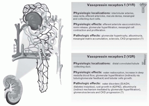
AVP is implicated in major clinical syndromes of alterations in water metabolism, namely nephrogenic diabetes insipidus (NDI) and the syndrome of inappropriate secretion of antidiuretic hormone (SIADH). In addition to water metabolism, it has been recently suggested that AVP may play a role in the initiation and the progression of chronic kidney disease (CKD) and in the most prevalent form of hereditary renal disease, namely the adult polycystic kidney disease (Fig. 8.2).59

NDI is characterized by impaired AVP-induced water reabsorption, resulting in polyuria and polydipsia.60 If water intake is inappropriate, patients with NDI may fail to thrive, suffer from mental retardation, and die early. NDI can be acquired such as following lithium treatment, hypokalemia, hypercalcemia, or ureteral obstruction, all of which lead to downregulation of AQP2.61,62,63,64 NDI can also be inherited (congenital).65 In fact, two gene mutations have been linked to congenital NDI: one in the AVPR2 gene encoding V2R (X-linked NDI) or mutations in the AQP2 gene (autosomal recessive or autosomal dominant NDI). More than 90% of patients with congenital NDI suffer from X-linked NDI. In both cases, patients cannot concentrate their urine despite normal or elevated plasma concentrations of vasopressin, leading to a massive loss of water through the kidney. So far, NDI is managed by salt restriction combined with hydrochlorothiazide diuretics to reduce urine output.66 However, no cure is available so far.

Abnormal water handling of central origin includes SIADH in which AVP levels are abnormally elevated and not suppressed when plasma osmolality concentration falls below the osmotic threshold for physiologic AVP secretion.67 SIADH is characterized by impaired water excretion in the absence of renal insufficiency, adrenal insufficiency, or any recognized stimulus for AVP secretion. Renal water retention and extracellular fluid expansion are compensated for by increased urinary Na+ excretion leading to life-threatening hyponatremia. The most common causes of the syndrome of inappropriate AVP secretion are neoplasia, neurologic disorders, congestive heart failure, liver cirrhosis, preeclampsia, and drugs such as thiazide diuretics or selective serotonin reuptake inhibitor antidepressants.67 Four patterns of AVP dysregulation in patients with SIADH have been observed.68 The most common pattern (in ˜40% of patients with SIADH) is the excessive and unregulated release of AVP, which is unrelated to plasma osmolality. In the second most common pattern (˜30% of patients), referred to as “reset osmostat,” AVP release continues to regulate water excretion at a lower plasma osmolality set-point. Although most tumors (e.g., lung carcinoma) manifest the first type of SIADH, some also present with the second type, thus the pattern of abnormal AVP secretion cannot be utilized to predict the cause of SIADH. A third rare pattern is characterized by an inability to stop AVP secretion at low plasma osmolalities, but the osmoregulation of AVP is otherwise normal. This pattern may be due to dysfunction of inhibitory hypothalamic neurons, leading to persistent low-grade basal AVP secretion. In the fourth pattern, a rare clinical picture of SIADH, the normal osmoregulation of AVP secretion is not altered (˜10% of patients) but AVP levels are low or undetectable. It is thought that a nephrogenic SIADH
(NSIADH) may be responsible for this picture. In fact, in some children, it appears to be due to an activating mutation of V2R. In other patients, it may be due to abnormal control of aquaporin-2 water channels in renal collecting tubules or production of an antidiuretic principle other than AVP.69 In a study with SIADH patients, treatment with V2R antagonists, commonly referred to as vaptans, increased serum Na+ concentration and decreased its excretion. Tolvaptan has been recently approved in the United States and Europe for the treatment of hyponatremia associated with SIADH, as well as cirrhosis and congestive heart failure. Recently, a dual vasopressin V1R and V2R antagonist, conivaptan, improves hyponatremia in rats with SIADH, suggesting a therapeutic potential for conivaptan in the treatment of SIADH.70


FIGURE 8.2 Vasopressin receptors: physiology and pathologic involvement in renal diseases. (Reproduced from Bolignano D, Zoccali C. Vasopressin beyond water: Implications for renal diseases. Curr Opin Nephrol Hypertens. 2010;19(5):499-504.)

AVP has also been shown to modify vascular tone in renal microvessels. Short-term infusion of AVP does not alter either renal blood flow or the GFR.71 Alternatively, chronic AVP administration increases the intraglomerular capillary pressure and GFR through tubuloglomerular feedback.72 In addition, a sustained increase in water intake and the consequent AVP suppression reduce proteinuria and the severity of glomerular and tubular damage in 5/6 nephrectomized rats.73,74 Thus, it seems likely that a chronic AVP-induced hyperfiltration may alter the glomerular barrier and start a series of events leading to enhanced protein loss and glomerulosclerosis. An increase in urinary albumin excretion represents an early predictor of glomerular damage in diabetes mellitus and a risk factor for cardiovascular complications in hypertension. Studies show that the Brattleboro rat, a model of central diabetes insipidus with complete lack of AVP, is protected from hyperfiltration, albuminuria, and renal hypertrophy after streptozotocin-induced diabetes mellitus.75 This suggests that AVP plays a role in hyperfiltration and glomerular damage induced by diabetes. These observations are also of relevance to humans. A marked increase in AVP plasma levels is well documented in diabetes mellitus.76 AVP through V1R induces contraction of cortical efferent, but not afferent, arterioles. Administration of a V1R selective antagonist to noninsulin-dependent diabetic patients modestly reduces albuminuria partly by decreasing intraglomerular capillary pressure.77 Thus, although V1Rs (but not V2Rs) are downregulated in diabetes mellitus, they could mediate part of the increase in albumin excretion. Interestingly, administering desmopressin, a selective V2R agonist, to healthy humans and patients with central diabetes insipidus significantly increases urinary albumin excretion, but this effect is absent in those with hereditary nephrogenic diabetes insipidus secondary to V2R mutations.78 These findings suggest that the AVP-induced rise in albuminuria depends on V2Rs. This is further confirmed by the observation that plasma copeptin levels correlate with microalbuminuria
in the PREVEND study.79 However, V2 receptors have not been found in glomeruli or proximal tubules, suggesting indirect effects. Recent evidence suggests a strong interaction between AVP and the renin-angiotensin system (RAS).80,81 In fact, chronic RAS blockade by angiotensin-converting enzyme (ACE) inhibitors or angiotensin receptor blockers (ARBs) prevents the desmopressin-induced albuminuria, indicating that RAS mediates the effects of AVP on glomerular hemodynamics.82 Recent studies using V1R knockout mice show that AVP regulates body fluid homeostasis and the GFR by activating RAS through V1Rs in MD cells, and subsequently the V2R-aquaporin 2 system.83 Simultaneous AVP and RAS blockade may represent a good therapeutic approach for delaying renal disease progression.

Another interesting observation is that mesangial cells express V1Rs, which mediate AVP-induced cell contraction.84 In addition, prolonged exposure to AVP promotes mitogenesis and proliferation of these cells, which ultimately leads to an increased accumulation of extracellular matrix, a pathologic feature found in various glomerular diseases.85,86 In fact, addition of AVP to cultured mesangial cells increases in a dose-dependent manner the synthesis and release of matrix proteins, such as type I and IV collagen, fibronectin, and transforming growth factor β.87 In addition, AVP inhibited the synthesis of matrix metalloproteinase (MMP)-2, which degrades matrix proteins including type IV collagen, and stimulated endothelin (ET)-1 secretion from mesangial cells, another mitogenic factor.88

In the remnant kidney model in rat, a model of progressive CKD in humans, V1R antagonists (but not V2R) prevent proteinuria and glomerulosclerosis in the initial phases of disease, but have limited effectiveness in established renal damage.89,90 Similar observations were reported in 5/6 nephrectomized, salt-loaded spontaneously hypertensive rats (SHRs), in which the increase in urinary protein excretion and the progression of nephrosclerosis were attenuated with a V1R antagonist but not with a V2R antagonist.91

AVP has also been linked to autosomal dominant polycystic disease (ADPKD), an inherited disorder characterized by the development within renal tubules of innumerable cysts that progressively expand to cause renal insufficiency.92 It has been shown that 3′-5′-cyclic adenosine monophosphate (cAMP) stimulates tubule cell proliferation and transepithelial fluid secretion, both of which contribute to enlarge renal cysts.93 AVP operates continuously in ADPKD patients to promote cAMP production in the distal nephron and collecting ducts via V2Rs, thereby contributing to cyst enlargement and renal dysfunction.94 Studies in animal models of ADPKD provide compelling evidence that blocking AVP’s actions dramatically improves disease progression.95,96 This prompted the initiation of an international clinical trial97 testing the efficacy of tolvaptan, a V2R inhibitor, in the treatment of ADPKD.98 Alternatively, a recent review discusses that the impact of simply increasing the amount of solute-free water drunk evenly throughout the day by patients with ADPKD on decreasing plasma AVP concentrations and mitigating the actions of cAMP on the renal cysts.99

In conclusion, in recent years, AVP has been implicated in the initiation and progression of many kidney diseases, playing a role beyond water metabolism. Further studies are required to determine whether AVP antagonists and/or AVP suppression by a high water intake can be useful for the treatment of nephropathies, from ADPKD to diabetic and nondiabetic CKD.

THE RENIN-ANGIOTENSIN SYSTEM


Historical Review

The discovery of renin goes back to 1898, when the Finnish physiologist Robert Tigerstedt and his student Per Gunnar Bergman found that extracts of rabbit renal cortex had a slowly developing and sustained pressor effect. Based on its origin, they named this substance renin.100 This effect was not observed with extracts of renal medulla and persisted despite removal of sympathetic activation. Subsequently, efforts to verify these experiments were unsuccessful until the 1930s when Harry Goldblatt and his colleagues demonstrated that clamping the renal artery in dogs produced chronic hypertension.101 This work converged with other subsequent experiments performed by leading scientists such as Juan Fasciolo and Bernardo Houssay to suggest the presence of a vasoactive substance produced by the kidney, other than renin.102,103 This substance was later isolated from the blood and was named hypertensin. Based on their physiologic properties, renin and hypertensin were clearly two different compounds, but the relationship between them was not established. Renin was later identified by an Argentine group as a proteolytic enzyme that acts on a plasma constituent to produce hypertensin as the final product of the enzymatic reaction.104 Subsequent research led to the characterization of the components of the renin-angiotensin system and Braun-Menéndez and Page gave the final nomenclature of the whole enzymatic system in 1958: the renin substrate was named angiotensinogen, hypertensin renamed angiotensin, and the enzymes that metabolize angiotensin were named angiotensinases.105,106


Components of the Renin-Angiotensin-Aldosterone System


Angiotensinogen

Angiotensinogen is a large molecular weight globulin primarily formed by hepatic cells. It is constitutively secreted into the circulation by the liver, therefore plasma levels are generally stable.107 Other sources of angiotensinogen have been identified and mRNA expression detected in tissues such as the kidney, heart, brain, vessels, placenta, and adrenal glands.108 It is currently accepted that intrarenal angiotensinogen is formed and secreted locally for several reasons: first, the molecular size of the molecule makes it unlikely for it to filter through the glomerular capillaries; second, intrarenal angiotensinogen mRNA and protein have been identified in proximal tubule cells109; third,
concentrations of angiotensinogen in the proximal tubule of anesthetized rats greatly exceeded the free angiotensin I and II concentrations110; and fourth, human angiotensinogen was not detected in urine of normotensive rats infused with the molecule.111


Renin and Prorenin

Renin is produced and stored in granular juxtaglomerular cells, which are modified smooth muscle cells found in the media of afferent arterioles.112,113,114 Genomic analysis of the renin gene identified a single locus in humans and rats designated Ren-1, whereas mice have two renin genes, designated Ren-1 and Ren-2.112 This duplicated renin gene in mice leads to production of substantial amounts of renin from submandibular and submaxillary glands.115 Renin is synthesized in an inactive precursor form, preprorenin. Cleavage of the signal peptide from the carboxyl terminal of preprorenin results in prorenin, which is also biologically inactive. Subsequent glycosylation and proteolytic cleavage leads to formation of renin, a 37 to 40 kDa proteolytic enzyme. Both circulating active renin and prorenin are released mostly from the kidneys; however, other tissues also secrete these substances.116,117 Because prorenin is the major circulating form, it is postulated that significant conversion of prorenin to renin follows secretion. Prorenin-activating enzymes have been localized to neutrophils, endothelial cells, and the kidney.112 In addition to juxtaglomerular cells, renin production has also been detected in the submandibular gland, liver, brain, prostate, testis, ovary, spleen, pituitary, thymus, and lung.112 Circulating renin, however, appears to be derived entirely from the kidney.


Angiotensin I

Angiotensin I is an inactive decapeptide formed upon cleavage of angiotensinogen by active renin in the circulation. Its rate of formation is highly determined by renin activity. Angiotensin I is easily hydrolyzed to angiotensin II given the widespread availability of ACE on endothelial cells of many vascular beds, including the lungs; the octapeptide angiotensin II is therefore formed by cleavage of the C-terminal dipeptide of angiotensin I.107


Angiotensin-converting Enzyme

The ACE, also known as kininase II, is a membrane-bound peptidase that catalyzes the conversion of angiotensin I (Ang I) to angiotensin II (Ang II), the primary active product of the renin-angiotensin-aldosterone system (RAAS). This enzyme is localized on the membrane of various cell types, mostly the endothelial cells. Other cell types include the epithelial cells of the kidney (e.g., the brush border of the proximal tubule cells) and the neuroepithelial cells. The ACE exists also in the plasma as a soluble circulating enzyme, but it is thought that the membrane-bound form is the physiologically active one.107 Other metabolic activities of ACE include the inactivation of the vasodilator peptides bradykinin and kallidin. Therefore, the functional activity of ACE results in enhanced vasoconstriction and reduced vasodilation. Differences between humans and various animal species regarding ACE localization in the kidney have been reported. Normal nonhypertensive human subjects show a widespread expression of ACE on the brush border of tubule epithelial cells and less expression on glomerular vascular endothelial cells, whereas the renal microvasculature of rats show more preponderant ACE expression compared to epithelial cells. These findings imply that the contribution of circulating angiotensin I to the local formation of angiotensin II in the kidney may be minimal.118

In the plasma, all conversion of Ang I to Ang II occurs by the activity of ACE with no species variation reported. However, non-ACE-dependent pathways exist at the tissue level and have species variation. In humans, tissue activity of chymase can allow for the local formation of Ang II in the heart, arteries, and kidney. In rats and rabbits, tissue activity of chymase is associated with the local degradation (instead of formation) of Ang II. Therefore, one must carefully evaluate experimental animal data when pharmacologic blockade of the renin-angiotensin system is used.119


Angiotensin Peptides

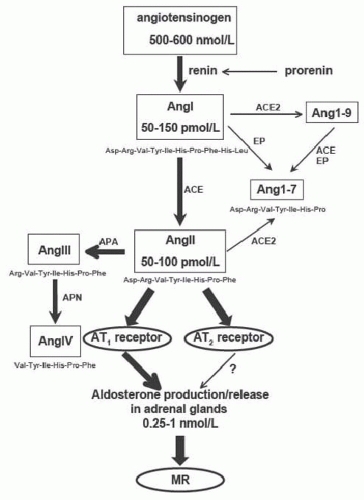
Ang II is the primary active product of the RAAS. It is an octapeptide derived from Ang I after cleavage of the C-terminal dipeptide by the ACE. Most of the physiologic actions of the RAAS on the vasculature and transport functions are mediated by Ang II action on angiotensin receptors, primarily type 1 (AT1) receptors. However, other angiotensin peptides with reported biologic activity have been identified, such as angiotensin III (Ang III) and IV (Ang IV), which are formed from Ang II by the sequential removal of amino acids from the N-terminus by aminopeptidases (Fig. 8.3). They are predominantly seen in the kidney and brain, where aminopeptidases A and N are prevalent.107,117 Ang III, also known as angiotensin 2-8, is a heptapeptide with suggested role in blood pressure maintenance in the brain. Ang IV (angiotensin 3-8), on the other hand, is a hexapeptide that possibly enhances Ang II signaling. The heptapeptide Ang (1-7) is currently considered one of the biologically active end products of the RAAS. It is formed from Ang I or Ang II in the kidney and heart by the action of tissue peptidases at the Cterminus. Once formed, it is rapidly hydrolyzed by ACE; in conditions of ACE inhibition or AT1 receptor antagonism, its concentration may increase severalfold. There are two major interactions between Ang (1-7) and bradykinin (BK): potentiation of BK by Ang (1-7) and mediation of vascular actions of Ang (1-7) by kinins. Both mechanisms are involved in the cardioprotective effects of ACE inhibitors. At the kidney level, the proposed role of Ang (1-7) is natriuresis and diuresis as opposed to Ang II, an effect blocked by AT1 receptor antagonists like losartan. In the vascular bed, it antagonizes the vascular effect of Ang II by acting as a competitive antagonist to AT1 receptors.


FIGURE 8.3 Schematic representation of the renin-angiotensin system showing plasma concentrations of some components as measured in anesthetized rats. EP, endopeptidase; APA, aminopeptidase A; APN, aminopeptidase N.


Angiotensin II Receptors

Circulating Ang II exerts its biologic effects by binding to specific receptors on the cell surface.112,121 At least four angiotensin receptor subtypes have been identified. AT1 receptors bind Ang II with higher affinity than Ang III and are selectively blocked by the biphenylimidazole compound losartan. AT2 receptors bind Ang II and III with similar affinity and are selectively blocked by tetrahydroimidazopyridines, such as PD123177.122 The type 3 angiotensin receptor AT3 has no known function and the type 4 (AT4) is thought to mediate the release of plasminogen activator inhibitor by Ang II, III, and IV.

Megalin is an abundant membrane protein heavily involved in receptor-mediated endocytosis. Megalin is a receptor for Ang II and Ang II internalization in some tissues is megalin-dependent. Megalin may play a role in regulating proximal tubule Ang II levels.123 Ang (1-7) exert its vasodilator and natriuretic actions presumably through its binding to a unique receptor, the Mas receptor.124

In rodents, two isoforms of AT1 receptors exist: AT1A (nephron) and AT1B (glomerulus), whereas in humans there is only one AT1 isoform. The AT1 receptor has widespread expression in the human adult and is found in the kidney, adrenal gland, heart, and brain. In the kidney, AT1 receptors are found in the glomeruli, proximal tubule brush border and basolateral membranes, thick ascending loop, proximal convoluted tubule, renal vasculature, the proximal and distal nephron segments, and in both cortical and medullary regions.125,126


Aldosterone

Aldosterone was identified by Simpson SA in 1953 and named electrocortin.127 Later studies characterized more the nature of this hormone and identified it as a mineralocorticoid synthesized and secreted by the zona glomerulosa of the adrenal cortex. The physiology of aldosterone action has been well established after several breakthroughs in research experiences, such as the identification of the mineralocorticoid receptor (MR) as the principal aldosterone receptor, the characterization of sites of aldosterone action in target tissues such as the ENaC, and the demonstration of post-receptor processes involved in physiologic responses to aldosterone.128,129,130 Aldosterone secretion is determined by several stimuli, the most important being Ang II and plasma potassium concentration. The MR is the principal receptor for aldosterone, but other target proteins can also bind aldosterone, such as the glucocorticoid receptor. Aldosterone has been implicated in the pathophysiology of several cardiovascular and renal diseases and will be discussed in details in other sections.129


Physiologic Actions of Angiotensin II


Systemic Effects of Angiotensin II

AT1 receptors have been shown to mediate many of the functions of Ang II in the regulation of blood volume, cell contraction, cell proliferation, aldosterone secretion, pressor and tachycardic responses, increased thirst, and hypertension secondary to renal artery stenosis. AT1 receptors are positively coupled to phospholipase C and mitogen-activated protein kinases (PI3 and MAP) and negatively to adenylate cyclase.122,125,131 The predominant function of the renin-angiotensin system is regulation of vascular tone and renal salt excretion in response to changes in the volume of extracellular fluid or blood pressure. Ang II represents the effector limb of this hormonal system, acting on several organs, including the vascular system, heart, adrenal glands, central nervous system, and kidneys. Through direct action on smooth muscle cells, Ang II significantly increases arteriolar resistance in renal, mesenteric, dermal, coronary, and cerebral vascular beds.132 Skeletal muscle and pulmonary vessels, on the other hand, are not affected because of Ang II-stimulated production of vasodilatory prostaglandins by endothelial and smooth muscle cells in these vascular beds.133,134 Ang II exerts indirect pressor effects via the central and peripheral nervous systems. Its effects on the central nervous system include increased sympathetic discharge and decreased vagal tone.135 Peripherally, Ang II augments the vasoconstrictive response to renal nerve stimulation in
dogs136 and its inhibition attenuates the pressor response to norepinephrine in humans.137 Experimental data suggest the presence of a local renin-angiotensin system in the vasculature that contributes to the regulation of vascular tone.112

A more recently recognized function of Ang II is its growth-promoting effects in smooth muscles of the vasculature, heart, and the kidney. Ang II has been shown to induce hypertrophy and mitogenesis in cultured vascular smooth muscles.138,139 This effect is at least, in part, mediated through autocrine production of growth factors such as platelet-derived growth factor (PDGF) and transforming growth factor-β (TGF-β).140 Some studies suggest that the renin-angiotensin system contributes to neointimal formation and restenosis after angioplasty.112 Ang II has been shown to have direct inotropic, chronotropic, mitogenic, and hypertrophic effects on isolated atria and ventricles.112 Amelioration of hypertensive cardiomyopathy by ACE inhibitors suggests that the renin-angiotensin system plays a role in cardiac hypertrophy.112

The predominant physiologic role of the AT2 receptor is to initiate vasodilation and natriuresis as a counterregulatory response to the vasoconstriction caused by activation of the AT1 receptor.141 Other functions include inhibition of growth and hypertrophy, and stimulation of apoptosis.142 This has been most clearly demonstrated in AT2 receptor knockout mice that have slightly elevated blood pressure in the basal state, but have an exaggerated increase in blood pressure in response to Ang II infusion compared to wild type mice.143,144 The intracellular signaling pathways coupled to the AT2 receptor are unclear. However, there is evidence that the AT2 receptor may be coupled to the production of a variety of renal vasodilator substances, counterregulating the pressor effects of Ang II via AT1 receptors.142 The most likely candidates are bradykinin and nitric oxide (NO)-stimulated cyclic guanosine monophosphate (cGMP),145,146 but other candidates include products of cyclooxygenase, such as PGE2 and PGI2.141


Renal Effects of Angiotensin II

Ang II serves at least three important functions in the kidney: regulation of blood flow and GFR, reduction of salt excretion through direct and indirect actions on renal tubule cells, and growth modulation in renal cells expressing AT1 receptors.

In conditions of decreased renal blood flow, GFR is preserved at nearly constant value over a wide range of perfusion pressures. This phenomenon is known as autoregulation of GFR. At low levels of renal perfusion pressures, Ang II contributes to this phenomenon.147 Micropuncture studies have shown that Ang II exerts substantial effects on the renal microvasculature and hemodynamics; however, the individual contribution of the systemic and local renin-angiotensin systems to the overall regulation is still controversial. Ang II constricts both the afferent and efferent arterioles, reduces single nephron glomerular filtration rate and plasma flow, increases the filtration fraction, and decreases glomerular filtration coefficient. Other studies have shown that Ang II infusion preferentially vasoconstricts efferent arterioles counteracted by endothelial-derived NO148 and constricts descending vasa recta (DVR) through Ca2+ signaling in pericytes.149 The disproportionate increase in postglomerular resistance results in a marked increase in glomerular capillary hydrostatic pressure (PGC), ultrafiltration pressure, and filtration fraction, thus preserving GFR in the face of declining renal plasma flow (RPF). The selectivity of the vasoconstrictive action of Ang II for the efferent arteriole results from stimulation of vasodilatory prostacyclin synthesis by the afferent arteriole and not a preferential action of Ang II on the efferent arteriole.150 In fact, Ang II increases both afferent and efferent resistance in the presence of a cyclooxygenase inhibitor.150 Under certain pathophysiologic conditions, afferent arteriolar constriction predominates, leading to a decrease in both RPF and GFR.151 Deep nephrons have higher postglomerular Ang II tone and also higher Ang II sensitivity than superficial nephrons. The better preserved GFR in deep cortex during Ang II action may contribute to maintaining the renal-concentrating ability by providing NaCl for reabsorption by the ascending limb of the loop of Henle.152

In addition to its vascular effects, Ang II induces mesangial cell contraction, which leads to decreased Kf in vivo.153,154 This effect, however, is attenuated by the concomitant production of prostaglandins by mesangial cells.155

Ang II is one of the most potent sodium-retaining hormones in the body. Increased tubular sodium reabsorption is enhanced by both direct and indirect tubular effects of Ang II. Physiologic concentrations of Ang II (10-12 to 10-10 M) stimulate proximal tubule NaCl and NaHCO3 absorption at the proximal tubule.156 Indirectly, Ang II stimulates ion transport in the proximal tubule by changing the peritubular milieu. Ang II can both decrease peritubular capillary hydrostatic pressure and increase peritubular oncotic pressure, resulting in an increased driving force for ion reabsorption. Directly, Ang II can stimulate transport in the proximal tubule through interaction with the AT1 receptors found on both the apical and basolateral membranes of the tubule cells.157,158 It also stimulates calcineurin phosphatase activity in proximal tubule epithelial cells through a mechanism involving AT1 receptor-mediated tyrosine phosphorylation of the PLC isoform, both linked to sodium transport in the proximal tubule.159 Specifically, Ang II stimulates the activity of apical membrane Na-H antiporters and basolateral membrane Na-HCO3-CO2 cotransporters160 and the activity of Na-K-ATPase by changing phosphorylation and conformation of Na-K-ATPase.161 The net effect is increased proximal tubule reclamation of Na and HCO3. Denervation of the proximal tubule results in attenuation of the Ang II-stimulated NaCl, but not NaHCO3 absorption, suggesting that Ang II enhances proximal Na transport indirectly by increasing presynaptic catecholamine release.159 Of note is that supraphysiologic concentrations of Ang II (10-9 to 10-7 M) inhibit NaCl and water reabsorption in the proximal tubule and also inhibit Na-glucose cotransporter translocation by inactivation of PKA and decrease of PI3-kinase activity mediated through the AT1 receptor.162,163 Ang II increases proximal tubule phosphate absorption by
direct stimulation of Na/Pi cotransport activity as a result of increase in the expression of brush-border membrane NaPi-IIa protein level and that stimulation is most likely mediated by posttranscriptional mechanisms.164

Intraluminal conversion of Ang I to Ang II can occur in the cortical collecting duct, resulting in enhanced apical sodium entry.165 The AT2 receptor regulates epithelial sodium channel (ENaC) abundance, consistent with a role for Ang II in regulation of collecting duct function via AT1 receptors.166,167

Ang II increases basolateral K-channel activity via the stimulation of AT1 receptors and the stimulatory effect of Ang II is mediated by a NO-dependent cGMP pathway.168

Other effects of Ang II on proximal tubule cells include enhanced gluconeogenesis and ammonia production.169 The effects of Ang II on distal tubule transport of Na and K are mediated through aldosterone release.170 In addition to proximal tubule epithelial cells, vasa recta and outer medullary vascular bundles express high density of AT1 receptors.121 ACE inhibitors increase descending vasa recta blood flow, whereas Ang II infusion markedly decreases medullary blood flow in rats.171 It is postulated that Ang II influences urinary dilution and concentration by modulating blood flow to the medulla.

Ang II induces hypertrophy of proximal tubule epithelial cells in vitro.172 It also exerts similar growth-promoting effects on mesangial cells.173 The signaling mechanism by which Ang II exerts this effect is not precisely known, but p27Kip1 is required for Ang II-induced hypertrophy.174 Downstream potential targets of Ang II are the extracellular signal-regulated kinases 1 and 2 (ERK1/ERK2) and Ang II activates ERK1/ERK2 via the AT1 receptor.175 Studies have shown that connective tissue growth factor (CTGF) might be an important mediator of Ang II-induced renal hypertrophy, which suggests that inhibiting the production of CTGF might be the new strategy in early prevention of renal fibrosis.176 Ang II can stimulate human kidney fibroblast (KFB) proliferation and enhance the expression of interleukin-6 in KFB. These findings suggest that Ang II might play a part in the mechanisms for modulating tubulointerstitial changes and inducing renal fibrosis.177

Previous in vivo studies in cardiomyopathic hamsters suggested that the expression of vasopressin (AVP) V2 mRNA is upregulated by Ang II. Ang II caused a significant increase in the AVP V2 mRNA in a dose-dependent manner mediated by PKA, whereas PKC suppresses the expression of V2 mRNA in the inner medullary collecting duct (IMCD) of the rat kidney.178

The role of Ang II in the pathogenesis of hypertension is complex. Several studies have demonstrated a crucial role of the intrarenal renin-angiotensin system in the development of hypertension and kidney disease.117 Animal studies have shown that blockade of the AT1 receptor resulted in increased plasma Ang II levels while it limited renal Ang II contents in response to Ang II infusion.179 This dissociation between systemic and local Ang II regulation has been demonstrated in several animal hypertension models including renovascular hypertension.180,181 Clinical studies have also shown that local intrarenal Ang II formation was central to the development of hypertension in humans.182

In addition to its effects on the maintenance of blood pressure, AT1 receptors may play a role in embryonic nephrogenesis. Blockage of the renin-angiotensin system with ACE inhibitors or AT1 inhibitors results in abnormal renal development that is characterized by both papillary and tubular atrophy and by interstitial fibrosis and infiltration. In addition, knockout mice lacking both the AT1A and AT1B receptor have similar renal abnormalities.183 Ang II also modulates mesangial cell growth, and induces proximal tubular cell hypertrophy in humans, effectively inhibited by irbesartan, an Ang II receptor antagonist.184

The AT2 receptor is expressed predominantly in fetal tissues, but in almost all tissues there is postnatal downregulation. AT2 receptor mRNA is expressed in the fetal and neonatal rat kidney, but disappears after the neonatal period and is not expressed in the normal adult. Although the AT2 receptor mRNA is not found in the adult kidney, both immunohistochemistry and Western blot analysis have detected AT2 receptor protein in the glomeruli, cortical tubules, and interstitial cells of the adult kidney.121,185 It was thought that AT2 receptors might play a role in the development of the kidney and urinary tract given the high levels of expression in the fetus. However, although early studies of AT2 receptor knockout mice showed no gross morphologic abnormalities of the kidney,143,144 a more recent study has demonstrated the presence of increased numbers of congenital anomalies of the urinary tract.186 The AT2 receptor may be implicated in some congenital abnormalities of the urinary tract or may be involved in the pathophysiologic response to ureteral obstruction by protecting against the formation of interstitial fibrosis.186,187 Ang II modulates the over-expression of AT2 receptors in renal ablation experiments through its own AT2 receptor and functional expression of this effect may represent a counterregulatory mechanism to modulate the renal damage induced by renal ablation.188

Ang II regulates renal parathyroid hormone-related protein (PTHrP), a vasodilator and mitogenic agent upregulated in kidney injury, and its type-I receptor (PTH1R) system via AT1 receptors.189


Local Effects of Ang II in Nonrenal Organs

Ang II exerts its actions on other organs, such as the adrenals and the brain. At the level of the adrenals, it stimulates aldosterone synthesis and secretion.170 Acute angiotensin administration stimulates the activity of 11β-hydroxysteroid dehydrogenase (HSD) type 2 in human kidneys and exerts a dual effect on the MR receptor (i.e., an indirect agonistic effect by increasing aldosterone availability and a direct or indirect antagonistic effect by stimulation of renal 11β HSD type 2 activity).190

At the central nervous system (CNS) level, it stimulates thirst and salt appetite191,192 and may increase secretion of
vasopressin and oxytocin from the posterior pituitary, and adrenocorticotropic hormone (ACTH), prolactin, and luteinizing hormone from the anterior pituitary.193


Regulation of the Systemic Renin-Angiotensin-Aldosterone System

Ang II in the systemic circulation is produced primarily from circulating angiotensinogen through the proteolytic action of renin. Most of the circulating renin is derived from the kidney, although other organs can secrete prorenin in the circulation. Angiotensinogen is mostly formed and secreted in the circulation by liver cells, allowing for systemic formation of angiotensin peptides, principally Ang II. The circulating concentrations of angiotensinogen are more than 1,000 times greater than those of Ang I and Ang II, therefore changes in plasma renin activity is the major determinant of Ang I formation (Fig. 8.3). Renin secretion is stimulated by various dynamic parameters such as renal perfusion pressure, tubular fluid sodium chloride concentration at the MD, sympathetic discharge to the kidney, and endocrine and paracrine hormones and growth factors.107

Stimulation of renin release by juxtaglomerular cells is mediated by increased intracellular cAMP, whereas a rise in cytosolic free calcium is inhibitory.194 Renin release responds inversely to changes in renal perfusion pressure.114 Elevation of intrarenal arterial pressure inhibits renin release and induces a “pressure” natriuresis. At least two mechanisms have been postulated. Increased afferent arteriolar wall tension secondary to increased renal perfusion elevates intracellular calcium in juxtaglomerular cells and inhibits renin secretion.114,194 Increased perfusion pressure also stimulates NO production and release by endothelial cells. NO, in turn, suppresses renin secretion.195,196 Conversely, decreased renal perfusion results in increased production of prostacyclin (prostaglandin I2), which enhances renin release.197 Mechanical strain leads to upregulation of the AT1 receptor and increased Ang II production in conditionally immortalized podocytes. The resulting activation of a local tissue angiotensin system leads to an increase in podocyte apoptosis, mainly in an AT1 receptor-mediated fashion.198

Decreased NaCl delivery to MD cells stimulates renin secretion, whereas increased urinary NaCl exerts an opposite effect.180 Schlatter and coworkers199 demonstrate that changes in luminal Cl concentration alter the rate of Na+-K+-2Cl transport in MD cells.199 The precise mechanism by which variation in the activity of this transporter translates to a signal that regulates renin release by adjacent juxtaglomerular granular cells is not entirely clear. Postulated mediators include adenosine, which inhibits renin secretion via activation of A1 receptors on juxtaglomerular cells, and alterations in interstitial osmolality, which may affect renin secretion directly.96 Experimental evidence also suggests that NO produced by MD cells and endothelial cells regulates renin secretion.195,200

The importance of renal sympathetic innervation in controlling renin secretion is well recognized.201 Stimulation of postjunctional β-adrenergic receptors increases renin release. The role of α-adrenergic receptors, on the other hand, is controversial.201 Ample evidence suggests that dopamine stimulates renin secretion by direct activation of dopamine A1 (DA1) receptors on juxtaglomerular cells.201,202

Several endocrine and paracrine hormones regulate renin secretion by the kidney. ANP has been shown to inhibit renin release from isolated juxtaglomerular cells.203 Other inhibitory hormones include AVP, endothelin, and adenosine (A1-receptor agonists).114,195 Regulation of renin secretion by Ang II is probably the most physiologically relevant.169 Ang II inhibits renin secretion and renin gene expression in a negative feedback loop. Treatment of transgenic mice bearing the human renin gene with an ACE inhibitor increases renin expression in the kidney by five- to tenfold.204 Similarly, ACE inhibition in rats augments renal renin mRNA expression, an effect that is reversed by infusion of Ang II.205 The effects of Ang II are believed to be direct and not dependent on changes in renal hemodynamics or tubular transport. Arachidonic acid metabolites produced in the kidney also play an important role in renin secretion.195 Intrarenal infusion of arachidonic acid increases (and indomethacin decreases) plasma renin activity in rabbits.206 Several studies have since confirmed that prostaglandins of the I series are potent stimulators of renin secretion.195,197 On the other hand, lipoxygenase products of arachidonic acid metabolism (12-HPETE, 15-HPETE, and 12-HETE) and cytochrome P450-mediated epoxides (14,15-epoxyeicosatrienoic acid) have been shown to inhibit renin release in renal cortical slices.207,208


The Local Renin-Angiotensin System

The renin-angiotensin system has been characterized as an endocrine, paracrine, and autocrine system. Contribution of systemically formed mediators to local control of dynamics within tissues is difficult to delineate. Recent evidence suggests that local formation is a major determinant of Ang levels in organs and tissues. In the brain, for example, Ang peptide levels are regulated in an autonomous manner.209 Local renin-angiotensin systems have been identified in several organs, including the kidney, heart, vasculature, brain, and adrenals.210 Although most organs have elements of the renin-angiotensin system, the adult kidney is unique in expressing all the components of the system.156,169

Compared with plasma levels, the renal Ang II contents are much higher despite suppression of renin secretion and release.126 Renin is principally produced by juxtaglomerular cells of the distal afferent arteriole, but has been shown to be expressed in the proximal tubule cells,126 whereas its substrate, angiotensinogen, is expressed by proximal tubule cells. ACE activity in the kidney has also been localized to the proximal tubule, with the highest concentration present on the brush border. Several studies have provided evidence for production of Ang II by the kidney, mainly concentrated in the proximal tubules,126 suggesting that the intrarenal renin-angiotensin
system is, indeed, functional.156 Some investigators have proposed that this local system plays a role in proximal tubule NaCl and HCO3 absorption, pathogenesis of essential hypertension, and expression of the phenotype of autosomal dominant polycystic kidney disease.156 Independent regulation of renal Ang II production has not been definitively demonstrated. Circulating Ang II stimulates renal angiotensinogen mRNA production and intact urine angiotensinogen suggests its presence along the whole nephron and that renin and ACE activity are available all through the nephron.126

Endogenous Ang II in both peritubular blood and luminal fluid is important for maximal expression of the stimulatory influence of this peptide on proximal tubule fluid uptake.211 Intraluminal conversion of Ang I to Ang II can occur in the cortical collecting duct, resulting in enhanced apical sodium entry.165

Renal degradation of Ang II is constitutively high, unaffected by chronic levels of arterial blood pressure, and is independent of long-term changes in levels.212

Low-density lipoproteins (LDLs) increase Ang II production by mesangial cells, which, in turn, results in increased O2 production, cell proliferation, and hypertrophy—these effects of Ang II are mediated by the AT1 receptor.213


Established Clinical Pathophysiologic Role of the Renin-Angiotensin-Aldosterone System in Humans

The RAAS has been implicated in the pathophysiology of several diseases of the cardiovascular system and the kidney, mostly hypertension and renal injury. The local renal renin-angiotensin system is activated in the renovascular type of hypertension in the stenotic kidney and accounts for most of the Ang II concentration in the renal tissue.214 In addition, it plays a crucial role in other forms of hypertension. Administration of a renin inhibitor to normal subjects and hypertensive patients showed sustained local renal vascular responses but time-dependent decreased drug activity at the systemic level.215 Several other studies demonstrated the importance of the intrarenal renin-angiotensin system in the pathophysiology of hypertension.216,217

Ang II promotes fibrogenesis and oxidative stress in the kidney by stimulating fibrogenic mediators, altering renal hemodynamics especially facilitating glomerular hypertension, inducing tubulointerstitial hypoxia, and enhancing free radical formation. Studies have shown that the degree of mesangial hypercellularity and expansion in IgA nephropathy correlated closely with glomerular expression of mRNAs for renin, ACE, chymase, and AT1 and AT2 receptors.217,218 The role of the intrarenal renin angiotensin system has been also established in the pathogenesis of membranous nephropathy.219 In diabetic nephropathy, the renin-angiotensin system plays a crucial role in disease progression: the intrarenal generation of Ang II is increased, despite suppression of the systemic RAAS. Details of the pathophysiologic mechanisms implicated in diabetic nephropathy are beyond the scope of this chapter.

The role of the RAAS in end-stage renal disease (ESRD) is even more complicated and implicates many parameters related to residual renal function, presence of renal replacement therapy and modality, and drug treatment of associated conditions. In addition, renal transplantation adds a new dimension to the complexity by introducing an immune component through graft rejection and immunosuppressive drugs. Despite the substantial decrease in renal blood flow and function in patients with ESRD on dialysis, studies have shown that those with remaining kidneys have an activated intrarenal renin-angiotensin system.117 On the other hand, renal transplant diseases, especially rejection and cyclosporine-induced nephropathy, were associated with increased activity of systemic and local RAAS in several studies.220,221

In conclusion, the RAAS is one of the most powerful pressure and volume regulatory systems in the body, involved in physiologic responses and pathophysiologic processes at both the systemic and local levels. Autonomous and independent control of local renin-angiotensin systems in various organs and tissues may exist and contribute to disease progression. New areas of interest in the RAAS are continuously emerging, facilitating the understanding of mechanisms of diseases and development of specific drug targets.


ATRIAL NATRIURETIC PEPTIDE


Molecular and Biochemical Properties of Atrial Natriuretic Peptide

The cDNA for human ANP was isolated in 1984 and, shortly afterward, the gene was localized to the short arm of chromosome 1.222,223 The chromosomal gene consists of three exons and two introns encoding for a mature mRNA transcript approximately 900 bases long.223

Translation of human ANP mRNA results in a 151-amino acid preprohormone.224 Pro-ANP, a 126-residue molecule, is formed after cleavage of the signal peptide sequence of prepro-ANP and represents the major storage form of the hormone in atrial granules.225 The circulating, biologically active form of ANP, often referred to as AN99-126 or ANP1-28, is a peptide comprising the 28 carboxy-terminal amino acids of the parent molecule.224 The amino acid sequence of ANP99-126 is highly conserved among mammalian species.224 A disulfide bond between cysteine residues 105 and 121 gives ANP99-126 its ring structure, which is essential for biologic activity.226

Pro-ANP (1-30), (31-67), and (68-98) are secreted from the heart and circulate in the plasma. Pro-ANP (1-30) and (31-67) increase sodium and water excretion and binding sites have been found in the proximal tubules and collecting ducts. Pro-ANP (31-67) inhibits the Na+-K+ pump at the medullary collecting duct through a prostaglandindependent mechanism and no effect on cGMP production. Pro-ANP (1-30) infusion in rats clearly increases urine output, sodium, and potassium excretion, the mechanism of which still needs to be elucidated.227


Secretion and Physiologic Regulation of Atrial Natriuretic Peptide99-126

Cardiac atria contain the highest concentrations of ANP and serve as the major source of circulating hormone.224 ANP99-126 secretion from cardiomyocytes occurs largely in response to atrial stretch resulting from increased atrial transmural pressure.228 Physiologic stimuli for the release of ANP99-126, include acute salt and volume loading, supine posture (head-down tilt), and head-out water immersion.228,229,230 An increased rate of atrial contraction has also been shown to stimulate ANP99-126 secretion.231,232 Ang II, vasopressin, epinephrine, and phenylephrine stimulate ANP99-126 secretion from the heart largely because of their systemic vasopressor effects.233 On the other hand, glucocorticoids and endothelin raise ANP99-126 levels possibly by acting directly on atrial myocytes.234 Leptin decreases ANP secretion via an NO-mediated mechanism.235 Physiologic and pathologic conditions in which elevated plasma levels of ANP99-126 have been detected are summarized in Table 8.3.

The local synthesis of natriuretic peptides is increased in the kidney and in the vasculature in obstructive uropathy.236 The activation of the renin-angiotensin system during low sodium intake antagonizes the biologic effect of ANP by interfering in the intracellular metabolism of cGMP.237


TABLE 8.3 Conditions Associated with Increased Levels of Circulating Atrial Natriuretic Peptide


Physiologic


Pathologic


Acute volume expansion


Congestive heart failure


Supine posture


Atrial tachycardias


Head-out water immersion


Myocardial ischemia


Mineralocorticoid escape


Acute and chronic renal failure


Exercise


Postobstructive diuresis


Neonatal period


Nephrotic syndrome (subset) Cirrhosis with ascites Severe hypertension Primary hyperaldosteronism Hypoxia Other (SIADH myxedema)


SIADH, syndrome of inappropriate antidiuretic hormone secretion.


Physiologic Actions of Atrial Natriuretic Peptide99-126

Three subtypes of ANP receptors (NPRs) have been identified.238,239,240 NPR-A and -B have intrinsic guanylate cyclase activity that catalyzes production of cGMP after ligand binding. cGMP then serves as an intracellular second messenger that mediates the biologic activities of ANP99-126.241 NPR-B appears to have 50-fold higher affinity for a related natriuretic factor originally purified from porcine brain, known as C-type natriuretic peptide (CNP).242 NPR-C, previously known as the (clearance) receptor, is devoid of guanylate cyclase activity and, therefore, does not confer biologic activity and is thought to mediate clearance of circulating ANP along with metalloendoprotease (E.C.3.4.24.11)242 and of other related hormones, such as brain natriuretic peptide (BNP).243 Selective downregulation of NPR-C in the kidney in response to dietary salt supplementation may contribute to local elevation in ANP levels and may be functionally significant in attenuating the development of salt-sensitive hypertension.244 NPR density is decreased in diabetic rats with significant increase in plasma ANP levels.245 ANP also antagonizes AVP-mediated increases in water permeability in IMCD cells.246

The major sites of action of ANP99-126 are the kidneys, adrenal glands, and vascular smooth muscle.247 Short-term administration of ANP99-126 in laboratory animals and in humans induces pronounced natriuresis, diuresis, alteration in renal hemodynamics and tubular function, suppression of renin release, inhibition of aldosterone secretion by the adrenal glands, and decreased vasomotor tone, resulting in transient drop in systemic blood pressure. From these actions it has been postulated that ANP plays an important physiologic role in protecting against extracellular volume overload.248


Renal Actions of Atrial Natriuretic Peptide99-126

ANP-induced increase in the GFR is well established.249,250,251 ANP99-126 increases efferent arteriolar resistance, resulting in increased PGC and filtration fraction.249 In addition, ANP99-126 relaxes mesangial cells in vitro, suggesting that it can increase filtration area by cGMP generation in podocytes252 and Kf in vivo.253 Indeed, when baseline Kf is low, as in water-deprived animals, ANP99-126 enhances GFR mainly by increasing Kf.253 If preexisting vascular constriction is present in isolated perfused kidney, ANP tends to vasodilate renal vessels and increase renal blood flow (RBF).254 In whole animals, however, ANP99-126 infusion causes either a decline or no change in RBF.254 The effects of ANP99-126 on RBF are influenced by its systemic actions on blood pressure and the renin-angiotensin system. ANP seems to increase NO production at both renal and cardiac levels, further explaining its natriuretic and diuretic effects.255 Finally, ANP has been reported to induce redistribution of blood flow from the cortex to the medulla and to increase vasa recta flow, leading to dissipation of the medullary solute gradient.256,257


ANP99-126 has both direct and indirect effects on tubular transport of Na, chloride, and water.258 In the proximal tubule, ANP99-126 antagonizes Ang II-induced Na reabsorption259 and, along with endothelin-3, inhibits the sodium-glucose transporter260 and, like urodilatin, inhibits Na+-ATPase activity.261 In the IMCD, it directly inhibits Na transport by binding to ANP-R1 receptors and influencing amiloride-sensitive Na channels and the activity of apical Na-K-2Cl cotransporters.262 Other mechanisms by which ANP99-126 induces natriuresis and diuresis include suppression of renin and aldosterone release,263 inhibition of the tubular actions of AVP,246 and dissipation of the medullary solute gradient by impairing the increase in intracellular Ca2+ concentration. ANP blocks both the stimulatory and inhibitory effects of AVP on Na+-dependent pHi recovery.264


Atrial Natriuretic Peptide Transgenic Mice

Transgenic mice over-express pro-ANP in hepatocytes. These transgenic animals have a hypotensive phenotype (20 to 30 mm Hg lower than control littermates) without compensatory tachycardia. GFR remained normal despite hypotension. Moreover, significant diuresis or natriuresis during steady state was not detected. Contrary to observations made after short-term infusion of ANP99-126, plasma renin activity also did not change while aldosterone levels were elevated.265,266,267


Physiologic Consequences of Interrupting the Atrial Natriuretic Peptide Pathway

Disruption of the gene that encodes pro-ANP results in knockout mice that lack expression of ANP. Homozygous ANP knockout mice fed a standard diet have both mildly elevated blood pressure (average increase of 8 mm Hg) and cardiac hypertrophy compared to wild type mice, suggesting that ANP has a physiologic role in maintaining the normotensive state. This hypertension appears to be sensitive to dietary salt intake as feeding the homozygous ANP knockout mice a diet with an intermediate salt content (2%) would further increase blood pressure by an average of 20 mm Hg compared to wild type mice.268 Knockout mice lacking ANP-R1 activity (known as GC-A null mice) have both elevated blood pressure and cardiac hypertrophy, similar to pro-ANP knockout mice, but the GC-A null mice have a salt-insensitive form of hypertension.267 It is unclear why there should be a difference in the phenotype of these two types of knockout mice. It is possible that other guanylyl cyclase receptors, such as guanylyl cyclase C, can help regulate blood pressure in the face of changes in dietary salt intake and compensate for the lack of GC-A receptors.268 ANP (-/-) mice have a blunted pressure-natriuresis response and suppressed expression of local renal renin-angiotensin system.269


Atrial Natriuretic Peptide-Related Peptides149,212

BNP is a 32-amino acid peptide with structural homology to ANP99-126. Although originally isolated from porcine brain,270 it is also secreted by cardiac ventricles and, to a lesser extent, from atria. The biologic effects of BNP infusion are similar to those of ANP99-126. Unlike ANP, BNP secretion seems to be constitutive and unrelated to myocyte stretch. The kidney-specific degradation of ANP provides a mechanism for preferential regulation of kidney function by BNP, independent of peripheral ANP concentration.270 In 1990, another homologous peptide, CNP, was isolated from porcine brain.271 CNP is produced in the brain, where it achieves concentrations much higher than those of ANP and BNP. In contrast, circulating levels of CNP are lower. CNP lacks natriuretic, diuretic, and hypotensive effects and probably acts in a paracrine fashion in the CNS. The physiologic significance of BNP and CNP remains unclear.


Clinical Use of Atrial Natriuretic Peptide and Atrial Natriuretic Peptide Analogs in the Diagnosis and Treatment of Kidney Injury and Congestive Heart Failure

Measurement of circulating ANP and ANP analogs is now a well established marker in assessment of volume overload and left ventricular dysfunction and for predicting mortality, in the presence or absence of renal dysfunction.272,273,274,275,276 A number of studies, reviewed recently,277 provide support for the use of ANP/ANP analogs in protection against and treatment of acute kidney injury (AKI).278,279,280,281 In these studies, low (but not high)-dose ANP infusions reduced the need for renal replacement therapy and hospital and intensive care unit length of stay, but did not appear to improve mortality in the management of cardiac surgery-associated AKI. Hyporesponsiveness to ANP in congestive heart failure may be due to reduced NPR-A expression.282 In dogs, maximizing cGMP action with type V phosphodiesterase inhibitors augments the natriuretic responses to exogenous ANP.283 Animal studies have shown the usefulness of encapsulated ANP gene transfected cells as a new tool for ANP gene delivery with possible implication for future therapy.284


URODILATIN OR RENAL NATRIURETIC PEPTIDE

Urodilatin is best described as a paracrine renal natriuretic peptide (RNP).285 It was first isolated from human urine in 1988.286 Its amino acid sequence is identical to ANP99-126, except for an additional four amino acids at the amino terminal. Despite its high degree of homology to ANP99-126, specific antihuman RNP polyclonal antibody has been generated and RNP levels can be measured by radioimmunoassay.287 To date, RNP has not been detected in the circulation and the kidney is presumed to be its site of synthesis and action.285

RNP binds to ANP receptors in the kidney and stimulates cGMP production.288 Its renal actions parallel those of ANP99-126 and include more potent hyperfiltration, diuresis, and natriuresis.288 RNP, like ANP99-126, inhibits sodium uptake by inner medullary duct cells by inhibiting entry of Na
through apical sodium channels.289 It appears that the natriuretic effect of RNP is more potent than that of ANP, possibly because it is resistant to degradation by renal cortical metalloendopeptidase.290 Systemic infusion of RNP results in effects similar to those of ANP99-126.

Several physiologic studies suggest that RNP functions as a paracrine hormone that regulates renal Na excretion. Drummer and coworkers291 demonstrated in the human that urinary excretion of RNP, but not plasma ANP concentration, correlates with circadian variation in sodium excretion over a 9-day period. Moreover, acute infusion of normal saline in healthy subjects, balloon dilatation of left atrium, and water immersion induce a significant increase in urinary RNP.292,293


GUANYLIN AND UROGUANYLIN

Guanylin and uroguanylin are cGMP-regulating agonists isolated in 1994, respectively, from rat intestine and human/opossum urine that appear to have natriuretic properties.294,295 In humans and mice, earlier studies reported that both genes for guanylin and uroguanylin are located close to each other on chromosomes 1 and 4, respectively, near the ANP A and B genes and probably arising from an ancestral uroguanylin/guanylinlike gene.296 Preproguanylin and preprouroguanylin probably derived from a common precursor gene, as they share approximately 35% homology.297 Bioactive uroguanylin can be found in the urine at higher concentrations than guanylin, suggesting that uroguanylin may be a hormonal link between the intestine and the kidney.298 Uroguanylin, prouroguanylin, and proguanylin peptides have been shown to circulate in the plasma of humans and other animals.299


CORTICOSTEROIDS

Corticosteroids are steroid hormones synthesized by the adrenal cortex. On the basis of their physiologic functions, corticosteroids are traditionally divided into two groups— glucocorticoids (cortisone and cortisol) and mineralocorticoids (aldosterone)300—based on their potency in electrolyte and metabolism regulation.

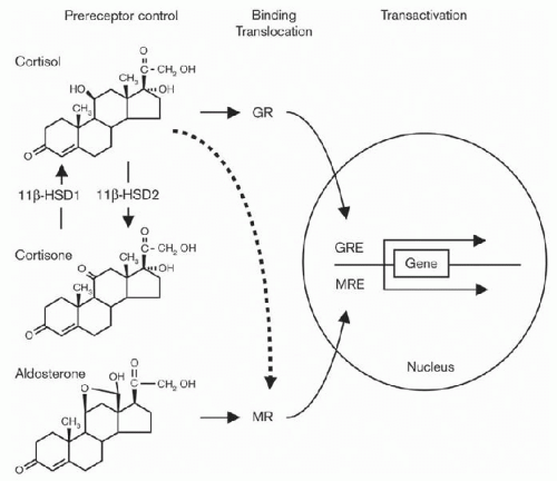
Corticosteroids bind to intracellular receptors: the mineralocorticoid receptor (MR) and the glucocorticoid receptor (GR). They are both part of the nuclear receptor family that also includes receptors for steroid and thyroid hormones, vitamin D3, and retinoic acids. These receptors translocate to the nucleus after ligand binding301 and regulate nuclear gene transcription at specific DNA response elements located in the five regulatory regions near the promoters of specific target genes (Fig. 8.4).302 Both glucocorticoids and mineralocorticoids modulate renal function. Aldosterone-sensitive tissues include the distal parts of the nephron (distal tubule, connecting tubule, and all along the collecting duct), the surface epithelium of the distal colon (where it increases sodium absorption and potassium excretion), and other specific nuclear-binding sites for aldosterone in the thick ascending limb of Henle’s loop and salivary and sweat glands. All MR-expressing tissues also express GR. In the kidney, evidence exists for distribution of the GR receptors along the whole parts of the nephron, except for the proximal tubule. No evidence exists for a glucocorticoid role in the colon.303,304,305

MR has the same affinity for both aldosterone and glucocorticoids. Glucocorticoids concentration in plasma is 100- to 1,000-fold higher than aldosterone and only 10% of it circulates as free, whereas all circulating aldosterone is free. MR selectivity exists to prevent complete occupancy of the MR receptor by glucocorticoids at physiologic concentrations. This is mainly mediated by the colocalization of the 11β-hydroxysteroid dehydrogenase 2 (11β-HSD2) enzyme in the distal nephron, along with MR. This enzyme transforms glucocorticoids (cortisol in humans) into metabolites (cortisone) that have weak affinity to MR.301,305,306,307 11β-HSD has two main isoforms. 11β-HSD1 acts predominantly as a reductase in vivo in many tissues, regenerating biologically active glucocorticoids (mainly cortisol in human and corticosterone in rodents) from their inactive forms (cortisone in human and 11-dehydrocorticosterone in rodents). 11β-HSD2, on the contrary, is a dehydrogenase that inactivates glucocorticoids.308 The major role of 11β-HSD2 is highlighted in clinical situations, such as the syndrome of apparent mineralocorticoid excess where it is inactivated or after ingestion of excessive amounts of licorice where glycyrrhetinic acid, a derivative of licorice, has been described to inhibit 11β-HSD2.309,310 Both of these conditions lead to hypokalemic hypertension with low renin and aldosterone levels. In cirrhosis also, there is MR activation by cortisol explained by a reduced activity of 11β-HSD2, which allows promiscuous activation of MR by the glucocorticoid cortisol as suggested by Frey.311

GRs have approximately equal affinities for aldosterone and endogenous glucocorticoids, but have the highest affinity for dexamethasone, a synthetic glucocorticoid.301 Because mineralocorticoids circulate at much lower concentrations than glucocorticoids, significant binding of mineralocorticoids to GR does not occur under physiologic conditions.


Nongenomic Actions of Aldosterone

The effects of aldosterone on its target cells have long been considered to be mediated exclusively through the genomic pathway and were characterized by a 45-minute lag period; however, evidence has been provided for rapid effects of the hormone that may involve nongenomic mechanisms.5,6,7,19,32 On the other hand, in a series of in vitro studies, aldosterone was shown to have a half maximal effect on both rapid (15 minutes) and delayed (120 minutes) Na flux.4,32 The Na/H exchanger has been identified as a target for nongenomic regulation. Aldosterone rapidly increases Na/H exchanger activity in a variety of cells, including distal colon and renal epithelial cell lines,3 but rapidly inhibits apical NHE3 and HCO3 reabsorption in medullary thick ascending limb (MTAL) and has a dose-dependent biphasic effect on the Na/H exchanger (low doses stimulate and high doses inhibit it).312


FIGURE 8.4 The intracellular concentrations of steroid molecules available for binding to glucocorticoid receptor (GR) or mineralocorticoid receptor (MR) depend on the free extracellular concentrations available for diffusion into the cytoplasm and the intracellular prereceptor control mechanism constituted by the 11β-hydroxysteroid dehydrogenase type 1 and 2 (11β-HSD1, 11β-HSD2) enzymes. Whereas 11β- HSD1 acts predominantly as a reductase and converts the 11-ketosteroid cortisone with virtually no affinity for MR and GR into the 11β-hydroxyglucocorticoid cortisol with a high affinity for both GR29,58 and MR, 11β-HSD220 is exclusively an oxidase and inactivates cortisol into cortisone, which allows protection of MR-expressing cells from promiscuous activation of MR by the glucocorticoid hormone cortisol. GRE, glucocorticoid-response element; MRE, mineralocorticoid-response element. (Frey FJ, Odermatt A, Frey BM. Glucocorticoid-mediated mineralocorticoid receptor activation and hypertension. Curr Opin Nephrol Hypertens. 2004;13:451-458.)

Aldosterone acts through nongenomic pathways to regulate many different ion transport proteins and signaling pathways in a variety of renal epithelial cells, such as proximal tubule cells derived from human renal cortex, MDCK-C11 cells (a cell line that exhibits properties of collecting duct intercalated cells), and principal cells isolated from rabbit cortical collecting duct and both M-1 and RCCD2 cortical collecting duct cell lines.6 Studies using isolated perfused tubules also demonstrate that aldosterone, via nongenomic mechanisms, regulates the transepithelial transport function of different nephron segments, such as proximal S3 segment,21 renal MTAL, type A intercalated cells of outer medullary collecting ducts, and principal cells in the connecting tubule and inner medullary collecting ducts.6,313 In renal epithelial cells, an elevation in cytosolic Ca2 serves as a second messenger for the nongenomic Na/H exchanger activation initiated by aldosterone.2 The existence of a nongenomic action of aldosterone is in general attributed to conditions where this action is observed over a short period of time (minutes) against the much longer time (hours, days) needed for a genomic action.313

Rapid nongenomic aldosterone effects are characterized by their rapid onset of action (within minutes) and an insensitivity to inhibitors of transcription (e.g., actinomycin D) and of protein synthesis. Aldosterone also acts via rapid nongenomic effects in vivo in humans at the renal vasculature. Antagonizing the endothelial NO synthase unmasks these effects. Therefore, rapid nongenomic aldosterone effects increase renal vascular resistance and thereby mediate arterial hypertension if endothelial dysfunction is present.314 Evidence suggests that there is a nonclassical membrane-bound aldosterone receptor.315 These data come from kinetic studies that demonstrate saturable, radiolabeled binding of aldosterone to cell surface membranes that have kinetics compatible with physiologic activity.316,317 In some cell lines, aldosterone can have very rapid physiologic effects that are not blocked by inhibitors of cell transcription and translation. For instance, in human mononuclear leukocytes, aldosterone can stimulate release of IP3 or calcium within 30 seconds of exposure.318,319 These membrane-bound receptors may explain some of the effects of aldosterone that occur prior to gene transcription, such as early stimulation of
sodium reabsorption320 or early stimulation of salt intake,321 possibly via phospholipase C/PKC signaling pathways.


Mineralocorticoid Actions in the Kidney

Aldosterone originates primarily from the zona glomerulosa of the adrenal glands. Some recent studies have suggested that the heart, vasculature, and brain can also synthesize aldosterone in response to local tissue injury, although extraadrenal synthesis in humans is still debated.6,7,8,11,17,322 It is the chief mineralocorticoid of the body. Its physiologic role as a regulator of sodium and volume balance also allows aldosterone the potential to play a pathologic role in the development of hypertension in patients with renal disease.322 It is also implicated in the pathophysiology of cardiac fibrosis and cardiac hypertrophy in end-stage heart failure.3,4,323

The major action of aldosterone in the kidney is regulation of Na, K, and H handling by the distal part of the nephron. Mineralocorticoid deficiency is associated with volume depletion, hyperkalemia, and mild metabolic acidosis. Conversely, mineralocorticoid excess leads to Na retention, hypokalemia, and metabolic alkalosis.

Ang II, high serum K+ levels, and ACTH stimulate aldosterone secretion from the adrenal gland.324 ANP and dopamine, on the other hand, suppress aldosterone secretion. Dietary sodium also modulates aldosterone release through its effects on the renin-angiotensin system. In addition to sodium and volume homeostasis, other triggers for aldosterone release have been cited, including hyperglycemia, adrenocorticotropic hormone (ACTH), and, more importantly in patients with CKD, angiotensin II and potassium.49


Sodium Reabsorption

One of the best-documented functions of aldosterone is its ability to increase Na reabsorption in the distal tubule and collecting duct.325,326,327 The rate-limiting step to sodium reabsorption across tight epithelia is the permeability of the apical membrane of the transporting cell. Aldosterone increases apical Na permeability of tight epithelia, such as those found in the mammalian distal tubule and descending colon, by increasing the activity of the amiloride-sensitive epithelial sodium channel (ENaC). ENaC is formed by three subunits (alpha, beta, and gamma) and, based on coexpression studies in Xenopus oocytes, these subunits assemble into a complex heterooligomer forming the amiloride-sensitive pore.328,329,330

Other characterized aldosterone-induced targets include the serum and glucocorticoid-regulated kinase-1 (SGK-1), an important mediator of renal sodium homeostasis; corticosteroid hormone-induced factor (CHIF), which regulates the activity of the sodium and potassium-dependent adenosine triphosphatase pump (Na-KATPase); the glucocorticoid-induced leucine zipper protein; a transcription factor; and the G protein K-Ras2.10,11,12 SGK-1 is thought to regulate Na+ flux by increasing ENaC activity at the apical surface of epithelial cells. Aldosterone treatment of a rat collecting duct cell line as well as an adrenalectomized rat model demonstrated a rapid induction of SGK-1 mRNA within 30 minutes of treatment.13,14 However, SGK-1 null mice only show mild abnormalities in sodium homeostasis, suggesting that other genomic targets are important for overall regulation of sodium transport.15 Wong et al. demonstrated that ET-1 is a direct aldosterone gene target in the kidney and colon in Sprague dawly rats and may play an important role in aldosterone-regulated ion homeostasis.323

Aldosterone also enhances Na reabsorption by increasing Na-K-ATPase activity in basolateral membranes of principal cells in mammalian collecting duct and distal tubule.327 Studies in toad bladder and mammalian nephron suggest that aldosterone upregulates Na-K-ATPase activity by at least three mechanisms: increased Na influx due to opening of amiloride-sensitive Na channels, induction of Na-K-ATPase subunit expression at the gene level, and induction of intracellular alkalosis, which occurs in tissues that contain aldosterone-sensitive Na/H exchangers.331 Other hormones also modulate aldosterone’s action on Na transport; for example, ANP is inhibitory and vasopressin is stimulatory (Table 8.3).332

Hypersecretion of endogenous mineralocorticoids or the administration of mineralocorticoids lead to transient sodium retention followed by a return to Na balance within a few days.333 The return to Na balance despite elevation of circulating mineralocorticoid levels is referred to as aldosterone or mineralocorticoid “escape.” During mineralocorticoid escape, increased Na reabsorption by the distal tubule and collecting duct remains unchanged, but is offset by decreased Na reabsorption in other nephron segments.332,334 The latter results from increased renal arterial pressure and elevated plasma ANP levels, both of which suppress proximal tubule transport of Na. Mineralocorticoid escape is also, in part, mediated by decreased Na and water reabsorption in the loop of Henle.334 Other factors, such as TGF-β and interleukin-1 (IL-1), may play a role in regulation of mineralocorticoid escape. These factors have been recently found to inhibit the action of aldosterone on the cells of the IMCD.335

Ang II binds to Ang II type 1 (AT-1) receptors in the kidney, which leads to glomerular hypertension, sclerosis, renal fibrosis, and cardiac remodeling, possibly through a TGF-β-mediated pathway.30,31,32 ACE inhibitors provide renoprotection by inhibiting the conversion of Ang I to Ang II and precluding the negative effects of AT-1 receptor activation. Studies have shown that Ang II can be generated by ACE-independent pathways such as chymase in the heart,33 which leads to “angiotensin escape.” Angiotensin escape is one of the factors frequently cited to explain aldosterone breakthrough, which may be secondary to the generation of non-ACE mediated angiotensin II.34 Potassium and adipocyte-released factors may also contribute to the phenomenon of aldosterone breakthrough. Continual dietary intake of potassium in the setting of reduced GFR may promote elevated potassium levels that subsequently trigger aldosterone secretion, as seen in the rat remnant kidney model.28 ACE inhibitors and ARBs are also known to reduce
potassium excretion, so prolonged use of such agents may enhance potassium retention and predispose a patient to aldosterone breakthrough. Recent studies in rat models of metabolic syndrome with early nephropathy have shown enhanced aldosterone secretion due to adipocyte activity that was not abolished by candesartan administration.35 These results suggest that adipocyte-released factors outside of Ang II may enhance aldosterone secretion and lead to increased proteinuria and podocyte injury in rats.35 Thus, potassium, angiotensin II, and adipocyte-released factors may all contribute to the increase in aldosterone secretion in patients on prolonged ACE inhibitors or ARB therapy. The exact definition of aldosterone breakthrough has been a subject of controversy, as there is no current consensus on its precise definition. One of the common definitions is a rise in plasma aldosterone concentration, often past baseline values, following an initial decrease after the initiation of ACE inhibitor or ARB therapy.322


Potassium Secretion

Mineralocorticoids are the predominant hormonal influence on K secretion by principal cells of the collecting duct and connecting segment of the distal tubule.335,336 Although mineralocorticoids always increase K secretion by these nephron segments, this does not necessarily translate into a kaliuresis because of the strong dependence of K excretion on distal Na delivery and urinary flow rate.337 For example, in conditions of decreased Na delivery and urinary flow to the distal nephron, the kaliuretic effect of aldosterone is either diminished or abolished. The mechanisms by which aldosterone stimulates K secretion by principal cells overlap with those responsible for its Na-retaining action. The late distal convoluted and connecting tubules (CNTs) and cortical collecting duct (CCD) of the distal nephron mediate, in large part, the final regulation of urinary K+ excretion.19 The traditional model by which K+ secretion is accomplished in these segments can be summarized as follows. Na+ enters the CNT and principal cell from the urinary fluid through the apical amiloride-sensitive ENaC and is then transported out of the cell at the basolateral membrane in exchange for uptake of K+ via the basolateral Na+-K+-ATPase. The high K+ concentration within the cell and lumen-negative voltage, established by electrogenic Na+ reabsorption, create a favorable electrochemical gradient for K+ to diffuse into the urinary space through apical K+-selective channels. Thus vectorial K+ secretion in these segments requires a favorable electrochemical gradient and an apical permeability to K+ increases in extracellular K+ concentration directly stimulate aldosterone production in zona glomerulosa cells of adrenal glands.6,59,338

Aldosterone-induced Na influx through the apical membrane leads to the generation of a lumen-negative potential difference that favors K secretion.326,327 In addition, although mineralocorticoids do not increase the density of active K channels in the apical membrane, they increase the conductance of apical and basolateral K channels independent of Na flux.327,339 Physiologically, it is difficult to understand how one hormone can regulate the concentration of two different solutes that have varying levels of dietary intake. Patch-clamp experiments have demonstrated that other nonaldosterone circulating factors exist that can regulate K channel activity. Infusion of aldosterone by osmotic minipump will increase the density of EnaC, but not of K channels.339 However, an increase in dietary K does increase the K-channel density.340 Currently, it is unknown what other circulating factor controls K secretion.

A large body of evidence suggests that SGK1 mediates, at least in part, the effect of aldosterone on renal K secretion.41,88,113,116 This notion is supported by studies performed in SGK1 knockout mice demonstrating that the phenotype of SGK1 deletion is similar to MR knockout mice and displays impaired renal K secretion in response to high dietary K intake.41,341


Renal Acidification

The role of mineralocorticoids in regulation of renal acidification is supported by several clinical observations. Syndromes of aldosterone deficiency are associated with metabolic acidosis because of reduced urinary acid excretion, whereas mineralocorticoid excess results in metabolic alkalosis. Aldosterone enhances urinary acidification through direct actions on epithelial cells in the collecting duct and indirectly by influencing various intrarenal and extrarenal factors.342 Aldosterone increases H secretion by type A intercalated cells in the collecting duct via two mechanisms: direct stimulation of the proton pump (H-translocating ATPase), and indirectly by stimulating Na influx, which creates a lumen-negative potential difference.342 As with K, the overall effect of aldosterone on renal acid excretion depends on Na delivery to the distal part of the nephron. Reduction of Na transport in the collecting duct, because of either decreased Na delivery or inhibition of distal Na reabsorption by amiloride, significantly attenuates the effect of aldosterone on net H excretion.343 Aldosterone regulates the expression of aquaporin, AQP3, with no effect on AQP1 and AQP2, in the collecting duct, independently of Na intake.344


Effect on Inflammation

Besides its classical effects on salt homeostasis in renal epithelial cells, aldosterone promotes inflammation and fibrosis and modulates cell proliferation. As outlined by recent reports, mineralocorticoid also mediates inflammation and fibrosis through NF-κB activation in liver, heart, and glomerular mesangial cells9,10,11,12,13 via a pathway involving the aldosterone early-induced gene, serum, and glucocorticoid-induced kinase 1 (SGK1).11,12,345

Studies in vitro revealed that aldosterone could induce proliferation of rat glomerular mesangial cells and promote collagen gene expression and synthesis via activation of extracellular signal-regulated kinase 1 and 2 (ERK1/2) mitogen-activated protein (MAP) kinase in renal fibroblast. Aldosterone
treatment of renal tubular epithelial cells increases calcium inflow and intracellular cyclic adenosine monophosphate levels. The results suggest that aldosterone plays a pivotal role in tubulointerstitial fibrosis by promoting tubular epithelial-mesenchymal transition and collagen synthesis in proximal tubular cells. The process is MR-dependent, and mediated by ERK1/2 mitogen-activated protein kinase pathway.346

Clinical trials have shown a potential role for MR blockers to further delay the development of end-stage renal disease by completing renin-angiotensin blockade. MR blockade produces a significant antiproteinuric effect and has minimal risk of causing hyperkalemia if the condition of the patient is closely monitored. As well as in the collecting ducts, mineralocorticoid receptors are distributed throughout the body, including the proximal tubule, thick ascending segment of the nephron, the heart, and the brain.14,20,21,31 Aldosterone is thought to mediate fibrosis by activating factors such as reactive oxygen species; TGF-β; and increasing collagen synthesis, the reduced form of nicotinamide adenine dinucleotide phosphate (NADPH) oxidase, and plasminogen activating factor I (PAI-1).7,20,22,23 The fibrotic effect of aldosterone is often enhanced by high dietary salt intake.24 Fibrosis and kidney damage induced in uninephrectomized rats placed on a high-salt diet and treated with angiotensin II were reduced by treatment with spironolactone and an aldosterone synthase inhibitor, FAD286.32,322

Quan et al. studied chronic kidney disease in rats subjected to subtotal nephrectomy.37 Rats with intact adrenal glands were found to have increased proteinuria, renal histopathologic changes, and decreased inulin clearance when compared with rats subjected to adrenalectomy. Furthermore, the difference in the severity of kidney disease between rats that had undergone adrenalectomy and those that had not was not abolished with corticosteroid administration, which suggested that some other factor from the adrenal gland was responsible for the renal defects.322

Experiments conducted on saline drinking, stroke prone, spontaneously hypertensive (SHRSP) rats have shown that inhibition of mineralocorticoid receptor activation may delay renal disease. Spironolactone, a mineralocorticoid receptor blocker, implanted into SHRSP rats results in a reduction in proteinuria. The expression of TGF-β, NADPH oxidase, and collagen I/IV was increased in the untreated diabetic rats, but was reduced in spironolactone-treated rats. Aldosterone was found to increase collagen expression in rat renal fibroblasts, which may contribute to fibrosis.20 Studies of humans have also shown that the deleterious effects of aldosterone are enhanced by high dietary salt intake, which is typical of the Western diet. A study involving 2,700 participants in the Framingham Offspring Study has shown that greater excretion of urinary sodium (a marker for dietary salt intake) correlates strongly with elevated urinary albuminuria and weakly (albeit nonlinearly) with serum aldosterone levels.52 In a subsequent study conducted on patients with resistant hypertension despite the use of three antihypertensive medications, elevated dietary salt intake correlated with higher levels of proteinuria in patients with elevated urinary aldosterone levels.53 This suggests that aldosterone may work in concert with dietary salt to accelerate kidney injury.322 Studies designed to delineate the factors responsible for the renal injury associated with aldosterone have also been performed in humans. Sato and Saruta measured the urinary excretion of collagen type IV (a turnover product of fibrosis) by patients with diabetes and found that those on spironolactone showed a significant decrease in urinary collagen excretion, which suggests that collagen may be one factor associated with aldosterone-mediated fibrosis.57 Because aldosterone is a major regulator of sodium and volume balance, it could also, theoretically, mediate its negative effects by increasing blood pressure.322


Glucocorticoid Actions in the Kidney

Glucocorticoids appear to be important for the normal maintenance of GFR.347 In both adrenalectomized animals and in humans with adrenal insufficiency, GFR is reduced compared to controls. Adrenalectomized rats given physiologic doses of glucocorticoids regain a normal GFR.348 Furthermore, short-term administration of pharmacologic doses of glucocorticoids has been reported to increase inulin clearance in both normal animals and humans.349,350 Micropuncture studies in normal rats indicate that glucocorticoids enhance GFR by increasing glomerular plasma flow.347,351 The latter results from selective vasodilation of both afferent and efferent renal arterioles.351 The mechanisms by which glucocorticoids alter the glomerular microcirculation remain obscure. Because amino acid infusion causes similar glomerular hemodynamic changes, Baylis and colleagues suggest that glucocorticoids may increase GFR through their effects on catabolism of proteins to free amino acids.347

Glucocorticoid actions on the proximal tubule include enhancement of gluconeogenesis, ammoniagenesis, and Na reabsorption.352 Lag in ammonium excretion resulting in acid retention is well described in subjects in a glucocorticoid-depleted state.353 In adrenalectomized animals, glucocorticoid replacement restores proximal tubule ammoniagenesis and the ability of the kidney to respond to the chronic phase of acidosis.354 Furthermore, in whole animals, glucocorticoid excess accelerates renal base generation, resulting in metabolic alkalosis.355 Glucocorticoids regulate ammoniagenesis possibly through altering glutamine uptake and metabolism by proximal tubule cells.355 Glucocorticoids increase proximal tubule Na reabsorption by at least two mechanisms: enhanced Na-K-ATPase activity and Na-H exchange.356,357 Several experiments suggest that glucocorticoids inhibit Na-dependent phosphate and sulfate reabsorption in the proximal tubule.352 These observations are supported by clinical reports of phosphaturia and lower serum phosphate levels in patients with Cushing disease and subjects given high doses of glucocorticoids.358

Both patients with Addison disease and adrenalectomized animals have decreased urinary concentrating ability,359 due in part to reduction in RBF, GFR, and hydroosmotic
permeability of the collecting tubule.360 In addition, adrenal corticosteroids contribute to urinary concentration by stimulating Na, K, and HCO3 transport in the thick ascending limb of Henle’s loop.361 It is not entirely clear whether glucocorticoids, mineralocorticoids, or both mediate the effects of corticosteroids on renal-concentrating mechanisms.

Systemic excess of glucocorticoids is known to cause hypertension. The effect of glucocorticoids may be in part mediated by the suppression of endothelial nitric oxide synthase (eNOS).2 Acute administration of glucocorticoids, however, may have beneficial effects on the cardiovascular system in part through nontranscriptional activation of eNOS.308


CATECHOLAMINES


Structure and Biosynthesis of Catecholamines

Norepinephrine (noradrenaline), epinephrine (adrenaline), and dopamine are collectively called catecholamines. These endogenous amines are derived from the amino acid tyrosine and the enzymes involved in their biosynthesis have been identified, cloned, and characterized362 and include: tyrosine hydroxylase, dopa decarboxylase, dopamine β-hydroxylase, and phenylethanolamine-n-methyltransferase. The first step in catecholamine synthesis involves the hydroxylation of tyrosine by tyrosine hydroxylase, which is regarded as a rate-limiting step. This enzyme is activated by stimulation of sympathetic nerves or the adrenal medulla and is subject to feedback inhibition by catechol compounds. Catecholamines are synthesized in nerve terminals of the postsynaptic neurons of the sympathetic nervous system and in the adrenal medulla. Most of the steps occur in the cytoplasm whereas some take place in storage vesicles.363 Storage of catecholamines in vesicles ensures their regulated release, protects them from enzymatic degradation, and prevents their leakage outside the cell. Termination of action of norepinephrine and epinephrine includes reuptake by nerve terminals, reuptake by nonneuronal cells, and metabolic transformation. Major enzymes involved in catecholamine metabolism include monoamine oxidase (MAO) and catechol-O-methyl transferase (COMT). The main source of epinephrine in the body derives from the adrenal medulla whereas noepinephrine originates mostly from nerve terminals.363 At the renal level, catecholamines derive from renal efferent nerves (norepinephrine and, to a lesser extent, dopamine), from the circulation (epinephrine and norepinephrine), from the adrenal medulla (epinephrine), and from renal proximal tubule cells (dopamine) via α1, α2, β1, and β2 receptors for epinephrine and norepinephrine and D1– and D2-like receptors for dopamine.364,365,366,367


Physiology of Catecholamine Action in the Kidney

Catecholamines play an important role in the regulation of RBF, GFR, renin secretion, and tubular transport. Their effects depend on site of action and receptor type. The kidney is one of the major long-term regulators of blood pressure (BP). This is achieved by the interplay of several hormonal, neural, and humoral factors that would regulate principally two parameters: sodium homeostasis and peripheral vascular resistance.368 In the following section, we discuss the renal functions of catecholamines separately on their receptors.


Alpha-adrenergic Stimulation in the Kidney

Both α1– and α2-adrenergic receptors have been localized to vascular smooth muscle and tubule cells of the nephron. Norepinephrine is the major agonist at these levels and adrenergic stimulation causes renal arteriolar vasoconstriction (increased afferent and efferent arteriolar resistance) by activating mainly α1 receptors on vascular smooth muscle cells.369 This α1-mediated renal vasoconstriction results in decreased RBF and GFR. Recent evidence suggested that norepinephrine increases afferent arteriolar sensitivity to angiotensin II through activation of α receptors and secondary increase in calcium sensitivity of mouse afferent arteriole.370

In the proximal convoluted tubule, where α1– and α2-adrenergic receptors are expressed in high density, norepinephrine increases Na and water reabsorption, in part, by stimulation of Na+-K+-ATPase activity.371,372,373 This activity is partially dependent on the cosecretion of neuropeptide Y that acts to synergize the stimulatory α-adrenergic effects of norepinephrine and to antagonize the inhibitory β-adrenergic effects. This is demonstrated by the fact that norepinephrine alone does not affect Na+-K+-ATPase activity in the proximal convoluted tubule unless neuropeptide Y or other β-adrenergic inhibitors are present.374 In isolated rat and rabbit proximal convoluted tubule cells, α1 and α2 agonists stimulate Na+-H+ exchange, the overall effect of which is enhanced Na+ and fluid absorption.375,376

In the loop of Henle, experimental evidence showed that α-adrenergic stimulation increases sodium and water reabsorption at the thick ascending limb level through α1– and α2-mediated activation.377,378 In the collecting duct, Krothapalli and Suki379 report that α2 agonists inhibit vasopressin-stimulated water reabsorption by inhibiting adenylate cyclase activity.379 Other investigators, however, challenge this observation.380


Beta-adrenergic Stimulation in the Kidney

β-adrenergic receptors have been identified in the glomerulus, juxtaglomerular apparatus, thick ascending limb of loop of Henle, distal convoluted tubule, and collecting duct.380,381 β1 stimulation enhances renin release from the juxtaglomerular cells of the afferent arterioles. Otherwise, there are few β receptors in renal vessels. Although β receptors have not been localized to the proximal tubule, physiologic studies suggest that β-adrenergic stimulation increases Na+ and fluid transport in this nephron segment independently of enhanced renin secretion and angiotensin II production.380 In the thick ascending limb, β-adrenergic receptor activation stimulates cAMP production and NaCl reabsorption.380 β agonists also
increase Cl-HCO3 exchange and H+-K+-ATPase activity in the collecting duct.371 The latter effect results in enhanced K+ reabsorption by type A intercalated cells (and an apparent decrease in K+ secretion).371 A potential mechanism of action involves β-adrenergic stimulation of cAMP production and subsequent conversion to adenosine.381


Dopamine and the Kidney

Dopamine is the immediate metabolic precursor of norepinephrine and epinephrine. Locally, dopamine is synthesized by proximal tubule cells via enzymatic decarboxylation of l-dopa by aromatic amino acid decarboxylase (AADC). l-dopa reaches the tubule cell after filtration and Na+-coupled reabsorption because renal proximal tubule cells lack tyrosine hydroxylase. The contribution of presynthesized and stored dopamine to local renal physiology is not yet clearly established.368,382 High salt intake increases AADC activity and dopamine synthesis in proximal tubule possibly by enhancing Na+-coupled uptake of l-dopa.383,384 Regulation of intrarenal dopamine concentrations can occur at several levels: synthesis, storage, or degradation. Dopamine is a substrate for both MAO and COMT. Synthesis and metabolism of dopamine differs between neural and nonneural cells. Dopamine synthesized in the proximal tubule does not undergo further metabolism to norepinephrine and epinephrine because of the lack of dopamine β-hydroxylase385; instead, it diffuses to peritubular space and tubular lumen where it acts locally on its receptors.

Dopamine receptors are members of the G protein-coupled superfamily of heptahelical receptors. At least five receptors have been identified, subclassified into D1– and D2-like subfamilies based on their molecular structure and pharmacology. Both of the cloned members of the D1-like receptor group (D1 and D5, also known as D1A and D1B in rodents) are coupled with the stimulating G protein, Gsα, and stimulate adenylyl cyclase. All three of the cloned D2-like receptors (D2, D3, and D4) are associated with the inhibitory G protein, Gi/G0, and inhibit adenylyl cyclase. Both families of receptors are expressed in the kidney. D1 and probably D5 are localized in the smooth muscle layer of renal arterioles, juxtaglomerular cells, proximal tubules, and cortical collecting duct. The D3 receptor is present in arterioles, glomeruli, proximal tubules, MTAL of loop of Henle, and the collecting duct. The D4 receptor is localized in the cortical collecting duct.386,387

Locally produced dopamine plays a central role in the regulation of sodium excretion. Circulating dopamine concentrations are in the picomolar range, hence not sufficiently high to activate dopamine receptors. The major signaling mechanism by which dopamine induces natriuresis is by inhibiting Na+-K+-ATPase in all the segments of the nephron via D1-like receptors, whereas D2-like receptors stimulate this enzyme. The end result of Na+-K+-ATPase inhibition is a dopamine-induced natriuresis. Several investigators suggest that the role of dopamine is to counterbalance the effects of antinatriuretic factors in the kidney.383 Interestingly, Kuchel and Kuchel388 point out that dopamine is the predominant catecholamine in fish, in which salt excretion is a priority. On the other hand, norepinephrine predominates in terrestrial animals, in which salt retention is essential for survival. In addition to inhibition of Na+-K+-ATPase activity, other studies suggest that dopamine suppresses Na+-phosphate cotransporter, antagonizes the stimulatory effect of Ang II on Na+-H+ exchange in cortical brush-border membranes mediated by cAMP and PKA,389,390 and stimulates renin synthesis in cultured rat juxtaglomerular cells.391 Studies in humans have shown that dopamine does not induce natriuresis in Na+-depleted subjects and that its natriuretic effect is more pronounced during conditions of volume expansion.368,392 This demonstrates again that the natriuretic and diuretic effects of D1-like receptors are dependent on sodium balance.

In the whole kidney, dopamine increases RBF and GFR through its D1 receptor-mediated vasodilatory effects.383,392 Supraphysiologic concentrations of dopamine, however, stimulate α-adrenergic receptors, which lead to vasoconstriction and decreased RBF. The natriuretic and vasodilating effects of dopamine have suggested a therapeutic role in patients with volume expansion, particularly when administered in low doses that do not activate adrenergic receptors. However, several studies have shown that dopamine has no major role as a therapeutic strategy in the prevention of further renal damage in acute kidney injury.393,394 Moreover, dysfunction of the renal dopamine system has been postulated to contribute to the pathogenesis of systemic hypertension.368,383 Results from at least two studies suggest that defects in renal generation of dopamine are common in patients with essential hypertension.395,396

The ability of the D1 receptor to induce both natriuresis and vasodilation makes D1 agonists, such as fenoldopam, potential therapeutic agents for the treatment of both hypertensive urgencies and acute renal failure. In healthy normotensive volunteers, fenoldopam has been shown to significantly increase renal plasma flow while only minimally reducing systemic blood pressure.397 In people with hypertensive urgencies, fenoldopam has been shown to reduce systemic blood pressure by 23% while increasing natriuresis by 200%, diuresis by 46%, and renal blood flow by 42%.398 Although the selective increase in renal plasma flow could be advantageous in the treatment of certain forms of acute renal failure, further studies must be done to define the specific utility of fenoldopam in this setting.


THE RENAL KALLIKREIN-KININ SYSTEM

In 1909, Abelous and Bardier reported for the first time the hypotensive effect of human urine when injected into the bloodstream of dogs.399 Further studies by Werle and colleagues from 1926 to 1939 attributed these effects to the kallikrein-kinin system (KKS) and described its basic components: kallikreins, kinins, kininogens, and kininases. The
major actions of this system are mediated by bradykinin, a peptide hormone that exerts potent proinflammatory and vasodilatory effects. Interestingly, all components of the KKS are also expressed in the kidney, especially in the distal convoluted and connecting tubule as well as in the collecting duct, and have been shown to regulate renal hemodynamic and tubular function. In addition, in the last few years, studies have linked the KKS to different pathologic states including the diabetic nephropathy as reviewed here.


Structure and Synthesis of Kinins

The KKS is a complex multienzyme system that can be divided into two types: (1) a circulating KKS that belongs to the coagulation system and (2) a tissue KKS that acts in a paracrine or autocrine fashion. The KKS leads to the production of kinins, namely bradykinin and lys-bradykinin (kallidin), from kininogens through the action of kininogenase, namely kallikrein (Fig. 8.5).399 Two types of kallikreins have been identified—a plasma and a tissue kallikrein—both of which are serine proteases that are encoded by different genes and differ in their distribution and regulation.399 Mice lacking tissue kallikrein show dramatically reduced levels of renal kinin, suggesting that tissue kallikrein is the main system involved in the kidney,400 although little is known regarding the putative role of plasma kallikrein under pathologic conditions. Renal tissue kallikrein is synthesized in large amounts by connecting tubule cells and is mainly secreted into the urinary fluid and to a lesser extent to the peritubular interstitium.399

In humans, two types of kininogens have so far been described: a high molecular weight (HK) form present in blood and a low molecular weight (LK) form present in various tissues. It is generally accepted that tissue kallikrein prefers LK but is capable of cleaving HK, whereas plasma kallikrein cleaves HK exclusively.399 Kininogens are synthesized in the liver and circulate in blood plasma at concentrations of 45 to 120 µg per mL for LK and of 65 to 115 µg per mL for HK, but are also found in other body fluids and organs such as kidney.401


FIGURE 8.5 Biosynthesis and metabolism of kinins. CPM, carboxypeptidase-M; ACE, angiotensin I-converting enzyme; NEP, neprilysin (endopeptidase 24.11); ECE, endothelin-converting enzyme; red, active peptides; blue, inactive peptides. (Adapted from Kakoki M, Smithies O. The kallikrein-kinin system in health and in diseases of the kidney. Kidney Int. 2009;75(10):1019-1030.) (See Color Plate.)

Novel functions of kininogens have been recently discovered. Derivatives of HK have been shown to be involved in the regulation of endothelial cell proliferation, angiogenesis, and apoptosis.402 In addition, some seem to possess potent and broad-spectrum microbicidal properties against both gram-positive and gram-negative bacteria, and thus may represent an alternative to conventional antibiotic therapy.402


Physiology of Kallikrein-Kinin System in the Kidneys

Tissue kallikrein is secreted by many cells throughout the body but some tissues produce particularly large quantities such as the kidney, lung, intestine, brain, and glandular tissues (salivary and sweat glands and pancreatic exocrine gland). This enzyme is activated intracellularly from a precursor, prokallikrein, to produce tissue kallikrein.403 The enzyme responsible for this conversion has not yet been identified.

Regulatory mechanisms of tissue kallikrein remain partly unknown. Aprotinin, a polypeptide purified from the lung, and kallistatin have been shown to inhibit the activity of renal and other tissue kallikreins.404 In addition, it has been shown that salt restriction stimulates the synthesis of renal kallikrein through an unclear mechanism, possibly implicating aldosterone.405 Interestingly, it was also recently shown that potassium intake triggers an increase in renal kallikrein secretion through an aldosterone-independent mechanism
involving membrane depolarization of kallikrein-secreting cells in the renal connecting tubules, followed by enhanced calcium influx.406,407 In addition to dietary sodium and potassium intake, hereditary factors may determine tissue kallikrein activity. In fact, a loss of function polymorphism in the human tissue kallikrein gene (R53H) has been identified with a frequency of 0.03 in Caucasians.408 Interestingly, these partially tissue kallikrein-deficient subjects develop a form of arterial dysfunction characterized by remodeling of the brachial artery, which is not adapted to a chronic increase in wall shear stress.408

Although of little relevance for the kidney, it is noteworthy that plasma kallikrein is regulated through a different, more complex, mechanism. Plasma kallikrein is synthesized and secreted by the liver as an inactive zymogen. It is activated by the intrinsic coagulation cascade whereby contact of plasma with negatively charged macromolecular surfaces initiates a proteolytic cascade that ultimately converts prekallikrein to plasma kallikrein.409

The half-life of bradykinin in plasma is short (˜30 seconds), suggesting that its actions are regulated locally through its production and degradation within tissues. In fact, both bradykinin and kallidin can be metabolized through two pathways.410 Kininase I (also called carboxypeptidase-N), as well as carboxypeptidase-M, remove the C-terminal arginine from the kinins to generate their des-Arg derivatives, which are agonists of B1R (see later). Kininase II (also known as ACE), neprilysin (endopeptidase 24.11), and endothelin-converting enzyme cleave off the two C-terminal amino acids (Phe and Arg) of the kinins, thereby inactivating them.411,412 It is noteworthy that kallidin can be converted into bradykinin by a plasma aminopeptidase.

Kinins exert their biologic effects by acting on two types of G protein-coupled receptors called bradykinin B1 receptor (B1R) and B2 receptor (B2R) (Fig. 8.6). Although B2R is ubiquitous and expressed throughout the kidney, B1R is mainly expressed after induction by endotoxin, cytokines, ischemia, and other noxious stimuli.411 Interestingly, treatment with an inflammatory signal (lipopolysaccharide) induces the expression of B1R mRNA in all renal segments except the outer medullary collecting ducts. B1R, once induced by inflammatory mediators and tissue damage, assumes some of the physiologic roles of B2Rs.413 Bradykinin and kallidin are equipotent and both have higher affinity for B2R. Interestingly, metabolites (des-Arg9-BK [DABK] and Lys-DABK) of bradykinin and kallidin, which result from the action of carboxypeptidases, have higher affinity for B1R.


FIGURE 8.6 Bradykinin intracellular signaling cascade. The thickness of arrows arising from the kinins indicates the relative potency of each peptide to elevate intracellular calcium concentrations. PIP2, phosphatidylinositol-4,5-bisphosphate; PI-PLC, phosphatidylinositolspecific phospholipase C; IP3, 1,4,5-inositol triphosphate; ER, endoplasmic reticulum; PL, phospholipids; PLA2, phospholipase A2. (Reproduced from Kakoki M, Smithies O. The kallikrein-kinin system in health and in diseases of the kidney. Kidney Int. 2009;75(10):1019-1030.)

Both receptors activate similar intracellular signaling cascades involving phospholipase C activation and intracellular calcium mobilization. In addition, through calciumdependent and -independent mechanisms, KKS increases NO and prostaglandin synthesis, both of which seem to mediate some of the effects of kinins.410 Alternative mediators of kinins also include endothelium-derived hyperpolarizing factor (EDHF), norepinephrine, substance P, cytokines, and tissue plasminogen activator.410

Experimental evidence suggests that kinins regulate renal blood flow and renal excretion of sodium and water.414,415 Studies on the role of the renal KKS, using congenitally kininogen-deficient Brown-Norway Katholiek rats and B2R knockout mice, amongst other models, revealed that this system starts to induce natriuresis and diuresis when sodium accumulates in the body as a result of excess sodium intake or aldosterone release, for example, by angiotensin II. Thus, it is hypothesized that the system works as a safety valve for sodium accumulation. In fact, mice lacking B2R and/or B1R develop salt-sensitive hypertension.416 Interestingly, humans with essential hypertension have low levels of urinary
kallikrein.405

Only gold members can continue reading. Log In or Register to continue

Stay updated, free articles. Join our Telegram channel

May 29, 2016 | Posted by in NEPHROLOGY | Comments Off on Hormones and the Kidney

Full access? Get Clinical Tree

Get Clinical Tree app for offline access