and Andrea Bischoff1
(1)
Pediatric Surgery, Colorectal Center for Children Cincinnati Children’s Hospital, Cincinnati, OH, USA
1.1 Introduction
So the conservative who resists change is as valuable as the radical who proposes it. It is good that new ideas should be heard, for the sake of the few that can be used; but it is also good that new ideas should be compelled to go through the mill of objection, opposition, and contumely; this is the trial heat which innovations must survive before being allowed to enter the human race. It is good that the old should resist the young, and that the young should prod the old; out of this tension, as out of strife of the sexes and the classes, comes a creative tensile strength, a stimulated development, a secret and basic unity and movement of the whole. By Will and Ariel Durant [1]
The history of the surgical treatment of anorectal malformations is a representative sample of the history and evolution of medicine. Centuries ago, medicine was related to religion and mysticism; the treatment of the different diseases and surgical conditions was performed by witches, barbers, or those who showed some “wisdom” in the community. It took many centuries for medicine and surgery to become scientific disciplines. Even in current days, the practice of medicine and surgery has a great element of art.
Because of its nature, an anorectal malformation is a particular defect that has been well known for many centuries. The explanation is very obvious; one does not have to be a doctor to make the diagnosis of an absent anal opening. That is perhaps one of the explanations for the existence of illustrations in history books, going back hundreds of years in different cultures and civilizations, related to the treatment of surgical conditions of the anus.
One’s goal in the study of history should not be to try to memorize names and dates, but rather to take advantage of the unique opportunity to look back and have a wide perspective of the evolution of our knowledge. Contemplation of the historical facts, hopefully without prejudices, allows us to recognize patterns of human behavior. Some of those patterns are creative and positive and should be imitated, and some others are to be abandoned. It allows us to see repetitive behaviors that disclose our limitations as human beings as well as the creativity when dealing with unknown facts. One can learn, for instance, that some of the “new discoveries” are not really new. Other times, an old concept is brought back, but with a different vision, and even when it is not essentially new, represents an advantage when compared to previous procedures. The dilemma of those who study history is always the commitment to “the truth.” We are limited by the literature that is available that may or may not be absolutely truthful.
Finally, we, the authors of this book, must confess that we are biased when describing the history of the surgical treatment of anorectal malformations. We are biased and impressed by the fact that the real, intrinsic anatomy of the anorectal malformations was really not known until 1980. Looking into the many historical publications that we reviewed, one can find diagrams that only show the imagination of the authors and the medical illustrators, but not the real anatomy. Those diagrams were followed by interpretations and erroneous conclusions about what should and should not be done in the treatment of these malformations. There are very few photographs showing the real anatomy, for instance, of the connection between the gastrointestinal tract and the urogenital tract. Some of the few real pictures of the intrinsic anatomy of these defects prior to 1980 belong to the publications of Dr. Douglas Stephens [2]. Yet, they are not representative of the whole spectrum of anorectal malformations.
The retrospective analysis of the history of anorectal malformations shows a very common human tendency to classify biological phenomena into types, groups, and categories. It is understandable that this is usually done for the specific purpose of communicating among ourselves and comparing our results. Yet, Mother Nature continues producing biological phenomena following a pattern of a spectrum without paying much attention to our classifications. Anorectal malformations are not an exception. In other words, anorectal malformations do not occur in artificially created groups, traditionally described as “high,” “intermediate,” and “low.” They occur as most biological phenomena, following a spectrum type of pattern. Over time and with careful analyses of presentation and results, it has become more and more clear that there are no “nevers” and no “always” when describing the variety of anorectal malformations.
1.2 The Early Times
The first reference of an anorectal malformation was found in Babylon, about 650 years B.C. It was written in stone, “When a woman gives birth to a baby with a closed anus the entire Earth will suffer from disease” [3].
Geracao and Aristotle wrote a book on the Generation of Animals; there, they described a cow that was born without an anus and defecated through the urethra [4].
Soranus de Ephesus was considered the father of obstetrics in ancient Rome. He wrote the book On the Care Of the Newborn. In that book, one can read that he instructed the women in charge of delivering babies how to trim off their fingernail of the little finger, to dilate the anus of those babies who did not pass meconium after birth [5].
Paul of Aegina (625–690) made the first description of an operation for imperforate anus: “If possible, the membrane that covers the anus must be divided with the finger. If this is not successful, then an incision must be done.” To avoid or to prevent the scarring or stricture of the new anus, he recommended a form of bougienage consisting of the local application of wine and balsam [6].
Perhaps the first illustration describing an anorectal procedure in pediatrics was found in a book entitled Cerrahiyei Ilhaniye, written in 1465 by Dr. Sharaphedin in Turkey [7].
In 1606, Guilhelmus Fabricius Hildanus described a case of a recto-bladder fistula. For that case, many doctors were consulted; they all saw meconium coming out of the urethra, and nobody wanted to do anything. The baby died on the 17th day of life [8].
Littre, in 1710, proposed (but did not perform) the opening of a colostomy in cases of anorectal malformation [9].
Frederik Ruysch (1683–1731) was immortalized in a famous painting showing the autopsy of a baby. He described the spontaneous rupture of an anal membrane after 5 days of life. The baby died soon thereafter [10].
The practice of a perineal incision followed by dilatations, in babies born with “imperforate anus,” was a method of choice until the later part of the nineteenth century. During that time, there were many anecdotal descriptions of babies with anorectal malformations that were treated that way, but the overwhelming majority of them died [11]. Some surgeons disagreed with the way of treating those patients, such as Dr. Bigelow, Professor of Surgery at the Massachusetts General Hospital in Boston 1857 [12]. He mentioned, “Based on the analysis of the results of those procedures, I believe that considering the state of the art in surgery for those anorectal defects, it is better to let those babies die.”
In 1753, M. Louis from Paris described the case of a little girl who had an orifice that was considered a cloacal malformation [13]. She was menstruating through the anus! That patient got married and told her secret to her husband. He convinced her to have sex with him, and she became pregnant. The lady had a “normal” delivery and was described as producing a “minor laceration” of the anal sphincter. The presentation of that case was considered in the Parisian courts, and it was decided by theologists and modernists that Dr. M. Louis somehow had acted in an illegal manner. The father of the baby was called, M. Louis was finally declared innocent, and the court allowed M. Louis to publish the case.
In 1771, Bertin [14] described a case of a baby that was passing feces through the urethra. He was convinced that the baby would die unless he had an operation. He approached the patient through the perineum and could not find the rectum. The baby died and Bertin concluded that the operation of choice for that particular case should have been a cystostomy.
In 1787, Benjamin Bell (1749–1806) from Edinburgh [15] described two successful operations in which the rectum was found to be located “high” in the pelvis. The procedure that he described consisted in the introduction of a sharp instrument in a blind fashion at the location where the anus was supposed to be located. This procedure was followed frequently by complications that included bladder perforation and opening of the cul-de-sac of Douglas, and in some cases, the rectum was never found. In his book entitled A System of Surgery, Bell described different types of anorectal malformations including “anal agenesis,” “anorectal agenesis,” “vesical fistula,” and “vaginal fistula.” Benjamin Bell was probably the first one to emphasize the need and importance of decreasing the pain during these procedures that were generally done using homeopathic techniques.
It was Antoine Dubois, in 1783, who apparently performed the first inguinal colostomy on the left side in a 1-day-old baby with imperforate anus. The patient died 10 days later [16]. In 1793, Duret, following the suggestion of Littre in 1710, was probably the first one to perform an inguinal colostomy in the sigmoid colon in a baby boy with imperforate anus; a week later, the patient was still alive [17].
In 1832, almost 100 years later, Martin decided to follow the suggestion of Bertin and to perform a cystostomy in a patient who was passing stool through the urethra. Unfortunately, the patient died [18].
Roux de Brignoles, in 1834, suggested that the fibers of the sphincter mechanism should be meticulously preserved during the perineal dissection [19].
Amussat, a prominent young surgeon, also in 1835, in Paris, described the case of a 2-day-old girl who was not passing meconium. He operated on the patient on the dining room table of the patient’s house, assisted by his collaborators. He found the blind rectum, and he is considered the first surgeon who decided to suture the wall of the rectum to the skin edges, which could be considered the first anoplasty. After 28 days, the baby was doing very well, without complications [20]. It was also Amussat who classified the anorectal malformations into five types: type 1, anal stenosis; type 2, anal membrane; type three, a blind rectum at a variable distance from the anal skin; type 4, a blind but also very “deficient” rectum; and type 5, the rectum communicated with other organs, such as the bladder, urethra, or vagina. He recommended dilatation for type 1, incision and excision of the membrane followed by dilatations in type 2, and suture of the rectum to the skin in type 3. In types 4 and 5, he recommended mobilization of the posterior part of the rectum and pulling it down to the perineum. In cases in which it was difficult to find the rectum through the perineal incision, he recommended making the incision larger and to totally or partially remove the coccyx.
In 1844, Stromeyer [21] suggested that in cases in which the rectum could not be found through the perineal dissection, the peritoneal cavity should be opened through the perineum, and the surgeon should look for the blind rectum with a finger. That idea was practiced in 1872 by Leiserink, and he described a “good result” [22].
In 1860, Bodenhamer [23] proposed a classification dividing these malformations into four types:
Type 1: Incomplete rupture of the “inner membrane” or anal stenosis
Type 2: Imperforate anus due to a persistence of the “anal membrane”
Type 3: Imperforate anus with blind rectum separated from the “anal membrane”
Type 4: The presence of a blind rectum separated from the anal canal
In 1866, Chassaignac [24] decided to follow the idea suggested by Martin de Lyon of opening a colostomy in order to introduce some sort of guide through the intestinal lumen of the colostomy, to facilitate finding of the blind rectal end. The perineum was then opened where the surgeon could feel the bulging of the guide. Chassaignac operated on a 7-month-old baby who had a previous colostomy and was able to create an opening in a “satisfactory” manner using that technique.
Delens, in 1874 [25], described a case in which he achieved good exposure in the perineum area by removing or mobilizing back the coccyx without resecting it. The next year, Polaillon described splitting of the coccyx in the midline, obtaining better exposure to be able to dissect the rectum in a deeper area [26].
In 1880, Neil McLeod was the first to suggest a combined abdominoperineal approach. He chose to start the operation through the perineum, and if the rectum was not found, to open the abdomen through a midline incision. With a finger, as a guide from inside the abdomen, the perineal incision should be created to reach the peritoneal cavity and the rectum pulled through [27].
In 1887 Vincent of Lyons performed a parasacral incision instead of a mid-sacral one. This was described by Maitre [28].
In 1894, Paul Delageniere suggested performing a lateral laparotomy to find the rectum and to reach the perineum through the abdominal cavity, using his finger as a guide and then pulling through the rectum [29].
In 1897, Rudolph Matas [30], a brilliant surgeon in New Orleans, mentioned that cutting, dividing, or destroying the sacrum had a negative effect because it damaged the muscle insertions as well as the innervation and blood supply of the pelvic structures. He suggested entering the pelvis through the third sacral foramen. He supported the idea of opening a colostomy. He also believed that the rectal ampulla could move down spontaneously; therefore, he proposed to open a colostomy and wait. In 1897, Matas wrote 22 conclusions related to the management of anorectal malformations. Some of which are still valid:
1.
“The most common types of anorectal malformations can be repaired through a perineal approach.” Interestingly, this conclusion is quite accurate.
2.
“There are no external signs to determine the internal anatomic malformations.” This conclusion is partially valid since now we know that we can learn a lot just by careful inspection of the perineum.
3.
“One should not depend on the introduction of guides through the vagina or the urinary tract to determine the presence or absence of intestine. The use of a needle to aspirate meconium is also dangerous because of the risk of peritoneal contamination.” This is still true.
4.
“The operation should be done as early as possible to avoid death consecutive to the passing of stool to the blood, peritonitis, intestinal obstruction, absorption of toxins, and migration of bacteria from the intestines.” Although now we are aware of many new, sophisticated pathophysiologic mechanisms, this concept is still valid.
5.
“The tolerance of the baby to the trauma is inversely proportional to the age in days after birth. And in addition, the baby without sepsis is as tolerant to trauma as the adult.” Again, he was right.
6.
“The ideal result in this kind of operation is the restoration of the passage of stool, creating an anus in a normal position with bowel control.” This, of course, is still valid.
7.
“The only way to obtain this kind of result is performing a proctoplasty as proposed by Amussat.” Obviously, this is mostly wrong.
8.
“In order to obtain the best possible results from the functional point of view, the operator must avoid the unnecessary injury of the sphincter mechanism, for that, the incision must be performed strictly in the midline.” He was right!
9.
“The old method of stab of the perineum without a proctoplasty was not justified.” He was right.
10.
“The initial peritoneal exploration of the pelvis through a perineal-sacral aperture was one of the greatest advances in the treatment of these conditions.” Of course, that is no longer true.
11.
“The peritoneal exploration through the perineum must be attempted systematically when the rectum is not found through the perineum.” This is no longer valid.
12.
“Those techniques that use a sacral resection or excision or osteoplasty to increase the exposure and to reach the peritoneum looking for the rectum are valid.” Obviously, we do not use that anymore.
13.
“The best approach is a midline incision through the coccyx and sacrum.” This is mostly true.
14.
“A predisposition to suffer prolapse must be expected in cases of resection of the sacrum.” Obviously, we do not touch the sacrum anymore.
15.
“A primary exploratory laparotomy is not indicated as a rule.” This is true.
16.
“The great majority of imperforate anus can be treated successfully through the perineum.” That is true.
17.
“The perineal anus can be created pulling the colon and connecting it to the perineum. But, in cases of emergency, one can connect the small bowel to the perineum.” Obviously, we do not do that.
18.
“The mortality from a colostomy is greater than the anoplasty and perineal-sacral approach.” This is obviously wrong by modern standards.
19.
“Primary colostomy in the groin, as a primary procedure, is only indicated when the baby is extremely sick. Under all of the other circumstances, the perineal incision must be the first one.” This statement is partially true.
20.
“An exploratory laparotomy is only performed after the rectum was not found through the perineum.” This statement is also partially true.
21.
“If, for some reason, the surgeon decided to open a colostomy first, he should always make every effort in a second procedure to open the anus in the perineum.” This is mostly true.
22.
“The perineal-sacral anus, when it is correctly done, is almost certain to have bowel control as time goes by.” That is, of course, mostly not true.
In 1899 and published in 1908, Mastin demonstrated that a permanent colostomy was compatible with growth and development [31]. He operated on a newborn baby and created a colostomy, and when he offered the family the opening of an anus, the family refused to have that operation done because the patient was doing very well and has adapted to the presence of the stoma, playing sports and growing and developing normally. In 1903, Mastin was called to take care of another case. He performed a perineal midline incision. He was able to find the bowel and perform an anoplasty that he sutured to the skin with catgut. He described that 4 years later, the patient had bowel control.
In 1915, Brenner [32] published an excellent paper in Surgical Gynecology and Obstetrics and described his experience with 61 cases. He described different degrees of development of the external sphincter. He suggested that the operations to repair imperforate anus should “last no more than 5–8 min!!” He performed a posterior incision, and he suggested opening a colostomy if the perineal approach was unsuccessful. He described that in males, the rectum opens more often into the bladder rather than the urethra, which is not true, since now we have well-documented evidence that the connection between the rectum and the bladder only occurs in 10 % of the male cases. He suggested that if the patient did not have external sphincter fibers, bowel control must be obtained by an axial rotation of the gut or using some muscle fibers from the gluteal region. Even though Brenner’s conclusions are not valid at the present time, his work is very significant, because of the number of cases and the meticulous description of them.
These ideas, like in many other historic events, illustrate how naive we tend to be. Therefore, we like to say that every time we try to cheat on Mother Nature, she teaches us a lesson.
In 1930, Owen Wangensteen and Carl Rice published a paper describing a method of radiologically determining the height of the blind rectal end to select the best surgical approach for patients with anorectal malformations [33]. The technique that they described is well known as an “invertogram.” It consisted of putting the newborn baby upside down for several minutes and taking an x-ray film of the pelvis to determine the location of the blind end of the rectum, as well as the distance from the blind end of the rectum to the anal skin. The blind end of the rectum can be seen because it is full of gas. That method still has some value. However, we use a variation of it in less than 5 % of all cases, in those in whom there is no clinical evidence of the location of the distal rectum. Yet, we have learned through the years that the same image that Wangensteen and Rice were able to obtain with the invertogram can be achieved by placing the patient in prone position with the pelvis elevated and taking a cross-table, lateral film.
We have learned many lessons from the external examination of the perineum of the babies, as well as other more sophisticated imaging methodology.
In 1934, William Ladd and Robert E. Gross [34] published a very comprehensive series of cases. Their publication also included good embryologic description. They also included a detailed table of associated malformations. This is extremely important since, as the reader will be able to see in this textbook, the frequency of the associated defects in cases of anorectal malformations is very significant and those associated defects have a vital role in the prognosis of these patients. Ladd and Gross’s publication is a beautiful one; it has very elegant drawings done personally by Dr. Robert Gross, illustrating the development of female malformations. The mortality in their series was 26 %.
In 1936, Stone [35] published a paper entitled “Imperforate Anus with a Rectovaginal Cloaca.”
In 1938, J. K. Berman [36] published a paper on 23 cases of anorectal malformations with 47 % mortality. He opposed the use of colostomies in his patients because of its high mortality and proposed an incision running from the perineal body to the coccyx in newborns, with local anesthesia. He used 0 size chromic catgut. He described only the pull-through of the bowel, leaving the fistula to the urinary tract untouched until the patient was older!!
In 1948, Rhoads et al. [37] (Fig. 1.1) published their experience with the first survivor of a primary abdominoperineal pull-through, without a colostomy. After that publication, many surgeons tried to perform that kind of operation, sometimes with success, but many other times with serious catastrophic results, and therefore, years after that, this approach was reconsidered. Lately, many others have been trying to approach newborn babies primarily without a colostomy. As will be seen in this textbook, that approach is sometimes justified, but not always.
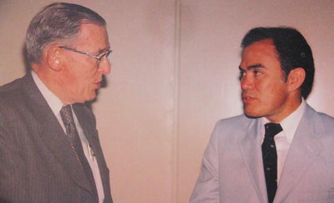
Fig. 1.1
Photograph – Dr. Peña with Dr. Jonathan Rhoad
In 1953, Douglas Stephens published his first landmark paper on the subject, in Australia [38]. Dr. Stephens has the unique distinction of being the first person who studied the anatomy of the pelvis in patients who died from an anorectal malformation. From his studies, he concluded that the key part of the sphincter mechanism to achieve bowel control in these cases was the “puborectalis sling.” It took time for his concept to be learned and accepted by the world community of pediatric surgeons, but within a few years, most pediatric surgeons recognized that was something to be considered seriously, and therefore, the “era of the puborectalis” began. From that time, most surgeons tried to design operations aimed to preserving the “puborectalis sling,” which was considered key for bowel control. Unfortunately, it is not easy to obtain cadavers of children born with anorectal malformations because most children with anorectal malformations survive, and therefore the number of specimens studied by Dr. Stephens was very limited. In retrospect, we believe that his conclusions are not valid because his studies were performed in a limited number of the most severe cases, not representative of what we call the spectrum of anorectal malformations. The cases (cadavers) that he studied we think are not representative of the most common types of malformations that we see. Yet, one of his recommendations is still valid: he recommended pulling the bowel down, as close as possible to the urethra. In addition, Dr. Douglas Stephens published a book [2] that represents the document with the largest amount of information related to the subject of anorectal malformations at that time.
Related posts:


Stay updated, free articles. Join our Telegram channel

Full access? Get Clinical Tree


