and Hubert Lepidi1
(1)
UER Médecine, Aix-Marseille Université, Marseille, France
Abstract
It would be logical to believe that the history of the clitoris is part of the history of the external female genitalia. However, this is not the case. Knowledge of the clitoris was acquired at a later date and remained inaccurate and incomplete for a long time. More surprising still, after having finally been the subject of remarkable anatomical studies, the clitoris was then ignored or unknown during certain periods! It was only in the twentieth century that the significance of this anatomical formation was acknowledged and integrated into an actual apparatus: the bulbo-clitoral apparatus.
1.1 General
It would be logical to believe that the history of the clitoris is part of the history of the external female genitalia. However, this is not the case. Knowledge of the clitoris was acquired at a later date and remained inaccurate and incomplete for a long time. More surprising still, after having finally been the subject of remarkable anatomical studies, the clitoris was then ignored or unknown during certain periods! It was only in the twentieth century that the significance of this anatomical formation was acknowledged and integrated into an actual apparatus: the bulbo-clitoral apparatus.
1.1.1 Foundations of Knowledge
Our prehistorical ancestors could already represent the vulva, such as revealed, on the one hand, by the numerous engraved, drawn or painted pubic triangles, discovered on prehistoric sites of the Palaeolithic era and, on the other hand, by the small statues, “Venus impudicae”, representing female bodies with generous and disproportionate curves, large breasts and exposed vulva. Anatomical details cannot be observed on these various vulvar figurations as the vulvas, represented by these proto-artists, are systematically summarised as triangles whose incomplete bisecting line represents the “large cleft”. Only in rare cases do the edges of the cleft, slightly convex, evoke labia majora.1 However, the anatomical precision does not go further and therefore knowledge of the clitoris and the labia minora cannot be prejudged. Generally, these are not representations (except in cases where the hips are outlined) but figurations, of which many “confine to abstraction”.
Therefore, we may question the meaning of these vulvas, especially when various numbers are represented, at the entrance to a cave or to a room or inside a cave:
Do they symbolise the reproductive system?
Do they symbolise the transition from darkness to light?
Are they an initiation symbol? (Worship glorifying the fertile woman? Worship of the goddess mother?)
As anatomists, we must assist historians in order to provide answers to these questions and must therefore question the general position of vulvar clefts, which have been represented and preferentially placed by the artists of this era, in a pre-pubic ventral position, whereas the men from the Magdalenian era could not be unaware of the fact that the vulvar cleft is almost invisible on an adult woman in a standing position, as it is located in the perineal region.
More close to us, old Egypt remains mysterious as regards knowledge of the labia minora and the clitoris. There is no ostracon specifically representing the external female genitalia. Neither is there any low relief specifically revealing this part of the female anatomy whereas the male apparatus is often represented (representations of ithyphallic god Min,2 in erection, and scenes of male circumcisions on the lower reliefs of tombs or temples, such as those of Karnak). However, a few small statues dating back from the Neolithic era have been found. They represent nude women3 but they are very rare, as opposed to the small phallic statues. Moreover, considering the size of these small statues, no conclusion can be drawn in relation to the anatomical knowledge of their authors, only that they had a more harmonious idea of the female body than their ancestors.
Later, Greek physicians made progress in their knowledge of the female genitalia, even if it is known that this knowledge was not direct. None of these famous physicians performed a vaginal examination or dissection on a woman’s body. It is well established that their knowledge was only based on reports from midwives and women (who became experts in the practice of vaginal self-examination)4 and perhaps also on what they observed during encounters with their own partners.
It is to be recalled that Hippocrates 5 provided a very good description of vaginal transudation, which occurs at the same time as sexual excitation, but was wrongly considered as “female sperm”. He used the term “to aidoïon”, i.e. “the shameful parts”6 to refer to the entire genitalia. He had good knowledge of the matrix and its neck. The vagina does not have a specific name denomination and is considered as part of the “aidoïon”. The vulvar cleft is known as “nature”. According to certain scholars having studied all of his work, he has knowledge of the clitoris but considers it as a simple protrusion, which he refers to as columella or uvula, on the basis of its resemblance with the uvula palatina.
As for Aristotle,7 he denies the existence of female sperm in relation to generation. For him, “when conceiving, the woman provides the matter and this matter consists of the menses”. In chapter XIV of the treatise on the generation of animals,8 he explains that “if some naturalists have believed that women release semen during the act of copulation, it is due to the fact that sometimes women feel as much pleasure as men and that they release a liquid excretion” but “this is an error”. “This liquid is not at all spermatic… It is a special fluid….In women who release this liquid, there is not the same amount of liquid as sperm but sometimes there is much more”. In this same chapter, Aristotle also mentioned clitoral excitation. He wrote “Which also shows that women do not release sperm, only during copulation, they feel pleasure from being touched in the same place as men, but, in their case, there is no liquid emission”.
Even Aristotle has provided an outlined description of what could correspond to the clitoris but with a number of improbabilities. Thus, it is possible to read in the history of animals: “This is how Nature has laid out the pathway followed by sperm in women: They have a canal, such as men have a sex, but it is inside the body. They suction through this canal by means of a small orifice positioned above the location where women urinate. Sperm falls into the uterus from this canal”.9 Overall, Aristotle describes an imaginary canal, but at the same time, he refers to the existence of an anatomical structure, located above the external orifice of the urethra.
As a good observer, Aristotle also describes the modifications of the external female genitalia during sexual excitation. He wrote that “by observing female rats, we can see that the humidified sex of the female swells when she approaches a male”.10
During the three following centuries, progress was made in the acquisition of gynaecological, anatomical and medical knowledge, and we can only marvel when reading the treatise “Diseases in women”, written by Soranos of Ephesus,11 leader of the Methodical School.12 The description of the female genitalia provided by this author in the first pages of his treatise13 is astonishingly precise, even if wild ideas remain, in particular concerning “the seminal tract”, which exits the matrix, crosses each ovary, skirts the sides of this apparatus up to the bladder and then accesses the neck of the latter.
According to him, “the wings (pterygomata) or labia of the vagina, thick and fleshy, are separated from one another by a cleft. Downwards, they end at the 2 thighs. Upwards, they end at what is called the clitoris (kleitoris) or myrton or nymph”. Soranos not only mentions this formation. He also specifies and explains: “The latter, which forms the beginning of the labia, consists of a waffle with a muscular aspect. This small formation is called the nymph because it is hidden underneath the labia such as young brides under their veil”. He even provides the following observations: “Slightly below the nymph, another protuberant wattle is hidden, it is the end of the neck of the bladder; referred to as the urethra”. Therefore, even if Soranos mixes up the internal and external ostia of the urethra, he positions the clitoris or nymph correctly, above the meatus, which is used for the emission of urine.
Rufus of Ephesus,14 a contemporary of Soranos, has good anatomical knowledge based on dissections performed on monkeys. He wrote, only in Greek, a book in which the entire nomenclature of anatomical terms of the time is assembled and specified. Thus, he confirms the term “clitoris”, which he connects to a verb with an erotic meaning: cleitoriaxo. He confirms that the labia majora or myrtle lips (myrtocheïla) must now be referred to as “pterygomata”. As for the term of nymph, it indicates the unit formed by the clitoris and the labia minora. The cleft is called “schisma”. All of the internal and external genitalia form the “shameful parts” or aidoïon.
Neither Soranos nor Rufus will achieve the celebrity of their successor, Galen,15 who is also a Greek physician based in Rome, where he will live until the end of the second century (the date of his death is discussed: 204 AD ?).
Galien recalls the bi-spermatic theory made by Hippocrates by adding the essential role (according to him) of blood “matter”, whose surplus overflows periodically as menstruations.
For Galen, the female genitalia is the reverse copy of the male genitalia. This copy is interiorised whereas man’s genitalia is exteriorised. The male’s penis corresponds, according to him, to the uterus. The scrotum occupies the location of the matrix. The female “testicles” are positioned on either side of the matrix. The prepuce of the penis becomes the vagina in women. The latter represents with the vulva, the female shameful part (aidaïon gunaikeïon). No place is made for the clitoris so that the progress made by Aristotle and the physicians of Ephesus as regards the anatomy of the female sex is truly forgotten.
Despite this fact, Galien became the greatest physician of Western civilisation, even more famous than Hippocrates, such that his findings influenced medical knowledge, especially in relation to human anatomy, until the fifteenth century.
One century and half later, Oribase,16 who was also born, such as Galien, in Pergamon, was put in charge, by the Emperor Julian the Apostate (331–363), of gathering all the knowledge of the eminent Greek physicians, since Hippocrates. Oribase completed this task by compiling a magnificent encyclopaedia (70 books), based on the work of his predecessors.
It refers to the female genitalia as pecten. It calls the vulva, “the big cleft”. The labia majora are “the wings” of this big cleft. The clitoris is obviously well known. He positions it correctly and describes it as “a muscular waffle, located in the centre”. He calls it myrtle or nymph so that the labia minora become “the wings of the myrtle”.
Simultaneously to Greek medicine, foreign medicines (Arab and Persian, in particular) have progressed, while profiting from the knowledge contributed by Greek and Latin work. Two physicians from the tenth century, who distinguished themselves among these foreign physicians, knew about the clitoris: a Persian physician, Avicenna 17 (980–1037), which calls the clitoris “el bathr” (penis) and an Arabic physician, Abulcasis 18 (936?–1013), which calls it “tentigo” (which is placed under tension) but also “softness of love”, a term which will be used by Colombo.
The main work completed by Avicenna, a book called Canon, includes 5 volumes and the first volume is a panorama of the anatomy and pathology of the various organs.
Abulcasis compiled a medical and surgical encyclopaedia, in 3 parts, and the last part was entirely devoted to surgery with a paragraph concerning clitoridectomy. Abulcasis’ reputation (who practised his art at Cordoue) went beyond the Arab world and the borders of Spain, and his work, a reference for teachers for more than one century, was to be republished several times and successively translated into Latin, English, French, Hebrew and the Provençal language.
Few physicians of the following centuries became known for their work and Galen’s influence lasted until the Middle Ages. Nevertheless, a few anatomical books were published and included superb prints, which were as beautiful as inaccurate. Among these books, the book written by Mondino De Liuzzi 19 entitled Anathomia was a real success. Moreover, many readers, on the basis of the author’s work, in which he mentions that, in 1316, he performed two dissections on female bodies, consider him as the “protodissector”, i.e. the first physician to have restored cadaveric dissection,20 which had disappeared since antiquity. Actually the first cadaveric anatomical dissection was performed in 1313, in Montpellier by Henri de Mondeville21 (who was also Mondino’s professor). Nevertheless, Mondino’s book, based on knowledge from human dissections and not from animal dissections, such as the work completed by Greek authors and Galen’s in particular, was going to initiate the future upheaval of anatomical knowledge of the human body.
Two other books using engraving were to be published a few years later and initiate the expansion of anatomical culture in all Europe of the early Renaissance. This was an indication of the development of this fundamental subject of medicine in the near future:
Firstly, a book written by Jacopo Berengario da Carpi,22 with a promising Latin title Isagogae breves perlucidae ac uberrimae in anatomiam humani corporis a communi medicorum academia usitatam, was published in Bologna in 1527 then a book written by Charles Estienne,23 a contemporary of Vesalius, who published the Dissection des parties du corps humain (Dissection of the parts of the human body), successively in 1545, in Latin, then in 1546, in French. This book, which included even more anatomical charts than Berengario’s book, used such as the latter, the technique of staging human bodies.
Sadly, this staged representation, although pleasant and elegant to look at, outweighed the anatomical reality and made the body structures very difficult to observe as they were too small, inaccurate and generally incorrect. The actual descriptions are inaccurate (e.g. that of woman’s “shameful member”, which seems to more or less refer to a description of the clitoris).
The fact remains that the three above-mentioned authors had established the foundations of modern anatomy, based on the direct observation of the human body and the reproduction of this reality in anatomical charts, which Vésalius rapidly enhanced and developed.
But what happened to the anatomy of the clitoris during the Middle Ages?
In spite of the relative small amount of anatomical discoveries made during this period, the assumed role of the clitoris in fecundation spread in the thatched cottage homes during the Middle Ages. The majority of physicians of the time adopted the theory of the existence of female sperm and considered that this secretion, called cyprine, was produced by the clitoris. The Catholic Church, however, which expressed reservations on the topic of carnal pleasure, had to accept the medical advice favouring fecundation: “before the sexual intercourse, the husband shall delicately rub the button of love with a finger moistened with perfumed oil, in a circular motion”.
1.1.2 Rebirth and “Discovery” of the Clitoris
The Renaissance, a period of renewal and all types of discoveries, was going to the era during which anatomical knowledge, including that of the clitoris, was developed. Little by little, prohibitions concerning cadaveric dissection were going to be finally reduced thus allowing a direct approach of the human body and thus of its knowledge.
It is in this new environment that the Belgian, Andreas Vesalius,24 who was going to become the most famous anatomist, was going to rapidly emerge and define the foundations of modern anatomy, in his masterly exercise at the faculty of Padua:
By transforming dissections on human bodies from exceptional “public place anatomies”, such as was the case, in Antwerp or Venice25 (Fig. 1.1) into a current practice, carried out in dedicated locations: initially, anatomical theatres, dismountable installations and then rapidly permanent structures26

Fig. 1.1
“Piazza dell’Anatomia” in Venice (a) and his accesses “sottoportego dell’ anatomia” (b): the stairs for going down from the bridge of anatomia (ponte dell’anatomia), and the porch that allows the access to the little river (rio). The first and exceptional public anatomies were carried in that place, hidden from sight! The cadavers and the anatomical remains were transported by gondola across the rio
By only transmitting to his students’ concepts verified through direct anatomical observations
By performing the dissection himself during his anatomy lessons, such as shown by the front page of his book
By being the first to write an exceptional book, De humani corporis fabrica, published in Basle in 1543, which provided a significant anatomical accuracy, enhanced by the presence of an exceptional iconography (charts prepared by students of the school of Titien, which had attended the cadaveric dissections) and, furthermore, which was printed (following the recent discovery of the printing technique by Gutenberg)
Nevertheless, in the De humani corporis fabrica book, libri septem, there is no detailed description of the external female genitalia and even less precisions on the clitoris, which Vesalius seems to ignore (a superb image of the entire genitalia shows a vulva, which is simply represented as 2 greater lips, with a nice representation of combed body hair) and even considers as an anomaly, which is only visible in hermaphrodites.
It is true that Vesalius was only able to study this anatomical region on a small number of specimens, which were only available for a very short time (autopsies).
It was thus necessary to wait for Vesalius’ successors as Professor of Anatomy at the University of Padua, Colombo then Fallopio, who conducted accurate studies on the clitoris, which clarified its function and structure.
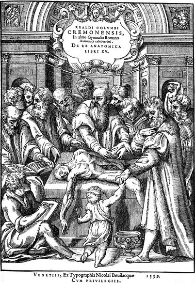
Realdo Colombo 27 was successively Vesalius’ assistant (1541), temporary substitute in 1543 and then permanent substitute as from 1544 (when Vesalius left to take up his position at the court of Charles Quint). He became the Professor of Anatomy at the University of Pisa in 1545 and wrote a remarkable book on anatomy De re anatomica, which was only published after his death in 1559.
Sadly, this book included no iconography, except for a superb front page (Fig. 1.2) by Veronese.28 The book (which was extremely documented) was a great success and its fame went beyond the borders of the Republic of Venice, such as represented by a frontispice painting “A Lesson of Anatomy”, commissioned by the famous English anatomist, John Banister, lecturer in anatomy of the Barber Surgeons’ Company (licensed by the king Henry VIII in 1540). On this painting, visible at the Glasgow’s University, Banister is performing an anatomical demonstration in front of his students while reading the book written by Colombo, which is open on his desk. Numerous discoveries are owed to Colombo, an exceptional anatomist, who performed many dissections (apparently, he has personally performed 1,000 dissections). He was also a talented physiologist. One of his most famous discoveries is that of pulmonary circulation29 (which was used as a basis, 30 years later, for the descriptions of the large and small circulation by William Harvey). We also owe him remarkable work on the clitoris, which he claimed to have discovered, whereas, such as we have noted, Greek (but so Arab and Persian) physicians and surgeons already had knowledge thereof, such as proved by their texts.


