Fig. 20.1
Lesions are primarily concentrated in the left lateral lobe, as in the following MRI image
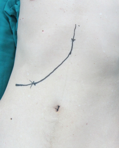
Fig. 20.2
Generally select a right costal margin incision for left lobe hepatectomy
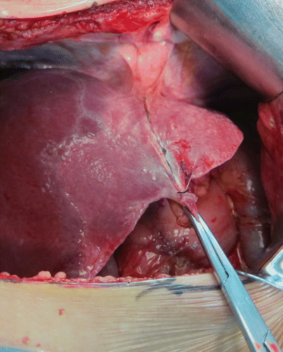
Fig. 20.3
Expose the lesion
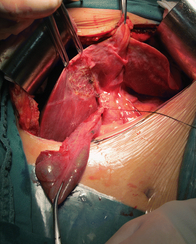
Fig. 20.4
Perform a cholecystectomy first
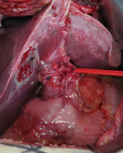
Fig. 20.5
Expose the hepatoduodenal ligament
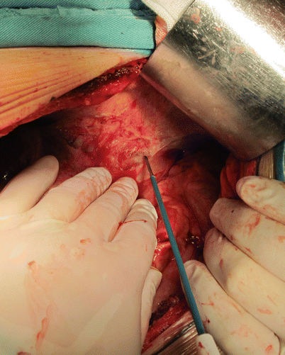
Fig. 20.6
Free the perihepatic ligaments, such as the left coronary ligament
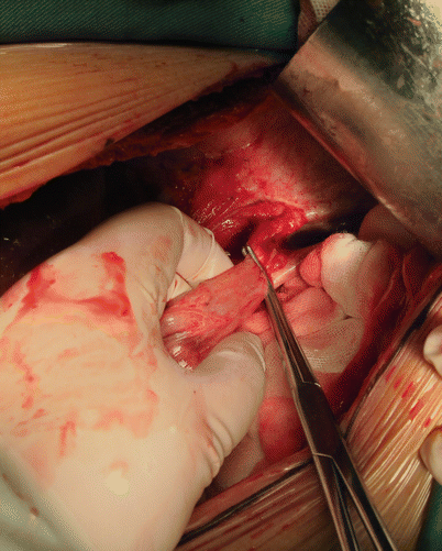
Fig. 20.7
Transection of the left triangular ligament
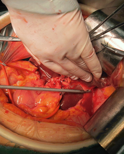
Fig. 20.8
Free and cut the hepatogastric ligament
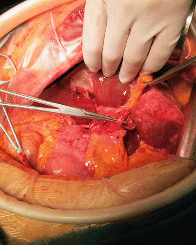
Fig. 20.9
Isolate and cut the left hepatic artery
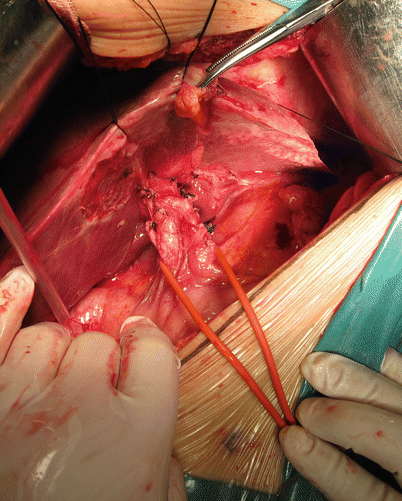
Fig. 20.10
Pre-blockage of the hepatoduodenal ligament
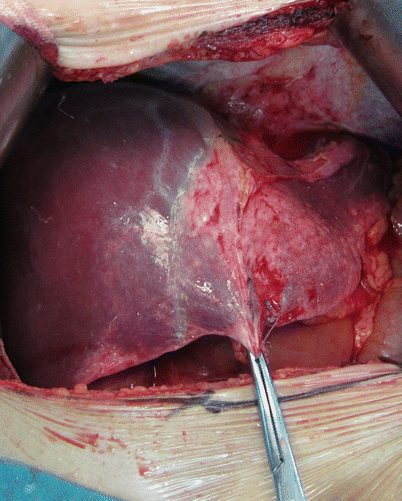
Fig. 20.11
Identify the liver tangent line
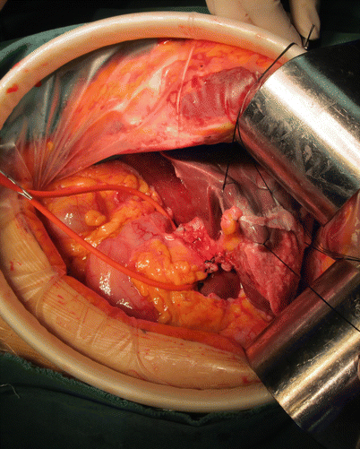
Fig. 20.12
Suture the traction line on both sides of the tangent line
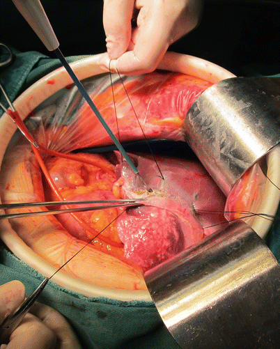
Fig. 20.13
Cut the liver capsule with an electric knife
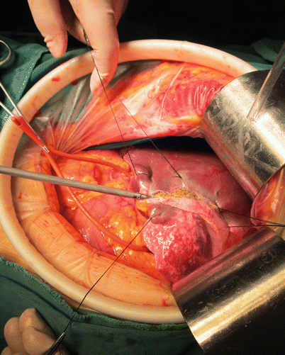
Fig. 20.14
Perform a hepatectomy with an ultrasonic scalpel with a small tube (e.g., artery, vein, and bile duct, with ultrasonic scalpel in direct contact)
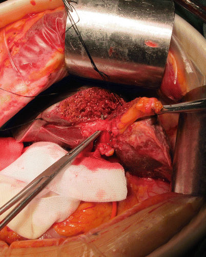
Fig. 20.15
Transect the left branch of the portal vein and left hepatic vein; expose and remove the left hepatic duct
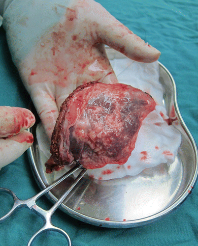
Fig. 20.16
Transect the specimen
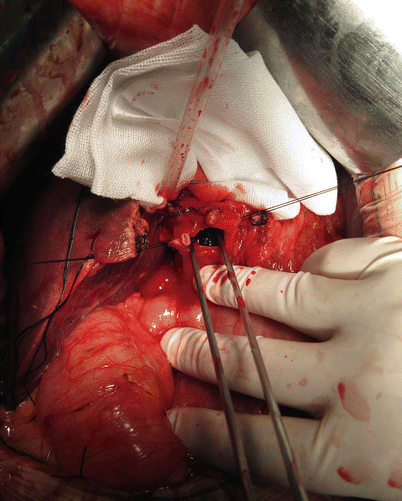
Fig. 20.17
Expose the common bile duct and remove the extrahepatic bile duct stones
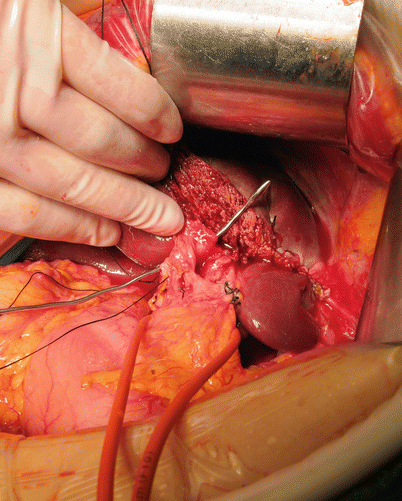
Fig. 20.18
Use a biliary bougie to explore for residual stones
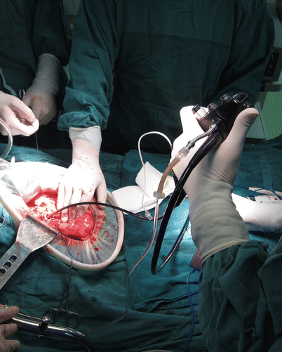
Fig. 20.19
Perform an intraoperative re-exploration with concomitant use of choledochoscope to check for residual stones
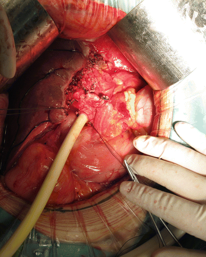
Fig. 20.20
After confirming that there are no residual stones, place a T-drainage tube in the common bile duct
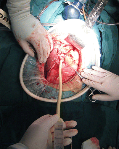
Fig. 20.21
Examine the bile ducts for bile leakage and hemorrhage. The drainage tube and the T tube are placed together, leading out of the body
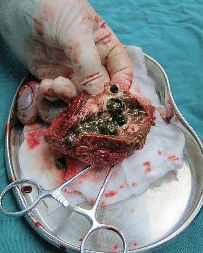
Fig. 20.22
After closing the incision, examine the specimens; here, numerous stones are visible
20.7 Postoperative Complications and Treatment
Complications for this procedure are roughly in line with other liver resections for the treatment of hepatolithiasis. The difference is to pay attention that the T tube remains unobstructed and to prevent its loss. In addition, because the clinical manifestations of the hepatolithiasis are varied, the operation can be more complex, resulting in more surgical complications. The incidences of various complications are as follows: incision infection, 22 %; pulmonary infection, 6 %; bile leakage, 5 %; hemorrhage, 3 %; and hematosepsis, 1 % [31]. If intrahepatic duct bile leakage is observed, percutaneous drainage is the most effective method [32].
20.8 Long-Term Prognosis
The clinical manifestations of hepatolithiasis are diverse and complex, with varying disease locations and associated lesions. These variations result in a high degree of difficulty with respect to diagnosis and treatment. Moreover, hepatolithiasis has a high postoperative recurrence rate. According to previous reports, surgical complications for one- and two-sided hepatolithiasis are 20.4 % and 38.5 %, respectively. The recurrence rates for these conditions after 5 years are 6.2 % and 16.7 %, respectively. Perioperative mortality is 0.4 %, and the overall survival rate 10 years after hepatic resection is 80.3 % [33, 34].
Related posts:


Stay updated, free articles. Join our Telegram channel

Full access? Get Clinical Tree


