Hand-Assisted
Paul E. Wise
Hand-assisted laparoscopic surgery (HALS) for colorectal procedures allows for a hybrid-type procedure between laparoscopy and open approaches to colorectal disease. These techniques use a hand-assist device that maintains a pneumoperitoneum to perform the procedure laparoscopically while a hand is inside the abdomen or when the hand or instruments (through the hand-assist device, some devices allowing this maneuver to occur with a hand in place) are being exchanged. The bases of these devices can often be utilized as a wound protector during specimen removal or as a wound retractor during any open aspects of the procedure.
HALS can help those surgeons not yet comfortable with more complex laparoscopic colorectal procedures to gain the skills needed to perform these procedures. However, studies have shown variable results as to whether HALS actually improves the learning curve for laparoscopic colorectal procedures.
HALS advantages:
Allows a less invasive approach when laparoscopy might not be an option, especially in a difficult situation such as fistulizing inflammatory diseases, large masses, or morbid obesity.
Allows tactile feedback to help identify small neoplastic lesions, to find a vascular pedicle in an obese patient or to palpate ureteral stents.
Allows the ability to provide hemostasis with the hand or to use an easily placed sponge to identify a bleeding source or clean up after a bleed.
Allows the hand to perform blunt dissection in the setting of benign inflammatory disease.
Allows the hand to provide retraction of heavier structures within the abdomen.
Decreases the rate of conversion to an open procedure when compared to laparoscopy (and may allow for avoiding a conversion from laparoscopy to open by utilizing HALS).
Shorter operative time versus laparoscopy in many comparative studies of colorectal procedures.
Shorter hospital stays versus open colorectal procedures.
Improved cosmesis over open colorectal procedures.
HALS disadvantages:
Cost of the hand-assist device increases cost over open colorectal procedures and perhaps over laparoscopy.
Operative times are longer with HALS than are with most open colorectal procedures, however, these times depend upon the underlying disease, patient, and surgeon variables.
HALS has increased incision size (and thus increased infection and hernia rates, depending on the incision utilized for the hand-assist device) over straight laparoscopy.
Cosmesis improvement is less than that with laparoscopy for colorectal procedures, especially in the case of total proctocolectomy, when compared to an open approach.
Indications for HALS total proctocolectomy (TPC) are the same as those described for open TPC and include the following situations:
Ulcerative colitis when no restoration is planned due to continence issues, patient comorbidities, the presence of a very distal rectal cancer, and/or patient preference.
Crohn’s colitis when no restoration is planned due to continence issues, patient comorbidities, the presence of a low rectal cancer or proctitis, patient preference, fistulizing perianal disease, and/or the presence of ileal disease.
Familial adenomatous polyposis (FAP) when no restoration is planned due to continence issues, patient comorbidities, the presence of a very distal rectal cancer, and/or patient preference.
Synchronous proximal and distal colorectal malignancies including very distal rectal cancer when no restoration is planned due to continence issues, patient comorbidities, and/or patient preference.
Hereditary nonpolyposis colorectal cancer (HNPCC, Lynch syndrome) in the presence of a very distal rectal cancer which would otherwise require abdominoperineal resection, or for a mid- to low rectal cancer that would require low pelvic rectal resection but when no restoration is planned due to continence issues, patient comorbidities, and/or patient preference.
Contraindications for HALS total proctocolectomy (TPC) are the same as those settings described for laparoscopic (or open) TPC and include the following:
Comorbidities that preclude a general anesthetic.
Comorbidities that preclude a laparoscopic approach due to intolerance to a pneumoperitoneum including intolerance to carbon dioxide and severe cardiovascular disease.
Portal hypertension due to cirrhosis.
Relative contraindications which depend upon the individual surgeon in dividing large tumors, large inflammatory masses, fistulizing Crohn’s disease, adhesions due to previous operations or inflammatory disease or desmoid disease, bleeding diathesis, and/or bowel distension due to obstruction or recent endoscopic evaluation.
Lack of surgical training and/or lack of availability of the necessary laparoscopic equipment, hand-device equipment, and/or operating room equipment.
After the initial assessment for indications and contraindications to HALS TPC, a similar preoperative evaluation to open TPC is recommended and includes the following:
Full endoscopic evaluation of the colon as well as upper endoscopy in the case of FAP and HNPCC.
Retrograde contrast radiography when the colon cannot be completely endoscopically assessed perhaps due to malignant or inflammatory stenosis.
Antegrade contrast radiography such as CT enterography or small bowel follow through may be preoperatively indicated.
Appropriate staging CT scanning, endorectal ultrasound or MRI for rectal cancer and laboratory evaluation and completion, if indicated, of any neoadjuvant treatment for malignancies.
Preoperative planning then includes the following:
Patient education, evaluation, and marking of the proposed ileostomy site by an enterostomal therapist.
Consideration of bowel cleansing with a mechanical (and/or antibiotic) bowel preparation. This preparation can improve the ability to manipulate the colon with the HALS and laparoscopic approaches but can increase bowel distension with any distal obstruction, so should be selectively used.
Standard preoperative use of intravenous antibiotic and deep venous thrombosis prophylaxis as per institutional and other guidelines for all high-risk and complex operative procedures, whether open or minimally invasive.
Ensuring that the laparoscopic instruments, hand-assisted device, operating room equipment, and appropriate assistants/personnel are available.
Positioning
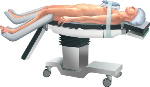
As with the laparoscopic TPC, patient positioning is split-leg in the modified lithotomy position to allow perineal access and for the surgeon to stand between the legs if desired. A position-ranging operating bed is necessary to allow for steep Trendelenburg, reverse Trendelenburg, and steep side-to-side positioning as gravity is used to move the small intestines away from the point of dissection to allow for an unobstructed view. The patient must be secured to the bed. This step may be facilitated by a bean bag attached to the bed with Velcro and wrapped around the well-padded patient, who is further secured around the chest and shoulders with three-inch tape (Fig. 25.1). There should be at least two mobile monitors to allow for adequate views from either side of the table. If the monitors cannot move to the foot of the bed to facilitate the view during the pelvic dissection, a third monitor should be available (Fig. 25.2).
Stay updated, free articles. Join our Telegram channel

Full access? Get Clinical Tree


