and Hubert Lepidi1
(1)
UER Médecine, Aix-Marseille Université, Marseille, France
Abstract
The frequent lack of knowledge of the anatomy of the clitoris, even among women considered as “experts”, is explained by the fact that the major part of this essential component of the external female genital apparatus is hidden and therefore cannot be observed.
The frequent lack of knowledge of the anatomy of the clitoris, even among women considered as “experts”, is explained by the fact that the major part of this essential component of the external female genital apparatus is hidden and therefore cannot be observed.
Only the distal end of the clitoris can be visualised during a vulvar examination, subject to slightly separating the folds consisting of the labia minora or nymphs, operation which also draws aside the labia majora.1 A median, convex and hemi-cylindrical protrusion is observed at the ventral end of the vulva. It occupies the dip formed by the labia majora before they meet at their ventral commissure. This protrusion corresponds to the body of the clitoris and the skin covering the clitoris is the clitoral prepuce. A cutaneous and nearly circular fold is present at the loose end of this prepuce: the clitoral hood. This hood overhangs and surrounds part of a more or less spherical and central formation, the glans clitoridis. Very often, the glans remains hidden at the bottom of the “preputial cylinder” and is not immediately visible. It can only be observed once the hood has been gently pulled back.
From an anatomical viewpoint, the clitoris belongs to the vulva2 (Fig. 4.2). It is thus located at the level of the anterior perineum,3 behind the ventral commissure of the labia majora (their point of union with the mons pubis).4 In order to complete the topographic arrangement, it is essential to recall that the glans clitoridis hangs over the external ostium of the urethra.
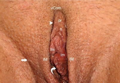
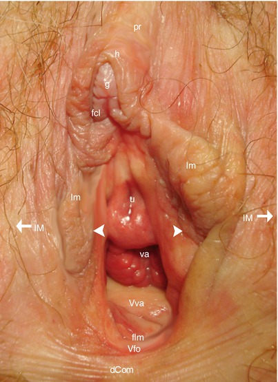
Fig. 4.1
Morphological aspect of the female pudendum (vulva) (after shaving). h hood of the glans, lM labium majus (greater lip), lm labium minus (lesser lip), pr female prepuce (praeputium clitoridis) covering the clitoral shaft, vCom ventral commissure (anterior commissure) of the lips, white arrowhead “labio-preputial” sulcus (continuation of the sulcus between majus et minus labia), white curved arrow penetrating into the pudendal cleft in order to accede to the vaginal vestibule, right white arrow genito-femoralis sulcus. NB: The labia minora are visible because the subject observed is a multipare female. Usually, closer young women, the labia minora are concealed within the pudendal cleft and the labia majora are approximated
Fig. 4.2
View of the opened pudendum (vulva) showing the external urethral orifice and the external genitalia. Each labium majus is tracted laterally (white arrow). dCom dorsal commissure of the labia, fcl frenum (frenulum clitoridis), flm frenum of the labia minora (frenulum labiorum pudendi), g glans clitoridis, h hood, lm labium minus (lesser lip), lM labium majus (greater lip), pr female prepuce (praeputium clitoridis), u external urethral orifice, va vaginal orifice (introïtus), Vfo vestibular fossa, Vva vestibule of the vagina, white arrowheads they show the Hart’s line (line of junction between labium minus inner skin and vestibule’s epithelium)
The hood of the clitoris is a cutaneous fold, which covers the glans clitoridis over the 2/3 of its periphery (top surface and lateral surfaces). It has various shapes (Fig. 4.3). The most common shape is a rounded hood resembling a roman arch. The hood may also have an ogival shape. It can even be shaped as a dihedron with a top edge (roof-shaped hood) (Fig. 4.9). It extends the skin covering of the body of the clitoris. The clitoral prepuce consists of this skin covering and the hood. The hood often has several folds. In most cases, the hood is not very thick but it can, even in young women, thicken and adopt a dysmorphic aspect: This hood is called by the French authors a crassiform hood (from the Latin word “crassus” meaning “fat”) (Fig. 4.4). The hood can slide along the surface of the glans, so that the latter can be retracted (such as with the male sexual apparatus). A cavity is therefore formed between the glans and the internal surface of the hood, the preputial chamber (Fig. 4.5). The bottom of this chamber is an arc-shaped groove, which stops, such as the hood, at the level of its inferior surface (due to the presence of the frenula): it is the neck of the glans clitoridis (equivalent to the balano-preputial groove of the male penis). It is at the level of this neck that the external epithelium of the glans is reflected on the internal coating of the hood (Fig. 4.6). The external surface of the prepuce is covered by a keratinised squamous epithelium. In the case of young girls, a mucous layer is generally present on the internal surface of the hood. In the case of women of an age to procreate, this layer is gradually replaced by a non-keratinised squamous epithelium.5 The presence of sebaceous glands (preputial glands) is observed at the neck of the glans, which explains the accumulation of smegma,6 especially if the hood is fully covering or if the level of hygiene is not satisfactory. With age, the prepuce tissues tend to undergo a ptosis and distend. This causes the formation of a sort of withered sheath, inside which the glans is often completely hidden (Fig. 4.3). The lateral parts of the hood extend beneath the glans and are gradually incorporated into the lateral surface of the labia minora. The fusion with the labia minora can be precocious (high fusion). It can also occur at a lower level, at the top-third to mid-third junction of the labia minora (low fusion). This case corresponds to a lateral extension of the hood, sliding on the lateral surface of the labium minus, before merging with the latter.7 Generally, there is no symmetry between the right and left levels of the junctions between the lateral extension of the hood and the homolateral labium minus.
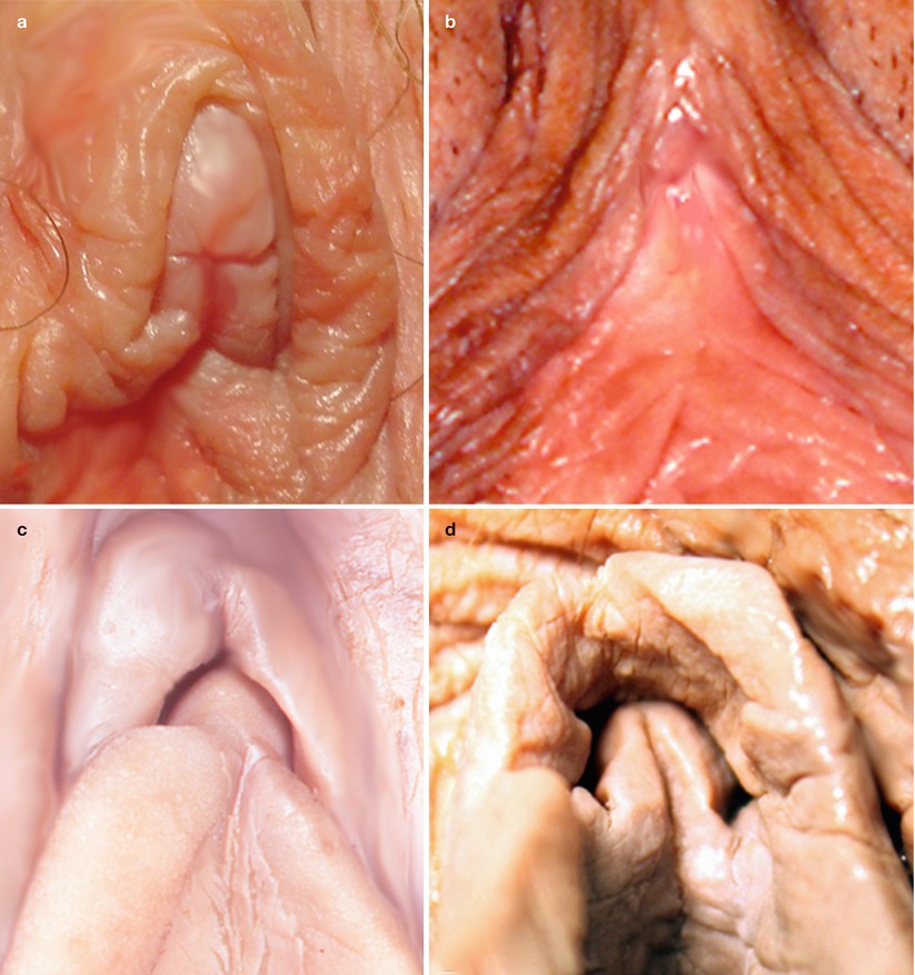
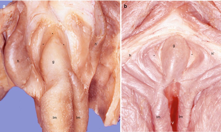
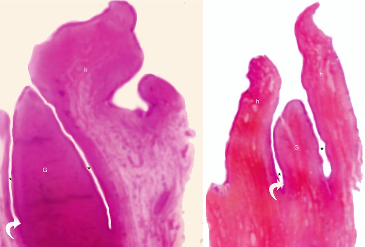
Fig. 4.3
Variations of the hood and their consequences on the cover of glans clitoridis. (a) Protruding glans; (b) barely apparent glans (ptosis of the preputial tissues in an old woman); (c) hidden glans (thick hood and large frenula); (d) hidden glans (only the frenula are visible)
Fig. 4.4
Aspects of two glans (a, b) after sagittal sections of their hoods. These sections allow to see the preputial chamber and the free part of each glans (a, b) better. g glans clitoridis, h clitoral hood (right half), h′ clitoral hood (left half), lm labium minus, ***asterisks locating the preputial chamber on each specimen, V vestibule. NB: On specimen a, the labia minora seem to be born directly from the glans (no frenula!)
Fig. 4.5
Relations of the glans to the hood (end of prepuce) on histological transverse sections, in the axis of the glans. G glans of the clitoris (glans clitoridis), h hood (end of the prepuce), asterisk preputial chamber (clitoro-praeputial sulcus) (The female preputial chamber is analogous to the male preputial sulcus), white curved arrow neck of the glans
The glans is the best known part of the clitoris due to its surface location and accessibility. Moreover, at present, it is rightly considered as the ideal site for female pleasure.
The glans clitoridis challenges neophytes by its resemblance to the glans of the penis, and at first sight, an observer will view the clitoris as a simple miniature penis with a glans whose aspect recalls that of a small-sized male glans! However, an accurate analysis rapidly shows the specific characters and multiplies differences contradicting this point of view.
The glans clitoridis is a protruding tubercle, which is more or less rounded (convex in all directions), and oblique rearwardly. It is often compared to a berry: the bilberry or a tiny acorn (with different colours). However, a slight flatness of the lateral surfaces and a dome-shaped top section are often observed, with a reduction in the diameters from the base to the end, such that the geometrical figure, which best represents the surface of the glans clitoridis, is that of an elliptic paraboloid (ends of a miniature missile or a mini-suppository). It can also have an oblong or cone-type aspect (with a rounded end or, on the other hand, a frankly acuminate end). Finally, it can have a levelled and flat end, shaped as a fish snout (Figs. 4.7, 4.8 and 4.9).
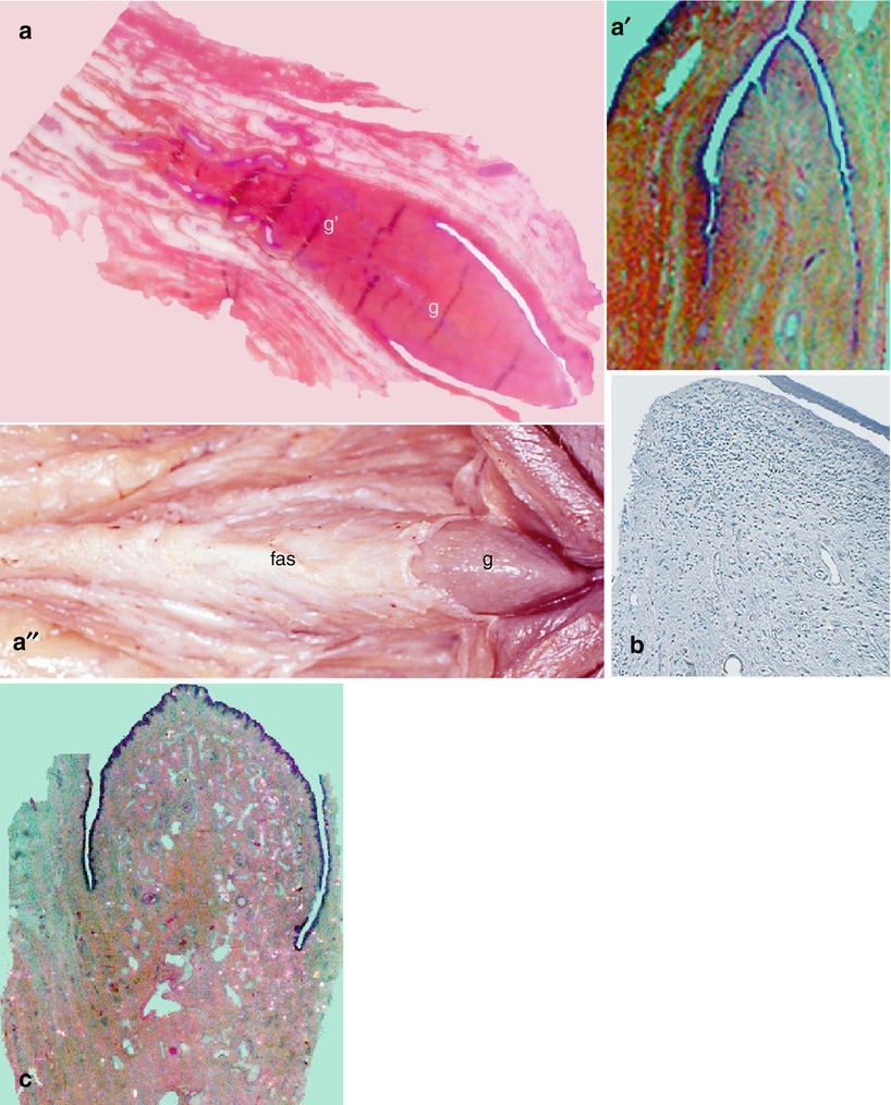
Fig. 4.6


The three principal morphological aspects of the glans clitoridis. (a, a′, b, c) Histological transverse Sections. (a″) Superior view of a dissected clitoris, after resection of the prepuce (skin of the clitoral shaft + hood of the glans). (a, a′, a″) Conoidal appearance; (b) paraboloidal appearance; (c) bulbous appearance. g free part of glans, g′ hidden part of glans, fas fascia of the clitoral body
Related posts:


Stay updated, free articles. Join our Telegram channel
Full access? Get Clinical Tree
