Esophagus


Esophagus


Among the most common types of biopsies encountered in daily practice are esophageal biopsies to evaluate for Barrett esophagus. Difficulties in evaluation include:


  • 1. Duplicated muscularis mucosae (note that the latter word is “mucosae” rather than “mucosa”—this term is commonly misspelled and mispronounced) in endoscopic mucosal resection (EMR)


  • 2. Distinguishing reactive changes from dysplastic ones


  • 3. Identifying “true” intestinal metaplasia on hematoxylin and eosin (H&E) and periodic acid-Schiff (PAS)/Alcian blue (AB) stains


  • 4. Multilayered epithelium


  • 5. Familiarity with the microscopic anatomy of the gastroesophageal (GE) junction

We will attempt to address these and other issues in the following sections.

It is worth remembering that, in most mucosal pinch biopsies, only epithelium and lamina propria are present; most lack submucosa and most only contain small wisps of muscularis mucosae. The lamina propria of the esophagus contains numerous lymphovascular channels, however, and carcinomas invading the lamina propria of the esophagus are thus staged as T1a. In contrast, neoplasms invading the lamina propria of the colon are staged as Tis (1).


BARRETT ESOPHAGUS AND ESOPHAGEAL NEOPLASMS

Esophageal adenocarcinoma presents an ever-growing burden to the US health care system with its incidence increasing from 3.5 to 25.6 cases per million from 1973 to 2006 (2). Since esophageal reflux disease has been shown on epidemiologic grounds as a strong risk factor for adenocarcinomas of the esophagus (3), evaluation of patients for the presence of histologic precursor lesions (Barrett esophagus and columnar epithelial dysplasia in this setting) is an indication for a large number of upper endoscopic biopsies.


Barrett esophagus is a change in the esophageal mucosa of any length that is visible at endoscopy, and contains intestinal metaplasia on biopsy (4). While in the United States, intestinal metaplasia is still (as of 2011) required for a diagnosis of BE, the American Gastroenterological Association (AGA) has defined BE as follows: “the condition in which any extent of metaplastic columnar epithelium that predisposes to cancer development replaces the stratified squamous epithelium that normally lines the distal esophagus” (5). This acknowledges the view of British (and Japanese) colleagues that either cardia or intestinal-type epithelium supports the diagnosis of BE (6) but the AGA has opted to retain the requirement for intestinal metaplasia in the United States as of 2011 (5). Because of the increased risk of malignancy, patients are subjected to periodic surveillance; esophagogastroduodenoscopies (EGDs) with biopsies are used to identify patients with dysplasia, who are at an even higher risk of developing carcinoma. Although life expectancy is not shortened directly as a result of BE (7), such a diagnosis carries significant economic, health insurance, and management implications.

Histologic requirement for intestinal metaplasia ensures that patients with hiatal hernias are not placed in the same risk category as those more likely to progress to adenocarcinoma. However, it is well known that intestinal metaplasia can often be found at the GE junction when no endoscopic lesion is apparent. This was first systematically studied by Spechler et al. who found that among 142 patients without endoscopically apparent Barrett esophagus, 26 (18%) had intestinal metaplasia (8). All these patients were white, and the male-to-female ratio was 1.9. In contrast, nonwhites accounted for 14% of the 114 patients without intestinal metaplasia and the male-to-female ratio was 0.8. The groups did not differ significantly in the frequency of symptoms or endoscopic signs of GE reflux. From these data, Spechler et al. concluded that adults frequently had unrecognized segments of specialized columnar mucosa (displaying intestinal metaplasia) at the GE junction, and raised the possibility that this might underlie the rising frequency of cancer of the GE junction in the United States and Europe (9-14). As such, the patients at risk for Barrett esophagus (overweight, middle-aged white males in the upper economic strata) are increasingly screened, despite the fact that it is not entirely clear who should be screened, how frequently, and how much sampling is required (15,16). In all likelihood, the soil in which intestinal metaplasia in the esophagus develops, at least in a subset of patients, is acquired cardiac-type metaplasia, as a consequence of reflux (17,18). The gastric cardia, itself, seems to be a very small zone in utero and in babies, which expands proximally as a consequence of injury (reflux). As such, pathologists currently evaluating esophageal biopsies or GE junction biopsies should probably report precisely what is seen and apply the term “Barrett esophagus” to biopsies that are from the tubular esophagus and show intestinal metaplasia.


WHAT IS COMPLETE VERSUS INCOMPLETE INTESTINAL METAPLASIA?

“Complete” intestinal metaplasia is the pattern typically seen in the stomach, in the setting of chronic injury such as from Helicobacter gastritis. In complete intestinal metaplasia, the mucosa mimics intestinal epithelium on routine H&E stains, such that a brush border is seen and Paneth cells may be present (Figs. 1.1 and 1.2, e-Figs. 1.1 and 1.2). On PAS/AB staining, goblet cells are stained blue, but the intervening absorptive cells do not have mucin. A slender strip of brush border is highlighted. In “incomplete” intestinal metaplasia, the metaplastic epithelium has features of both gastric and intestinal epithelium, namely goblet cells with interposed cells having neutral (magenta-colored on PAS/AB staining) cytoplasmic mucin, in the manner of gastric foveolar cells (Figs. 1.3 and 1.4, e-Figs. 1.3-1.5). This type of metaplasia has also been called specialized-type metaplasia and is the type that is typical of Barrett esophagus. It is believed that incomplete intestinal metaplasia is more prone to enter the dysplasia-carcinoma sequence than complete intestinal metaplasia. Occasionally, complete metaplasia is seen in Barrett mucosa. We do not report this subtype of metaplasia, but note it as an (imperfect) adjunct for suggesting whether the biopsy is more likely derived from the gastric cardia or the tubular esophagus, as in some cases. Gastric intestinal metaplasia is more likely to be of the complete type, whereas esophageal intestinal metaplasia is more often of the incomplete type.


FIGURE 1.1 Complete intestinal metaplasia. This type of metaplasia occurs predominantly in the stomach in the setting of Helicobacter pylori infection, as in this case. Note Paneth cells on the bottom center and lower left. No intervening foveolar-type mucin is seen here.


FIGURE 1.2 Complete intestinal metaplasia, PAS/AB. Note discrete, alcianophilic goblet cells, the presence of a brush border and the absence of intervening foveolar-type mucin.


WHAT CELL TYPES CAN RESULT IN “BLUE” ON ALCIAN BLUE STAINING?

Some gastric foveolar cells stain blue with the Alcian blue stain, as do cells in pancreatic acinar metaplasia/heterotopia (Figs. 1.5 and 1.6, e-Figs. 1.6-1.8) and esophageal submucosal glands. This is not equivalent to intestinal
metaplasia. The cells in “multilayered epithelium” (19), which is discussed further below, and which may be a precursor to intestinal metaplasia, are also Alcian blue reactive.


FIGURE 1.3 Incomplete intestinal metaplasia. The metaplastic epithelium has features of both gastric and intestinal epithelium, namely goblet cells with interposed cells having foveolar-type mucin.


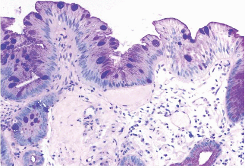
FIGURE 1.4 Incomplete intestinal metaplasia, PAS/AB stain. Note discrete purple goblet cells and the intervening magenta foveolar mucin cap.


WHAT OTHER TYPES OF METAPLASIA ARE FOUND AT THIS SITE?

Pancreatic acinar metaplasia (Fig. 1.7, e-Fig. 1.9) is commonly seen at the GE junction and may be heterotopic, rather than metaplastic, as noted in Volume 1, Chapter 1. We have also occasionally seen respiratory type
metaplasia/heterotopia in GE junction biopsies (e-Fig. 1.10) as well as oncocytic change of the submucosal glands (e-Figs. 1.11 and 1.12). Anecdotally, these changes seem to be of no clinical significance.


FIGURE 1.5 Foveolar epithelium. Note the foveolar mucin cap. Some cells appear larger than others and may provoke concern for the presence of intestinal metaplasia (see Fig. 1.6).


FIGURE 1.6 Foveolar epithelium (PAS/AB stain). Note alcianophilic staining of consecutive foveolar cells as opposed to discrete goblet cell staining in true intestinal metaplasia (compare with Figs. 1.2 and 1.4).


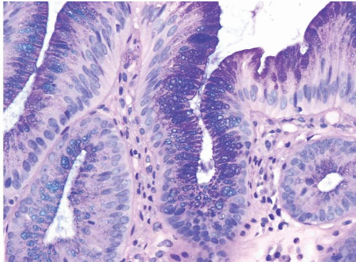
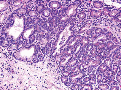
FIGURE 1.7 Pancreatic acinar metaplasia/heterotopia. This feature is commonly seen at the GE junction.


GOBLET CELLS AT THE GE JUNCTION


Definition of Barrett Esophagus

According to the definition of the American College of Gastroenterology (ACG) (4), Barrett mucosa is a change in the esophageal epithelium of any length that:


  • a. Can be recognized at endoscopy


  • b. Is confirmed to have intestinal metaplasia by biopsy

As a result, we pathologists cannot make a diagnosis of Barrett mucosa unless we know the endoscopic findings. Furthermore, regardless of the endoscopic findings, Barrett mucosa can only be diagnosed if there are goblet cells, whether ACG (4) or AGA criteria (5) are used.

The differential diagnosis of Barrett esophagus is with gastric carditis showing intestinal metaplasia, one that requires clinicopathologic correlation with endoscopic findings.

Members of the Gastrointestinal Pathology Society, led by H. Appelman and J. Greenson early in the 21st century, developed the following informal guidelines to help report findings in biopsies from the GE junction:


  • a. Gastric-type mucosa with no goblet cells: If it is cardia, the chances are overwhelming that it has inflammation. Most mild carditis has no known cause, although some observers believe the etiology is reflux, but some cases are attributable to Helicobacter pylori. In the absence of organisms, the appropriate diagnosis is “carditis of unknown etiology.” If the mucosa is oxyntic, it may be derived from a hiatal hernia.


  • b. Goblet cell containing mucosa from endoscopic tongues (e-Figs. 1.13-1.15) that the endoscopist believes is Barrett esophagus: Diagnose as “Barrett mucosa,” with the appropriate dysplasia designation (none, indefinite, low grade, high grade).


  • c. Goblet cell containing mucosa, but there are no endoscopic tongues although there may be a prominent, endoscopic “Z-line” (the squamocolumnar junction): Diagnose as “goblet cells at the cardia.” Some studies suggest that immunohistochemistry can help assign a designation to these, but such studies are often not practical (see below).


  • d. Goblet cell containing mucosa, but there is endoscopic uncertainty (i.e., the endoscopist is not sure (a) if there are tongues or (b) if there is only a prominent Z-line): Diagnose as “goblet cell containing mucosa, either Barrett mucosa or goblet cells at the cardia,” with the appropriate dysplasia designation. If the endoscopist is uncertain as to whether there is Barrett mucosa, we pathologists cannot be certain.


The presence of Barrett esophagus does not, by itself, cause any symptoms for the patient, other than those caused by the associated GE reflux. In fact, patients tend to have attenuation of their symptoms once the Barrett metaplasia supervenes. The major importance of Barrett esophagus lies in its status as a preneoplastic condition that predisposes affected tissue to developing esophageal adenocarcinoma.

The pathogenesis of Barrett esophagus is not clear. Gastroesophageal reflux disease (GERD) is regarded as an important risk factor for the development of BE. An Australian study by Smith and colleagues reports that subjects with self-reported monthly and weekly episodes of acid reflux are at a three- to fourfold and 30-fold increased risk of being diagnosed with BE, respectively, when compared with control subjects (with unknown BE status) randomly selected from the same geographic region (20). Avidan et al. report similar findings in their study, which included data from esophageal manometry and pH-metry, with significantly more reflux episodes in patients with BE when compared with those with no BE and nonerosive GERD (21). Likewise, BE is more common among subjects with hiatal hernia when compared with patients with no BE and nonerosive GERD (21-23). While some studies document an association between BE and smoking and alcohol (20,21), others do not (23,24).

The “cell of origin” of columnar epithelial cells in Barrett esophagus is debated, but possibilities include columnar cells from the normal esophageal glands, migration of columnar cells from the adjacent gastric cardia, and columnar differentiation of pluripotent epithelial stem cells in areas of mucosal injury (the latter is considered to be the most likely cause.) Some observers have discussed “multilayered epithelium” as a precursor to Barrett esophagus (19). This epithelium is characterized by four to eight layers of basally located squamous cells and associated superficial columnar, mucinous epithelium (Figs. 1.8 and 1.9, e-Figs. 1.16 and 1.17). Mucin properties and immunohistochemical characteristics are similar to those seen in the columnar mucosa in BE and to gland duct epithelial cells, raising the possibility that multipotential cells within these ducts may give rise to this particular type of mucosa (9). At present, it is not our clinical practice to report on the presence of multilayered mucosa as its implications for surveillance remain unclear although it has been associated with GERD (25).

Endoscopically, Barrett mucosa appears as tongues and patches of reddish, salmon-colored mucosa (in contrast to the normal, pearly graypink color of the squamous epithelium) that extend from the GE junction for varying distances up into the tubular esophagus (e-Figs. 1.13-1.15). Short-segment Barrett esophagus is defined as ≤3 cm of metaplastic columnar epithelium; long-segment Barrett esophagus is described as >3 cm of metaplastic columnar epithelium. Whether segment length correlates with neoplastic risk is not entirely clear, but some authors report a higher adenocarcinoma risk in patients with long segments of BE (>3 cm, 0.57% per year) than in those with short segments (<3 cm, 0.26% per year) (26).


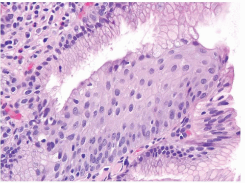
FIGURE 1.8 Multilayered epithelium. This epithelium is characterized by four to eight layers of basally located squamous cells and associated superficial columnar, mucinous epithelium.

Immunohistochemical stains for cytokeratin (CK) subsets has been used to separate cardiac intestinal metaplasia from Barrett epithelium. Ormsby et al. (27,28) evaluated CK7 and CK20 immunoreactivity patterns in resection specimens with long-segment Barrett esophagus, and compared them to distal gastric resection specimens with intestinal metaplasia.
Virtually, all cases with long-segment Barrett esophagus were characterized by superficial and deep CK7 immunoreactivity (Figs. 1.10, 1.11 and 1.12, e-Figs. 1.18 and 1.19) in the intestinalized mucosa, with only superficial CK20 staining in the intestinalized zones. In contrast, distal gastric intestinal metaplasia was characterized by patchy, superficial, and deep CK20 staining in areas of incomplete intestinal metaplasia; strong, superficial, and deep CK20 staining in areas of complete intestinal metaplasia; and patchy or absent CK7 staining (e-Figs. 1.20-1.22) in either type of gastric intestinal metaplasia. As an extension of this work, this same group of
researchers evaluated the utility of CK subsets in distinguishing intestinal metaplasia of the distal esophagus from intestinal metaplasia found in the gastric cardia, in endoscopic biopsy specimens (27,28). Patients with intestinal metaplasia of the cardia all had a normal-appearing squamocolumnar junction, and biopsies of <5 mm were obtained from the esophagogastric junction, with the endoscope in a retroflexed position. The “Barrett CK7/20 pattern” (described above) was found in all cases of Barrett esophagus, but was absent in all 13 cases of cardiac intestinal metaplasia. However, other observers have questioned the utility of CK7/20 staining. The CK7 is certainly the more useful of the two, and it is critical to only stain areas with a full gland profile with intestinal metaplasia. Piazuelo et al. found CK7 staining helpful in distinguishing esophageal from gastric intestinal metaplasia (29). A study of carcinomas believed to arise in the esophagus versus the cardia, however, failed to find CK7/20 differences between these two types of cancers (30).


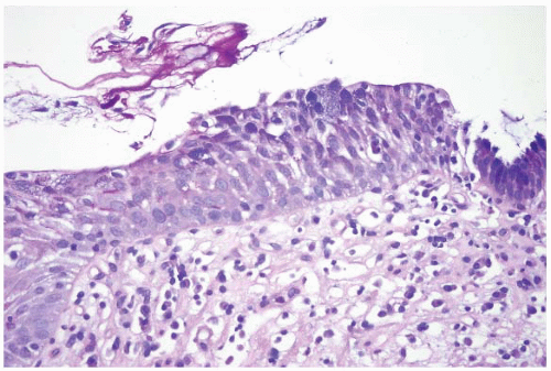
FIGURE 1.9 Multilayered epithelium (PAS/AB stain). Note superficial alcianophilic cells, corresponding to mucinous epithelium.


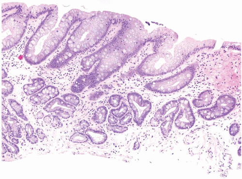
FIGURE 1.10 Biopsy from an “irregular GE junction.” It was unclear whether this tissue originated from the tubular esophagus or the GE junction. Immunohistochemical stains for CK7 and CK20 supported origin from the tubular esophagus, and thus supporting an interpretation of Barrett esophagus (see Figs. 1.11 and 1.12). This biopsy was considered negative for dysplasia due to surface maturation and abundant mucin compared with deeper portions of the crypts. There is abundant lamina propria between the glands.


FIGURE 1.11 CK7 staining in Barrett esophagus. Same case as Figure 1.10; note superficial and deep staining of the epithelium.


FIGURE 1.12 CK20 staining in Barrett esophsgus. Same case as Figures 1.10 and 1.11; note superficial CK20 staining only.

Using mucin core (MUC) polypeptides to better characterize cardiac versus esophageal, intestinal metaplasia has also been explored. In one study a significantly higher number of Barrett esophagus cases (p < 0.05) showed goblet cell staining for MUC1 (55%) or MUC6 (32%), compared with patients with carditis with intestinal metaplasia (MUC1, 14%; MUC6, 7%) or antritis with intestinal metaplasia (MUC1, 6%; MUC6, 0%). Barrett esophagus also showed a higher frequency of MUC1 and MUC6 positivity in nongoblet columnar cells, compared with carditis and antritis cases with intestinal metaplasia. Only cases of Barrett esophagus showed a combined MUC1 and MUC6 staining (sensitivity 23%, specificity 100%). The sensitivity and specificity of MUC1 staining for BE are 55% and 96%, respectively, and of MUC6 staining are 30% and 96%, respectively. Interestingly, normal gastric cardia mucosa also showed a significantly higher prevalence of MUC2 and MUC3 expression in glandular epithelium (29% and 38%, respectively) compared with the antrum (0% for both markers) (p < 0.05).

Das-1 antibody (modestly so-named by Kiron M. Das, who described it) (31) is also believed to stain Barrett metaplasia strongly, with less staining in gastric intestinal metaplasia.


TABLE 1.1 American College of Gastroenterology (ACG) and American Gastroenterologic Association (AGA) Guidelines for Follow-up of Barrett Esophagus


Dysplasia Grade


Documentation


Follow-up, ACG/AGA


None


Two EGDs with bx


3 y/3-5 y


Low grade


Highest grade on repeat, expert confirmation


1 y until no dysplasia/6-12 mo


High grade


Repeat endoscopy within 3 mo to exclude cancer and expert confirmation if biopsies taken from flat mucosa. EMR recommended if biopsies are taken from an area of mucosal irregularity.


Every 3 mo/Every 3 mo


EGD, esophagogastroduodenoscopy; bx, biopsy; y, year; mo, months; EMR, endoscopic mucosal resection.


Adapted from Wang, KK. Updated guidelines 2008 for the diagnosis, surveillance and therapy of Barrett’s esophagus. Am J Gastroenterol. 2008;103(3):788-797 and American Gastroenterological Association medical position statement on the management of Barrett’s esophagus. Gastroenterology. 2011;140:1084-1091.


Others have used CDX2 staining to either label areas of intestinal metaplasia (it is a mystery that these observers cannot simply look for goblet cells, but there is a great deal of pressure for university-based colleagues to publish papers) (32) or to note that some cases lacking goblet cells express these markers anyway in order to make the point that the United States should drop the requirement for goblet cells to diagnose Barrett esophagus (33). Others have suggested that hepatocyte antigen (Hep Par-1, Carbamoyl Phosphate Synthetase 1) is similarly helpful in detecting intestinal metaplasia or processes that are intestinalized in the absence of goblet cells (34). It seems more practical to simply search for goblet cells since that can be done consistently in any laboratory without the added costs of immunolabeling.

In daily practice, we do not use any of these ancillary stains and they have not caught on in general practice. In reality, patient follow-up remains inconsistent, despite suggestions from both the ACG and the AGA for intervals (Table 1.1).


DYSPLASIA IN BARRETT ESOPHAGUS

To help diagnose dysplasia in Barrett esophagus, criteria established at a consensus meeting follow (35). However, after noting these, attention will be drawn to cases that defy classification based on such criteria.


GRADING DYSPLASIA IN BARRETT ESOPHAGUS: ALGORITHM

The following algorithm is based on four mucosal features in Barrett esophagus. The algorithm presupposes that the biopsy in question is taken from the esophagus containing compatible endoscopic features of Barrett esophagus and that intestinal metaplasia is found.


  • 1. Surface maturation compared to the underlying glands


  • 2. Architecture of the glands


  • 3. Cytologic features


  • 4. Inflammation and erosions/ulcers

Each feature may vary, but all are combined to arrive at a diagnosis.

“Surface maturation” is assessed at low magnification and confirmed at high magnification. In nondysplastic Barrett esophagus, the proliferating nuclei in the most basal layers of the glands are larger, more hyperchromatic, and more stratified than those at the surface. In contrast, the surface nuclei are generally arranged in a monolayer with polarized basal nuclei. Characteristically, the glands in Barrett esophagus are mildly atypical, especially when viewed in comparison to adjacent nonmetaplastic gastric-type glands (either fundic or cardiac). Thus, an eye-catching feature when scanning a biopsy at low magnification is the tinctorial comparison between the deep portions of the biopsy and the surface. The possible patterns may be any of the following: (a) the glands have proportionally larger nuclei, (b) the glands and surface have similar nuclear size, or (c) the surface has proportionally larger nuclei.

The “architecture” of the tissue is also best assessed at low magnification. The glandular architecture of a biopsy is the relation between the glands and the lamina propria, and also encompasses the outline of the glands. Architectural abnormalities encompass both increased numbers of glands and changes in their shape. In the nondysplastic setting, the glands tend to be round with little budding, and are surrounded by abundant lamina propria. Crowding of normal-appearing glands is considered a mild architectural abnormality. Crowding of abnormal glands is a feature of dysplasia. Cribriform glands, cystic dilation, and necrotic luminal debris are considered severe architectural abnormalities.

“Cytologic features” are assessed at high magnification in zones selected as abnormal during the assessment of surface maturation and architecture. It must be recognized that some degree of nuclear enlargement and atypia is inherent in Barrett metaplasia in the absence of dysplasia, especially in the basal zone and in the columnar epithelium adjacent to squamous mucosa. In summary, cytologic atypia in Barrett esophagus can be due to (a) dysplasia, in which case it should be cytologically and architecturally unequivocal; (b) reactive changes, particularly associated with inflammation; and (c) inherent changes in the deeper glands of Barrett esophagus, in which case the changes are mild and mature towards the
surface, or (d) any of the above and without certainty (a situation which would merit an interpretation of indefinite for dysplasia [IND]). Dysplastic cells are generally hyperchromatic. Once the cells have been interpreted as dysplastic, assigning a low- or high-grade category reflects a matter of degree along a morphologic continuum, a point emphasized in the 1988 criteria (36). Also included in the assessment of cytologic features is the relationship of one nucleus to another, referred to as nuclear polarity. In “normal polarity,” the long axis of the nucleus remains perpendicular to the basement membrane, and the nuclei are aligned parallel to one another. “Loss of nuclear polarity” refers to the loss of this perpendicular orientation and a random, “jumbled” appearance of the nuclei, in relation to the basement membrane and one another (Fig. 1.13).


FIGURE 1.13 High-grade dysplasia showing loss of nuclear polarity. Note the loss of perpendicular orientation and a random, “jumbled” appearance of the nuclei, in relation to the basement membrane and one another.

“Inflammation and erosions/ulcers” are a potential component that add difficulty and are assessed at both scanning and high magnification. They can obscure a truly malignant lesion or impart worrisome cytologic alterations that are attributable to a reparative process.


APPLYING THE ALGORITHM TO CLASSIFY DYSPLASIA


Barrett Esophagus: Negative for Dysplasia

In Barrett esophagus without dysplasia (Figs. 1.10 and 1.14, e-Figs. 1.1-1.5, 1.18, and 1.23), the surface appears more mature than the underlying glands. That is, the nuclear-to-cytoplasmic ratio of surface cells is lower
than that of the deeper glands. The architecture is normal, with abundant lamina propria between glands. The cytologic features are normal, noting that mitoses may be present in deeper glands, as well as nuclear stratification. The individual nuclei should have smooth nuclear membranes, and nucleoli, if present, should be small with smooth outlines. Nuclear polarity should be maintained in deep and superficial aspects of the biopsy. If inflammation is a component, reparative features may be present. In this setting, nuclear membranes should remain smooth, although the cells may display nuclear-to-cytoplasmic enlargement and nucleoli may become more prominent but retain smooth contours. The surface should show maturation compared to the deeper glands, but there may be some loss of surface mucin. A common pitfall in the diagnosis of Barrett esophagus is the presence of carryover intestinal mucosa from the duodenum since typically the duodenum is biopsied first with the same forceps used to biopsy the esophagus, and tissue from the intestine can contaminate the container labeled as “esophagus” (e-Figs. 1.24-1.28). These contaminants are typically small and seen detached from the esophageal mucosa. Some cases have a hypermucinous pattern reminiscent of hyperplastic polyps in the colon (e-Figs. 1.29 and 1.30).


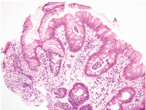
FIGURE 1.14 Barrett esophagus, negative for dysplasia. Although there is some superficial nuclear elongation there is no hyperchromasia, nuclear irregularity, or mucin loss. There is abundant lamina propria between the glands.


Barrett Esophagus: Indefinite for Dysplasia

Using the algorithm, the IND category included cases that had deeper cytologic changes suggestive of dysplasia, but which showed surface maturation. But, other observers have used the indefinite category as a
“waste can” (36). Cases with IND could have normal architecture or some degree of glandular crowding (Figs. 1.15, e-Figs. 1.31-1.34). On cytologic evaluation, lesions could have hyperchromasia, nuclear membrane irregularities, and increased mitoses in the deeper aspects, and these matured to the surface. Loss of nuclear polarity was not a feature of IND. In the presence of inflammation, more striking architectural abnormalities were to be included in the indefinite category. This interpretation can also be applied when tangential embedding does not allow assessment between the glands and the surface. Some cases display peculiar hypermucinous features, and it is unclear whether they are neoplastic or reparative (e-Figs. 1.35 and 1.36). We often offer an explanation for resorting to this category when it is assigned.


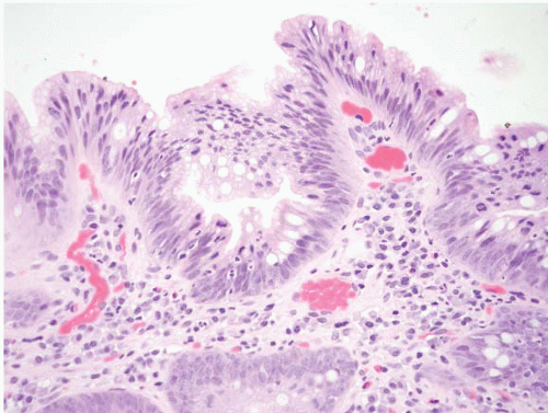
FIGURE 1.15 Barrett esophagus, indefinite for dysplasia. There is superficial nuclear enlargement and hyperchromasia associated with intraepithelial neutrophils. It is unclear whether these changes are truly neoplastic or reactive.


Barrett Esophagus: Low-grade Dysplasia

In Barrett esophagus with low-grade dysplasia (Figs. 1.16, 1.17 and 1.18, e-Figs. 1.37-1.56), the surface appears similar to the underlying glands at low magnification, or displays only slight maturation. The architecture may be mildly to markedly distorted with glandular crowding, although lamina propria should be identifiable between glands. The cytologic features are important and the changes should extend at least focally to the surface. This lack of surface maturation need not be diffusely present within all tissue fragments and, in fact, areas of abrupt transition between nondysplastic and dysplastic epithelium are a clue that the changes are indeed neoplastic as opposed to reactive. Superficially located nuclei are irregular,
hyperchromatic, mildly enlarged, and may show some degree of stratification and mucin loss. Mitotic figures may be seen at or close to the surface. More than mild nuclear enlargement is allowed if the other features support an interpretation of low-grade dysplasia. Loss of nuclear
polarity and nucleoli are not features of low-grade dysplasia, though nuclear stratification, similar to that seen in colonic adenomas, is within the spectrum of low-grade dysplasia and may be present at the surface (e-Figs. 1.57 and 1.58). Inflammation is typically minimal; cases with abundant inflammation and the other features of low-grade dysplasia are usually best classified in the IND category. If tangential embedding precludes evaluation of the surface, low-grade dysplasia can be diagnosed in the absence of inflammation if (a) there are dysplastic features in the deep aspects and (b) the features of high-grade dysplasia (see below) are lacking.


FIGURE 1.16 Barrett esophagus, low-grade dysplasia. The superficial epithelium on the left hand side appears similar to that within the deeper glands with nuclear enlargement, hyperchromasia, and loss of mucin. There is an abrupt transition between the nonneoplastic (right) and neoplastic (left) surface, a helpful clue.


FIGURE 1.17 Barrett esophagus, low-grade dysplasia. The superficial epithelium shows mucin loss, nuclear enlargement, and hyperchromasia. There is no loss of nuclear polarity.


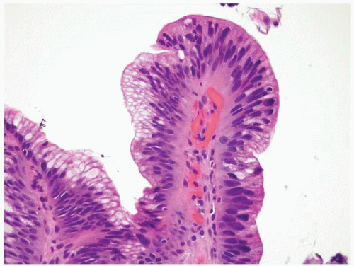
FIGURE 1.18 Barrett esophagus, low-grade dysplasia. The superficial epithelium shows mucin loss, nuclear enlargement, and hyperchromasia. There is no loss of nuclear polarity. Note nondysplastic epithelium to the left hand side and compare the sizes of the nuclei to those at the tip of the villiform area.


Barrett Esophagus: High-grade Dysplasia

As in low-grade dysplasia, in high-grade dysplasia, surface maturation is lacking (Figs. 1.19, 1.20 and 1.21, e-Figs. 1.59-1.84). The architecture may show crowding of cytologically abnormal glands, or be markedly distorted with prominent glandular crowding and little intervening lamina propria. If the cytologic features are sufficiently dysplastic, lesser architectural distortion is acceptable. Most cases similar to dysplasia in the uterine cervix, do not display prominent nucleoli, which tend to be present in either marked repair or when invasion has begun. Both of these latter situations are associated with ulcers. Nuclei may have either delicately clumped, dark heterochromatin and inconspicuous nucleoli, or prominent, irregular nuclei with irregularly clumped chromatin and irregular nucleoli. Markedly enlarged hyperchromatic cells are a feature of high-grade dysplasia and these may
extend to the surface. Loss of nuclear polarity is seen in high-grade dysplasia. Mitoses are readily identifiable. Inflammation is typically minimal. There is some evidence to suggest that high-grade dysplasia, accompanied by an ulcer, is a worrisome feature for an associated, unsampled, invasive carcinoma, and we suggest additional biopsies and/or endoscopic ultrasound when we see this pattern (27).


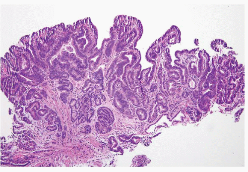
FIGURE 1.19 Barrett esophagus, high-grade dysplasia. This low power magnification shows total lack of surface maturation with crowding of cytologically abnormal glands.

Certain features, when seen in association with high-grade dysplasia, should provoke concern for the possibility of an underlying, unsampled carcinoma. These include (a) cribiform architecture, (b) dilated tubules containing
necrotic debris, (c) associated ulceration (e-Fig. 1.69), (d) neutrophils within dysplastic glands (Fig. 1.22), and (e) extension of neoplastic cells into the overlying squamous epithelium in a Pagetoid pattern. Zhu and colleagues demonstrated that identification of one of these features in association with high-grade dysplasia is associated with carcinoma in a subsequent resection
specimen in 39% of the cases. This figure increased to over 80% when two or more of these findings were present. In contrast, cases lacking any of these findings had no carcinoma in the resection specimen. Involvement of the overlying squamous epithelium with neoplastic glands in a Pagetoid pattern was associated with cancer in 100% of the cases (37). Other than Pagetoid spread pattern, these features, in fact, overlap with features of intramucosal carcinoma. Likewise, identification of single Paget cells (intraepithelial glandular neoplastic cells) in a biopsy specimen is invariably associated with an underlying adenocarcinoma containing at least a focal, poorly differentiated component (e-Figs. 1.85-1.87) (38). Overall, we believe that sufficient cytologic alterations “trump” architectural pattern.


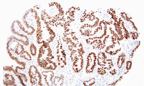
FIGURE 1.20 Barrett esophagus, high-grade dysplasia. A p53 immunostain shows prominent positivity.


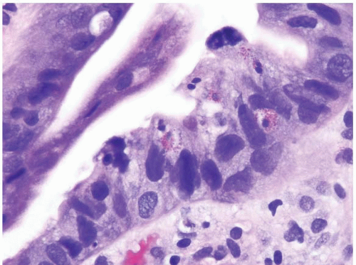
FIGURE 1.21 Barrett esophagus, high-grade dysplasia. This high power magnification shows loss of nuclear polarity and smudged, irregular, large nuclei.


FIGURE 1.22 Barrett esophagus, high-grade dysplasia. Luminal neutrophils located within glands with high-grade dysplasia should provoke concern for an associated, unsampled carcinoma.

In the past, esophagectomy was typically offered as a treatment in patients with high-grade dysplasia. Nevertheless, using modern techniques, endoscopic treatment has become the standard (5,16). Surveillance epidemiology and end results (SEER) data show that patients with high-grade dysplasia and early carcinomas have the same mortality whether managed endoscopically or surgically (39). More current practice is to sample aggressively for occult invasive carcinoma and perform close follow-up or endoscopic intervention. Knowing local practices may also influence interpretation of such cases.

In addition to EMR, available endoscopic treatments include multipolar electrocoagulation, argon plasma coagulation, photodynamic therapy (PDT), radiofrequency ablation, and cryotherapy. There is most experience with PDT but radiofrequency ablation is emerging as the preferred technique since it appears to have fewer complications than the others (16,40,41). A source of trepidation in the past has been that these techniques would fail to ablate dysplastic mucosa underneath squamous mucosa (buried BE) (Figs. 1.23, 1.24 and 1.25, e-Figs. 1.88-1.97). Surely dysplastic mucosa can be encountered underneath squamous mucosa, but is usually associated with surface dysplasia, at least in patients who have had PDT (42).


INTRAMUCOSAL CARCINOMA

The distinction between high-grade dysplasia and the earliest intramucosal carcinoma (defined as invasion through the basement membrane into the lamina propria or muscularis mucosae but not beyond) remains difficult. In general, these cases begin to demonstrate an effacement of lamina propria architecture and a syncytial growth pattern, extensive back-to-back microglands, cells with prominent nucleoli, and an intermingling of single cells and small clusters within the lamina propria (Figs. 1.26, 1.27 and 1.28, e-Figs. 1.98-1.115). Typically, desmoplasia ranges from absent to incompletely developed at this stage, hence its recognition is difficult and subjective. In carcinoma that has invaded more deeply (into the submucosa), desmoplasia and a clearly infiltrative growth pattern become readily apparent (Fig. 1.29 and 1.30, e-Figs. 1.116-1.120), although tangentially embedded and scarred tissue can pose diagnostic problems. In the upper
gastrointestinal tract, invasion into the lamina propria is more significant than in the colon, because colon lamina propria lacks significant lymphatic access. In the colon, invasion into the lamina propria is biologically equivalent to high-grade dysplasia, whereas in the esophagus, invasion into the
lamina propria can lead to metastatic disease (e-Figs. 1.102, 1.108, and 1.109). Interobserver variability can be a factor when diagnosing intramucosal carcinoma in a small biopsy (43), but this distinction is less important than it was in the past since both high-grade dysplasia and intramucosal carcinoma can be managed endoscopically (5,16).


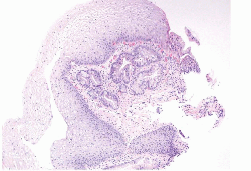
FIGURE 1.23 Burried Barrett epithelium (pseudoregression pattern). This photomicrograph shows Barrett mucosa buried underneath squamous epithelium, a finding associated with previous endoscopic treatment for Barrett esophagus but that can also be encountered in patients who have never had ablation treatment.


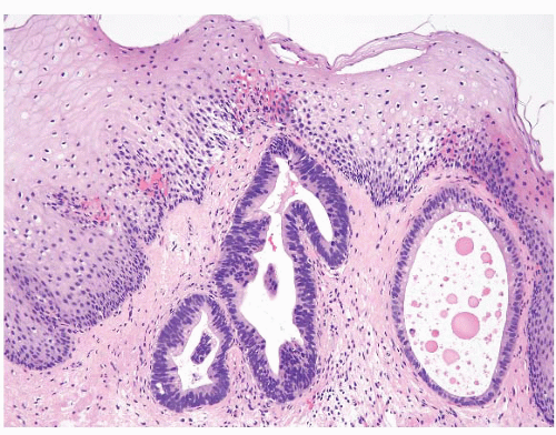
FIGURE 1.24 Buried dysplastic Barrett epithelium (pseudoregression pattern). Barrett mucosa with dysplasia is seen here “buried” underneath squamous epithelium following endoscopic treatment for Barrett esophagus.


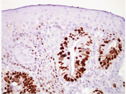
FIGURE 1.25 Buried dysplastic Barrett epithelium, p53 stain. There is prominent nuclear labeling.


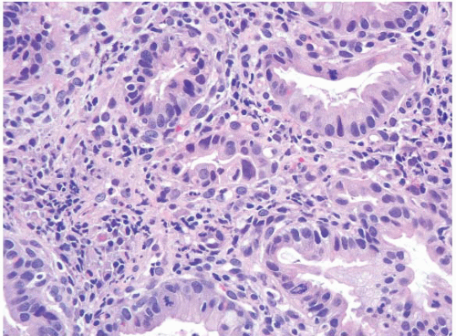
FIGURE 1.26 Intramucosal carcinoma. Note small clusters of epithelial cells within the lamina propria in association with glands showing high-grade dysplasia.


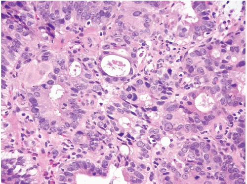
FIGURE 1.27 Intramucosal carcinoma. Note prominent nucleoli, back-to-back glands, and intraluminal debris.


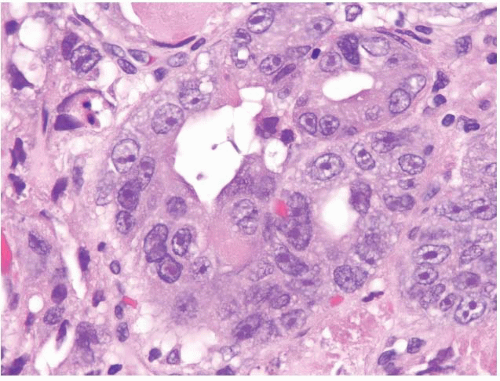
FIGURE 1.28 Intramucosal carcinoma. Higher magnification of Figure 1.27. Note the prominent nucleoli.


FIGURE 1.29 Invasive carcinoma. In carcinoma that has invaded into the submucosa, desmoplasia and a clearly infiltrative growth pattern may become more readily apparent, as in this case.


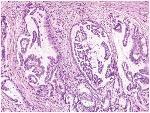
FIGURE 1.30 Invasive carcinoma. This lesion also features desmoplasia. This particular example shows a papillary growth pattern.


VARIANT DYSPLASIA PATTERNS

Some examples of high-grade dysplasia display abundant acute inflammation (e-Figs. 1.121-1.124). They are distinguished from reparative lesions by their more prominent hyperchromasia and their relative paucity of nucleoli. If biopsies have been processed with Bouin or similar fixatives, these examples display, at their surfaces, similar nuclear membrane irregularities to those seen in more typical dysplasia.

Only gold members can continue reading. Log In or Register to continue

Stay updated, free articles. Join our Telegram channel

Jun 18, 2016 | Posted by in GASTROENTEROLOGY | Comments Off on Esophagus

Full access? Get Clinical Tree

Get Clinical Tree app for offline access