Colon


Colon


Since colorectal cancer is the second leading cause of cancer-related deaths in the United States, much of the pathologist’s day is spent reviewing biopsy specimens obtained in the course of screening for this disease. A variety of nonneoplastic polyps and other neoplasms can be encountered in this process. The American Cancer Society estimated that about 141,210 Americans were diagnosed with this malignancy, and approximately 49,380 will die of the disease in 2011. Colorectal cancer is the third most common cancer in both men and women. Seventy-two percent of the cases arise in the colon and twenty-eight percent in the rectum. Incidence has been declining over time, largely thanks to the use of screening colonoscopies and removal of neoplastic polyps. Updated information on colorectal cancer is available from the Center for Disease Control (http://www.cdc.gov/cancer/colorctl) and The American Cancer Society (http://www.cancer.org/index).

The risk of developing colorectal cancer increases with age (>90% of cases diagnosed at 50 years or older). Other risk factors include inflammatory bowel disease (IBD), a personal or family history of colorectal cancer or colorectal polyps, and various syndromes. Incidence and mortality rates are 35% to 40% higher in men than women.

According to the American Cancer Society, beginning at age 50, men and women at average risk for colorectal cancer should be screened with one of the following: high-sensitivity fecal occult blood testing or fecal immunochemical test annually, stool DNA test (screening interval uncertain) and/or a flexible sigmoidoscopy every 5 years, colonoscopy every 10 years, CT colonography every 5 years, or double-contrast barium enema every 5 years (1). If adenomas are found on sigmoidoscopy, full colonoscopy is suggested. Accelerated screening is recommended for highrisk patients (1-3) (Table 4.1).

In contrast to the stomach, where the pathologist must focus on background mucosal pathology (or lack thereof) when polyps are found, most colon polyps arise in isolation. Many polyps that cannot be classified as adenomas are simply inflammatory polyps (“zits” of the colon). When a simple inflammatory polyp is encountered, most of the time it is an isolated incidental finding in a site prone to local trauma (such as the ileocecal valve), but occasionally it is a marker of undiagnosed inflammatory

disease. Thus, biopsy of the surrounding flat mucosa should be performed if the patient is symptomatic. However, endoscopists are good at recognizing mucosal pathology grossly in the colon (in contrast to the stomach, in which the endoscopic appearance correlates poorly with the presence of inflammatory processes), and they usually biopsy flat mucosa near a polyp, if the flat mucosa appears abnormal. Before discussing the adenomas and other neoplastic polyps, it is worthwhile to consider some nonneoplastic lesions that produce endoscopic polyps.


TABLE 4.1 Screening Recommendations for Colorectal Cancer and Polyps


Risk Category


Screening Method


Age to Begin Screening


Average risk


One of the following:


  1. Fecal occult blood testing or fecal immunochemical test annually


  2. Stool DNA test (interval uncertain)


  3. Flexible sigmoidoscopy every 5 y


  4. Fecal occult blood testing or fecal immunochemical test annually and flexible sigmoidoscopy every 5 y


  5. Double-contrast barium enema every 5-10 y


  6. Colonoscopy every 10 y


  7. CT colonography every 5 y


50 y


Family history


Colonoscopy every 5 y


Age 40 or 10 y before cancer was diagnosed in the youngest affected family member, whichever is earlier


Hereditary


Colonoscopy every 2 y


20-25 y until age 40, then every 1 y


Nonpolyposis colorectal cancer


Genetic counseling


Consider genetic testing


Familial adenomatous polyposis


Flexible sigmoidoscopy or colonoscopy every 1 y


Genetic counseling


Consider genetic testing


Puberty


Ulcerative colitis


Colonoscopy with multiple biopsies for dysplasia every 1/2 to 1 y


8-10 y after the diagnosis of colitis


Adapted from Smith RA, Cokkinides V, et al. Cancer screening in the United States, 2011: A Review of Current American Cancer. Society Guidelines and Issues in Cancer Screening. CA Cancer J Clin. 2011;61:8-30; Rex DK, Johnson DA, et al. American College of Gastroenterology Guidelines for Colorectal Cancer Screening 2008. Am J Gastroenterol. 2009;104:739-750; Kornbluth A, Sachar DB, et al. Ulcerative Colitis Practice Guidelines in Adults: American College of Gastroenterology, Practice Parameters Committee. Am J Gastroenterol. 2010;105:501-523.


NONNEOPLASTIC POLYPS


Elastosis/Elastofibromatous Change

There is a subset of polyps of the gastrointestinal (GI) tract in which the submucosa and muscularis mucosae exhibit a focal or diffuse increase of elastic fibers. This elastosis or elastofibromatous change is most commonly manifested as a colonic polyp and usually is found during screening colonoscopy (Figs. 4.1 and 4.2, e-Figs. 4.1-4.5). Gastric and small intestinal cases are less frequent and are associated with ulcers or an inflammatory process (4). Hobbs et al. (4) summarized the literature and found reports of 13 GI elastotic lesions with a topographic distribution similar to their own series of 13 cases. Histologically, elastosis appears as finely granular and/or fibrillar amphophilic material, sometimes with a fibrous component (elastofibromatous change). The changes occasionally appear centered around blood vessels and often are mistaken for amyloid but are negative for Congo red stain and strongly positive for elastic stain. These lesions are probably incidental and of no clinical consequence.


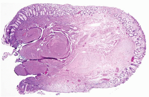
FIGURE 4.1 Colonic elastosis. This example shows a polypoid fragment of tissue with submucosal increase in elastic fibers.


FIGURE 4.2 Colonic elastosis. An elastic stain highlights elastic fibers within the submucosa.


Filiform Polyps

Filiform polyps (also called post inflammatory polyps) are essentially a subtype of polyps associated with prior mucosal injury, so they are not unusual in patients with inflammatory bowel disease (IBD). But, they can be found in patients who have had any type of prior ulceration and can sometimes have a dramatic gross appearance in patients with IBD, sometimes simulating a neoplasm and termed filiform polyposis or giant filiform polyposis (Fig. 4.3, e-Figs. 4.6-4.8). They consist of fingerlike projections of submucosa covered by mucosa on all sides. They reflect healing of undermined mucosal and submucosal remnants and ulcers and are typically multiple. To the endoscopist, they appear as long, thin, cylindrical projections (5). They are diagnosed by noting their composition: two protruding layers of mucosa plastered together with only one intervening layer or no intervening layer of muscularis mucosae. This construction reflects regrowth of mucosa over an area of ulcer that has damaged the muscularis mucosae. Note the lack of muscularis mucosae in the filiform polyp depicted in Figure 4.4 (e-Figs. 4.9 and 4.10).


MUCOSAL PROLAPSE CONDITIONS

This category of lesions encompasses a host of processes in diverse GI tract sites, but prolapse changes often yield an endoscopic polyp when they occur in the colon. Well-defined colonic prolapse conditions include solitary rectal ulcer syndrome, localized colitis or proctitis cystica profunda, inflammatory cloacogenic polyp, prolapsing folds associated with diverticular disease of the colon, and fibrin cap polyps of the colon. So-called
myoglandular polyps are presumably part of the same spectrum (6,7). In some GI sites, prolapse is virtually physiologic. For example, the ileocecal valve is prone to prolapse and, if there is abundant submucosal fat in the prolapsed area, it is termed “lipoma of the ileocecal valve,” but is probably not neoplastic. These conditions are all benign and, occasionally, are mistaken (both clinically and microscopically) for carcinomas.


FIGURE 4.3 Giant filiform polyposis. Note the fingerlike projections of submucosa covered by mucosa on both sides. This particular case showed impressive active inflammation and presented as a transverse colon mass in a patient without a prior history of IBD.


Solitary Rectal Ulcer Syndrome

Solitary rectal ulcer syndrome describes a pattern of mucosal changes (encompassing polyps lacking ulceration) that is localized to the terminal rectum and imparted by mucosal prolapse. It occurs at all ages, with a
peak incidence between 20 and 40 years. The classic history is of a young woman who strains when defecating. There may be hamatochezia, pain, tenesmus, and sometimes lower abdominal pain. Inability to evacuate the rectum, or a “foreign body” sensation, is described. At endoscopy, ulcers are seen in 20% to 70% of patients, usually on the anterior or anterolateral rectal wall, but a mass-like lesion can also be found, which raises the possibility of a neoplasm. Sometimes defecation studies are used to evaluate these patients because they are believed to have difficulty coordinating the smooth muscle during the defecation process, such that the puborectalis sling does not relax at the proper time.


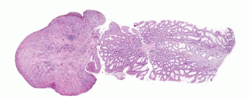
FIGURE 4.4 Filiform polyp. Note the lack of muscularis mucosae in this impressive low power image.

The pathologic changes on biopsies consist of hypertrophy of the muscularis mucosae, with splaying of fibers which course into the mucosa and are seen throughout the lamina propria. The proliferated smooth muscle is accompanied by variable fibrosis, and the glands become entrapped and distorted. As the process continues, there is surface ulceration, and glands can herniate into the submucosa, accompanied by wisps of lamina propria (a theme in adenomas as well) (8-13). Thus, lesions can have a “polypoid phase” or an ulcerated phase (Fig. 4.5, e-Figs. 4.11-4.16). Often crypts become “diamond-shaped” (14,15) (e-Fig. 4.14). Frequently such polyps have serrated features (e-Fig. 4.15), which has resulted in confusion with sessile serrated adenomas (SSA)/polyps (SSA; discussed below). Studies published when understanding of the molecular underpinnings of SSA was poor included attempts to seek evidence of mismatch repair (MMR) defects in prolapse polyps (16) (such studies are negative but SSA have intact MMR proteins, so
their results are misleading in the first place). Since prolapse polyps are typically left sided (in contrast to SSA) and feature prominent smooth muscle proliferation, most cases can be assigned to one or the other category. In general, sessile serrated adenomas/polyps lack smooth muscle proliferation in the lamina propria, although some examples have associated perineuriomas/benign fibroblastic polyps (discussed below) in their lamina propria (17-22).


FIGURE 4.5 Mucosal prolapse. Note fibromuscular stranding into the mucosa with associated surface erosion.

A caveat in diagnosing mucosal prolapse polyps is that mucosal prolapse changes adjacent to carcinomas are the same as those of isolated mucosal prolapse, so we suggest multiple biopsies of large “solitary rectal ulcers” to exclude sampling error (23). “Colitis cystica profunda” is part of the same spectrum of disease and implies that glands have prolapsed into the submucosa. Additionally, occasional patients with prominent distal rectal mucosal prolapse can present with an apparent polyposis (24) (e-Figs. 4.17 and 4.18).


Inflammatory Cloacogenic Polyp

Inflammatory cloacogenic polyp refers to a mucosal prolapse polyp arising at the anorectal transition, thus having both squamous and columnar mucosa (12,25) (Fig. 4.6, e-Figs. 4.19-4.22). Patients with such polyps present with hematochezia, and the polyps are typically found on the anterior wall of the anal canal. These polyps display a tubulovillous growth
pattern surface ulceration, displaced clusters of crypts into the submucosa, and abundant fibromuscular stroma that extends into the mucosa.


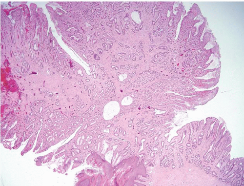
FIGURE 4.6 Inflammatory cloacogenic polyp. This is a mucosal prolapse polyp arising at the anorectal transition. Note both squamous and columnar mucosa, tubulovillous growth pattern, surface ulceration, and abundant fibromuscular stroma that extends into the mucosa.


Diverticular Disease Associated Polyps (Polypoid Prolapsing Mucosal Folds)

Diverticular disease results in two principal alterations to the mucosa. There is the pulsion diverticulum itself, in which the mucosa and its investing muscularis mucosae penetrate through the colonic muscularis propria wall. Around this diverticulum, the smooth muscle hypertrophies and contracts such that the associated mucosa becomes thrown into redundant folds (Fig. 4.7, e-Fig. 4.23). These prominent folds can prolapse and form polyps (26), which become variably inflamed (27). The prolapse changes in these polyps are like those elsewhere, consisting of reparative epithelial features and herniation of smooth muscle into the mucosa, and of the mucosa into the submucosa. Thus, biopsies show crypt elongation, crypt distortion, and ingrowth of muscularis mucosae into the lamina propria; thus, polypoid prolapsing mucosal folds (PPMUFs) are analogous to the classic more distal prolapse lesions (6). Occasionally, a diverticulum inverts and presents as a polyp. Biopsy of such a “polyp” can lead to bowel perforation (e-Fig. 4.24) if there is accompanying muscularis propria. This is similar to the occasional removal of an inverted appendix. Finding muscularis propria on mucosal biopsies should prompt a dialogue with the endoscopist to help exclude an iatrogenic perforation.


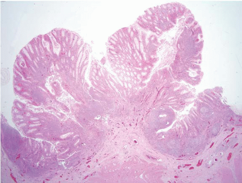
FIGURE 4.7 Diverticular disease associated polyp (polypoid prolapsing mucosal folds). Smooth muscle hypertrophies and contracts around diverticula such that the associated mucosa becomes thrown into redundant folds, prolapsing and forming polyps. The mucosa is commonly inflamed, as seen in this example.


Cap Polyposis

This unusual condition is often included with prolapse syndromes but is not necessarily related to mucosal prolapse. It is a rare benign colorectal condition in which patients have numerous polyps in the distal colorectum that are covered by an inflammatory “cap” of granulation tissue (28,29) with normal intervening mucosa. Overall, most patients are adults with a median age in their early 50s but pediatric cases have been reported. There seems to be a female predominance. Patients present with mucous diarrhea, rectal bleeding, and tenesmus. This can be sufficiently severe to result in a protein losing colopathy with peripheral edema. Many patients are reported to have long-standing constipation and straining to defecate. On colonoscopy, patients have 1 to >100 distal polyps covered with thick purulent exudates. Some authors show images with appearances of prolapse polyps (28), whereas the original report showed peculiar polyps with hyperplastic epithelium showing abundant mucus production and surface exudate with pristine intervening mucosa (Fig. 4.8, e-Figs. 4.25 and 4.26). In some instances, protein loss and symptoms can be sufficient to warrant distal colectomy and some patients have rectal prolapse (30). Some authors have reported response to 1) immunomodulation, 2) Helicobacter pylori eradication, and 3) avoiding straining while defecating (28,31-37).


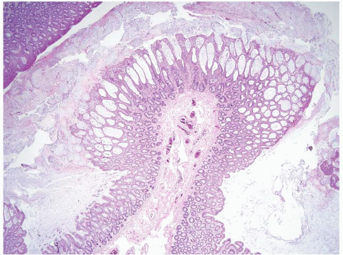
FIGURE 4.8 Cap polyposis. This case shows polypoid hyperplastic epithelium with abundant surface mucus and normal intervening mucosa.


Juvenile Polyps

Juvenile polyps are the most commonly encountered colorectal polyps in children, and in one-third to one-half of instances, more than one juvenile polyp is found (38-45). Sporadic juvenile polyps usually have a spherical lobulated surface, which is often eroded (Fig. 4.9). These polyps are considered hamartomatous so, in the colon, they display colonic-type mucosa and have irregularly shaped and dilated glands, accompanied by lamina propria that is expanded with edematous granulation tissue (e-Figs. 4.27-4.30). Dysplasia (e-Figs. 4.31-4.33) is rare in sporadic juvenile polyps.

Juvenile polyposis has been recognized since 1975 (46), and criteria for diagnosis consist of (a) more than five juvenile polyps of the colorectum, (b) juvenile polyps throughout the GI tract, or (c) any number of juvenile polyps in a patient with a family history of juvenile polyposis. The incidence is about 0.5 to 1 per 100,000 in Western countries. The polyps may be found anywhere in the GI tract (and sometimes have a very nonspecific appearance). They can be subtle equivocal lesions or large polyps on long stalks (e-Fig. 4.34) and the patients are at increased risk for colorectal, gastric, duodenal, and pancreatobiliary carcinomas. In addition, juvenile-type polyps are a component of several genetic syndromes (42,47-53). The syndromes include classic juvenile polyposis, Cowden syndrome, and Bannayan-Riley-Ruvalcaba syndrome. Germline mutations in DPC4 (also known as SMAD4) and BMPR1A predispose an individual to juvenile polyposis, and both genes are involved in transforming growth
factor β (TGFβ) superfamily signaling pathways. In Cowden syndrome and Bannayan-Riley-Ruvalcaba syndrome, juvenile polyps are a less consistent feature. Cowden syndrome patients are at risk for breast and thyroid cancers. Mutations of the tumor suppressor gene, PTEN, have been found in the germline of both Cowden syndrome and Bannayan-Riley-Ruvalcaba syndrome patients. Despite different underlying genetic mechanisms, these and other syndromes share the same phenotypic feature of juvenile polyps. A combined syndrome of juvenile polyposis and hereditary hemorrhagic telangiectasia (Osler-Weber-Rendu disease) has also been described in patients with SMAD4 mutations (54). Hereditary hemorrhagic telangiectasia is an autosomal dominant disorder characterized by vascular malformations of visceral organs usually caused by mutations in ENG (endoglin) or ACVRL1 (ALK1). Though both syndromes have distinct features, a subset of patients with SMAD4 mutations shows a combined phenotype. As a result, patients with juvenile polyposis with SMAD4 mutations should be evaluated for the presence of occult arteriovenous malformations of visceral organs. These syndromes are summarized in Table 4.2.


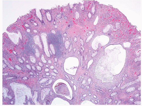
FIGURE 4.9 Juvenile polyp. Note colonic-type mucosa with irregularly shaped and cystically dilated glands with intraluminal acute inflammatory cells, accompanied by an expanded and inflamed lamina propria that contains granulation tissue.

When juvenile polyps are biopsied from syndromic patients, smaller ones are identical to the typical sporadic ones. However, larger ones display an increase in the relative amount of epithelium compared to stroma, are multilobulated with rounded or fingerlike lobes, and are more likely to display true dysplasia (Figs. 4.10 and 4.11). Both types of juvenile polyps are prone to surface erosions, with attendant reactive epithelial changes. In general, the pathologist is cautioned against diagnosing dysplasia when active inflammation and erosions are features. One study suggests that the crypt to stroma ratio correlates with the underlying genetic defect; Polyps with SMAD4 germline mutations tend to have a crypt:stroma ratio >1, while those with BMPR1A mutations tend to have a crypt:stroma ratio <1 (55).

Dysplasia is more likely to be encountered in syndromic juvenile polyps than in sporadic ones. In an early study, 12% of patients with juvenile polyposis had dysplasia/neoplasia, and 5% had carcinoma resected (39). In the last 10 years at Johns Hopkins, we have encountered about 100 patients with juvenile polyposis in our material and about 25% had dysplasia in their polyps and about 6% had associated carcinomas; our consult material may be enriched for patients with neoplasia but it is frequent enough that all patients are advised to undergo surveillance with polypectomies or colectomy depending on the number of polyps that they harbor.

Morphologically, dysplasia in juvenile polyps results in a pattern essentially indistinguishable from sporadic adenomas (e-Figs. 4.31-4.33). Occasionally, there is a clue that the underlying lesion is a juvenile polyp; an expanded lamina propria or especially dilated glands may be present. However, in some cases, it can be impossible to distinguish juvenile polyps complicated by dysplasia from adenomas. Similarly, since the polyps appear inflammatory, the appearance can suggest an inflammatory polyp in the setting of ulcerative colitis or Crohn disease. However, the background flat mucosa between polyps is normal rather than inflamed (e-Fig. 4.35).


TABLE 4.2 Summary of Syndromes Associated with Multiple Juvenile Polyps


Syndrome


Components


Genetic Defect


Cancer Risk and Other Implications


Juvenile polyposis


More than five colorectal juvenile polyps or juvenile polyps throughout the GI tract, or any juvenile polyp in a patient with family history of juvenile polyposis


Autosomal dominantgermline mutations in SMAD4/DPC4


Colorectal, stomach, small intestine, and pancreatobiliary


Cowden syndrome


Hamartomas involving organs from all three germ layers; benign skin tumor (trichilemmoma), thyroid abnormalities, fibrocystic disease, juvenile polyps, uterine leiomyomas, macrocephaly, mental retardation, and dysplastic gangliocytoma of the cerebellum


Autosomal dominantgermline mutations of PTEN/MMAC1 gene


Breast and thyroid cancer


Bannayan-Riley-Ruvalcaba syndrome


Microcephaly, lipomatosis, hemangiomatosis, speckled penis, polyps; overlap with Cowden syndrome


PTEN germline mutations


Presumed at risk for cancers


Combined syndrome of juvenile polyposis and hereditary hemorrhagic telangiectasia


Juvenile polyposis and vascular malformations (mucocutaneous telangiectases, epistaxis, solid organ and CNS arteriovenous malformations)


Germline mutations in SMAD4/DPC4


Same neoplastic risk as patients with juvenile polyposis. Patients also at risk of internal bleeding as a result of arteriovenous malformations.


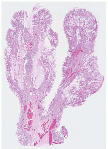
FIGURE 4.10 Syndromic juvenile polyp. Compared to sporadic juvenile polyps, syndromic examples display an increase in the relative amount of epithelium compared to stroma and tend to have fingerlike projections.

The polyps encountered in Cowden syndrome have less specific features than those of classic juvenile polyposis. As noted above, Cowden syndrome is the most common subset of the PTEN (phosphate and tensin homolog gene on chromosome 10 hamartoma tumor syndrome. The classic patient manifests skin trichilemmomas and keratoses. Patients are at increased risk for cancers of the thyroid, breast, and endometrium. GI polyposis is a common finding and numerous (>50) polyps can be encountered. The pathologist should consider this possibility when a patient has nonspecific inflammatory polyps without associated prior mucosal injury. In a Mayo clinic study of 13 patients with Cowden syndrome
who had undergone colonoscopy, patients had hamartomatous polyps, inflammatory/juvenile-type polyps, ganglioneuromas, adenomas, and two of the 13 had adenocarcinomas (56). Most of the patients had three or more types of polyps in their samples. Examples of polyps encountered in a patient with Cowden syndrome are shown in e-Figures 4.36-4.39.


FIGURE 4.11 Syndromic juvenile polyp. At high magnification, this juvenile polyp contained extensive low-grade dysplasia (see Figs. 4.31, 4.32 and 4.33) resembling tubular adenoma but the architecture of this polyp is clearly different from that of a sporadic tubular adenoma. Diagnosing such a lesion on a small superficial biopsy would be impossible.

In our somewhat limited experience, the polyps in patients with Bannayan-Riley-Ruvalcaba syndrome (polyps, macrocephaly, lipomas, hemangiomas, thyroid problems, penile freckling) have the same appearances as those encountered in juvenile polyposis (e-Figs. 4.40 and 4.41), an observation in line with literature reports (51).

Remember that adult patients often have inflammatory polyps that are presumably a result of prior mucosal injury and that can perfectly mimic juvenile polyps (Fig. 4.12, e-Fig. 4.42). We report these in a descriptive fashion as “inflammatory polyp, juvenile type.”


Peutz-Jeghers Polyps

Peutz-Jeghers polyps are most common in the small intestine and are further discussed in Chapter 3, but their manifestations in the colon are similar to those in other sites. These polyps are associated with perioral and intraoral (mucosal) pigmentation (e-Fig. 4.43) and generally are not especially numerous in resection samples. They are characterized by sitespecific (colonic) mucosa with arborizing smooth muscle. Unfortunately, because mucosal prolapse is so common in the colon, it is difficult to prospectively diagnose Peutz-Jeghers syndrome on the basis of a colonic polyp in isolation and diagnosing colonic examples is sometimes only possible
in the context of clinical history. Overall Peutz-Jeghers polyps differ from prolapse polyps by featuring zones of disorganized mucosa partitioned by cords of smooth muscle, whereas prolapse polyps feature thin strands of smooth muscle replacing lamina propria and investing individual crypts. On small superficial samples, however, it can be impossible to confidently separate the two types of polyps. When we encounter a colon polyp with features of a Peutz-Jeghers-type polyp (Fig. 4.13, e-Figs. 4.44-4.49), we issue a descriptive diagnosis and suggest correlation with other stigmata of Peutz-Jeghers syndrome. Rare examples show dysplasia (e-Fig. 4.50). As discussed in Chapter 3, there are data to suggest that even patients with single “sporadic” Peutz-Jeghers polyps may be at risk for Peutz-Jeghers syndrome-associated malignancies and that isolated examples are the exception (57). Patients with this syndrome are at risk for breast and pancreas cancer, and for female (adenoma malignum of cervix and sex cord tumors with annular tubules of the ovary) and male (sertoli tumors of testis) genital tract tumors. The syndrome associates with mutations/deletions in an involved gene, LKB/STK11, in approximately 80% to 94% of cases.


FIGURE 4.12 Inflammatory polyp, juvenile type. Some inflammatory polyps in adult patients are presumably a result of prior mucosal injury and can perfectly mimic juvenile polyps. Note the cystically dilated glands and expanded and inflamed lamina propria.


Cronkhite-Canada Polyps

Unfortunately, a Cronkhite-Canada polyp is a type that is virtually impossible to diagnose prospectively based on microscopic features in isolation.

Cronkhite and Canada reported a series of patients in 1955 who had polyposis, pigmentation, alopecia, and onychotrophia (58). There have been few subsequent reports of this condition (other than case reports), although Burke
et al. were able to amass polyps from nine patients from the consultation files of the Armed Forces Institute of Pathology (AFIP) for histologic analysis (59), and Ward has provided excellent reviews of the literature (60-62).


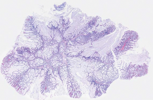
FIGURE 4.13 Peutz-Jeghers polyp. This example shows colonic mucosa partitioned by cords of smooth muscle that invests groups of glands.

Cronkhite-Canada syndrome is characterized by diffuse polyposis occurring in patients with unusual ectodermal abnormalities, including alopecia, onychodystrophy, and skin hyperpigmentation. Europeans and Asians are most frequently affected, with a mean age of onset at 59 years. Several hundred cases of Cronkhite-Canada syndrome have been reported worldwide, with 75% of these reports originating from Japan. The male-to-female ratio is 3:2. Potentially fatal complications—such as malnutrition, GI bleeding, and infection—often occur, and the mortality rate has been reported to be as high as 60%. Neither a familial association nor a genetic defect is known.

The most common presenting symptoms include diarrhea, weight loss, nausea, vomiting, hypogeusia, and anorexia. Paraesthesias, seizures, and tetany (apparently related to electrolyte abnormalities) have also been reported. Nail dystrophy—with thinning, splitting, and separation from the nailbeds—are the typical nail features. Both scalp and body hair alopecia may be present. Diffuse hyperpigmentation of the skin, manifested by light-to-dark brown macular lesions, is seen most frequently on the extremities, face, palms, soles, and neck. Microscopic examination of biopsied skin reveals abnormally increased melanin deposition with or without increased melanocyte proliferation.

Cronkhite-Canada syndrome is distinguished by the diffuse distribution of polyps throughout the entire GI tract, except for the characteristic sparing of the esophagus. The question of whether polyps in Cronkhite-Canada syndrome possess malignant potential remains controversial.


A number of complications may occur with Cronkhite-Canada syndrome and can contribute to poor outcomes in patients with this disease. These include potentially fatal GI bleeding, intussusception, and prolapse. Electrolyte abnormalities, dehydration, protein-losing enteropathy, and other nutritional deficiencies due to malabsorption can complicate the course of the disease. Cronkhite-Canada syndrome patients are prone to recurrent infections, but it is not known whether this is related to malnutrition or is a primary immunologic deficiency.

The Cronkhite-Canada polyp is characterized by its broad sessile base, expanded edematous lamina propria, and cystic glands (59) (Fig. 4.14, e-Figs. 4.51-4.53). Similar features are found in the lesions of juvenile polyposis. The only distinguishing feature reported between Cronkhite-Canada and colonic juvenile polyposis was the pedunculated growth of the latter, a feature that did not hold for gastric lesions. Therefore, the diagnosis of Cronkhite-Canada polyps, especially in the stomach, requires correlation with the presence of the ectodermal changes characteristic of this syndrome. Additionally, if the endoscopist has biopsied the flat mucosa between the polyps, the flat mucosa is normal in patients with juvenile polyposis, whereas it is abnormal in Cronkhite-Canada syndrome. Finding dysplastic changes favors juvenile polyposis as dysplasia is essentially never seen in Cronkhite-Canada polyps, but dysplasia occasionally complicates juvenile polyps as discussed above. See additional discussion of Cronkhite-Canada syndrome and illustrations in Chapters 2 and 3.


FIGURE 4.14 Cronkhite-Canada polyp. These are characterized by a broad sessile base, expanded lamina propria, and cystic glands.


OTHER CONDITIONS PRODUCING NONNEOPLASTIC POLYPS


Endometriosis

Endometriosis is well known to present in a variety of sites and affects the GI tract in up to 40% of patients with pelvic endometriosis; about one-third of these patients have mucosal lesions amenable to biopsy (63). The key to diagnosing endometriosis is to think of it! The sigmoid colon is the most common site. A similar phenomenon, termed deciduosis (ectopic decidua), has been found in pregnant women (64).

On gross examination, endometriosis appears as it does in other sites, as firm areas, which may contain cysts filled with brown fluid. The appearance of deciduosis is less specific—it manifests as whitish plaques.

Most examples of endometriosis affect the serosa or muscularis propria and are accompanied by abundant fibrosis and adhesions, though submucosal examples are also reported. Endometriosis of the colon resembles examples found elsewhere, consisting of endometrial-type glands, stroma associated with hemosiderin deposition, and a fibroblastic response (Fig. 4.15, e-Figs. 4.54-4.61). The endometrial-type epithelium changes with the menstrual cycle. A stromal decidual reaction may be found in endometriotic foci in pregnant patients (e-Figs. 4.62 and 4.63). Endosalpingiosis can also be encountered (e-Figs. 4.64 and 4.65).

Deciduosis differs from endometriosis by lacking glands and consists only of large polyhedral cells arranged in sheets in the serosa or outer
muscularis propria, although deciduosis has rarely been reported in the mucosa of the GI tract. Such cells can sometimes be mistaken for malignant (often epithelial) lesions.


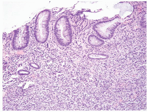
FIGURE 4.15 Colonic endometriosis. This rare example involved the colonic mucosa. Note horizontally oriented endometrial-type glands and associated stroma.

When endometriosis is found, it can mimic Kaposi sarcoma (e-Figs. 4.58 and 4.59). Reactive epithelial changes can also simulate colonic neoplasms (e-Fig. 4.58).

If deciduosis raises a concern for carcinoma or melanoma based on its large, pink, polyhedral cells, immunohistochemical stains for keratin, carcinoembryonic antigen (CEA), epithelial membrane antigen (EMA), and S100 protein can be performed and can be expected to be negative. The large decidualized cells typically express vimentin and may show desmin or muscle actin expression. When the stroma of endometriosis is unaccompanied by glands, a CD10 (e-Fig. 4.61) stain can be reassuring to exclude Kaposi sarcoma.


Other Conditions

Since polyps are simply protuberances, any number of nonneoplastic lesions can manifest as such. Amyloidosis can present as a colon polyp (Fig. 4.16) and colonic xanthomas can occasionally be encountered as incidental findings. Foreign material (including ingested fragments of bone) can lodge in the colon and produce a polyp. In the case of lodged ingested bone versus ossification, the ingested bone shows nonviable nuclei (e-Figs. 4.66-4.69), whereas metaplastic bone is viable (e-Fig. 4.70).


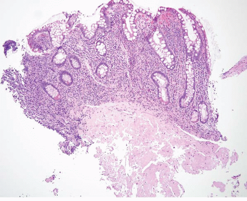
FIGURE 4.16 Amyloidosis. Cases of colonic amyloidosis can give the endoscopic impression of a polyp. Note submucosal amorphous, eosinophilic material.


ADENOMAS

It is estimated that more than half of the Western people will develop a benign colorectal tumor (adenomatous polyp) during their lifetime and that approximately 10% of such tumors will progress to malignancy (65). Familial adenomatous polyposis (FAP) patients have germline defects in the adenomatous polyposis coli (APC) gene, the gatekeeper of colorectal neoplasia (65), and have hundreds to thousands of colon adenomas and essentially all develop colon cancers if no prophylactic colectomy is performed (e-Figs. 4.71-4.73). Adenomas of the colorectum generally pose few diagnostic problems to the pathologist, and endoscopists usually recognize and remove them readily (e-Figs. 4.74-4.76). The situations that can be difficult involve differential diagnosis between reparative conditions with epithelial changes simulating adenomas and adenomas themselves, and dysplasia in the setting of IBD.

Adenomas have dysplasia by definition (Figs. 4.17, 4.18 and 4.19, e-Figs. 4.77-4.80). It is usually low-grade with regular nuclei showing maintained nuclear polarity (their long axes are perpendicular to the basement membrane). When this is the case, we do not offer a grade of dysplasia, but simply diagnose an adenoma and comment on cauterized margin status if the specimen is not tiny or fragmented. Typical adenomas display elongated “pencillate” nuclei that are similarly hyperchromatic throughout, in contrast to normal mucosa that shows reduced nuclear hyperchromasia at the surface. Apoptotic bodies are usually prominent in sporadic adenomas (Fig. 4.17). Adenomas may contain foci of clear cell change (66) (Fig. 4.20, e-Figs. 4.81-4.83), squamous-like morules akin to those seen in

the endometrium (67) (Fig. 4.21, e-Figs. 4.84 and 4.85), and Paneth cell differentiation, none of which matter in an adenoma, but which may inform some of the variation in the appearances of invasive carcinomas (66) (e-Figs. 4.86-4.90). Prominent intraepithelial lymphocytes and reduced numbers of
apoptotic bodies are encountered in colorectal adenomas from patients with hereditary nonpolyposis colorectal carcinoma (Lynch syndrome, see below) (e-Fig. 4.91) (68) but it is not known whether finding these features prospectively predicts the diagnosis. Of course, high-grade neuroendocrine (small cell) carcinomas may arise in the background of ordinary-appearing adenomas, but this is rare and discussed below.


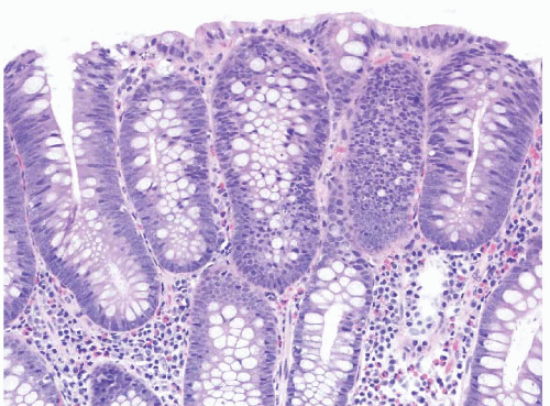
FIGURE 4.17 Tubular adenoma. This example shows low-grade dysplasia with hyperchromatic, pencillate nuclei. Apoptotic bodies are evident in the center of the field.


FIGURE 4.18 Tubular adenoma. Note the pencillate, hyperchromatic nuclei that remain perpendicular to the basement membrane. Compare with the normal mucosa on the right, which shows reduced nuclear hyperchromasia and more abundant mucin at the surface.


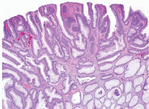
FIGURE 4.19 Tubular adenoma. Some larger examples undergo separation of the crypts at the surface, giving the impression of villi. Tubulovillous adenomas should have long, well-formed fingerlike villi.


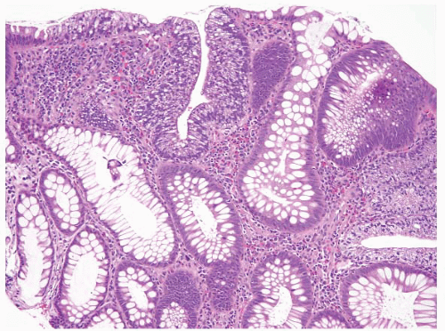
FIGURE 4.20 Tubular adenoma. Some examples contain foci of clear cell change as is seen here in the center of the field.


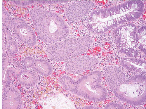
FIGURE 4.21. Tubular adenoma. This example shows squamous-like morules akin to those seen in the endometrium.

Adenomas have their initiation point near the surface of the mucosa, such that tiny adenomas only affect the upper half of the mucosa and grow in a “top-down” fashion (69) (e-Figs. 4.77 and 4.78). Genetically altered cells in the superficial portions of the mucosa spread laterally and downward to form new crypts that first connect to preexisting normal crypts, and eventually replace them. This is a useful feature to remember when separating adenomas from both reactive lesions and colitis-associated dysplasia, both of which seem to display “bottom-up” growth. Also, adenomas usually display prominent apoptosis (Fig. 4.17) and may contain scattered neutrophils. The presence of prominent apoptosis can be a helpful diagnostic feature.

When endoscopists biopsy “polyps,” sometimes the pathologist sees nothing to account for a polyp. If recut sections are performed in such instances, about 10% of such cases can be shown to harbor an adenoma on additional sectioning (70) and if tissue blocks are re-embedded and recut, up to 20% can be found to harbor adenomas (71). Each laboratory should probably determine its own protocol for further evaluating sampling in which the endoscopist notes a polyp and no lesion is seen on evaluated slides. In our hospitals, we perform no recuts if a separate sample from the same patient has an adenoma, whereas perform additional sectioning if this might alter the patient’s follow-up and thus recut the sample/s of “polyps” that are negative for adenoma.

Adenomas may undergo striking mucosal prolapse changes, which can cause a host of diagnostic problems. That is, neoplastic glands can herniate into the submucosa and, similarly, strands of muscularis mucosae can proliferate into the lamina propria and simulate submucosa. When neoplastic glands from adenomas prolapse into the submucosa, this can occasionally impart an appearance similar to that of invasive carcinoma, especially if the glands become obstructed and inspissated mucus dissects into the surrounding connective tissue, termed “pseudoinvasion” (72) (Figs. 4.22 and 4.23, e-Figs. 4.92-4.102). The following clues are used to separate this artifact from true invasive carcinoma: (a) In “pseudoinvasion,” lamina propria is “dragged” along with the neoplastic glands into the submucosa and thus invests the misplaced glands; (b) there is often accompanying hemosiderin; (c) glands tend to be rounded (gland angulation is more commonly seen in carcinoma); and (d) the proliferation in the submucosa has the same cytoarchitectural features as the adenoma that is clearly in the mucosa—even high-grade dysplasia may be among these cytologic features (e-Fig. 4.102) (72).

When an adenoma has high-grade dysplasia (Figs. 4.24 and 4.25, e-Figs. 4.103 and 4.104), we mention this in our report but point out that the lesion
still has no metastatic potential to forestall overly aggressive treatment if the reader of the issued report is not familiar with this basic principle. Criteria to diagnose high-grade dysplasia in colon adenomas are not established, an interesting phenomenon since finding high-grade dysplasia in an adenoma is a reason to intensify post polypectomy follow-up (73).The 2006 guidelines for management after diagnosis of adenomas specifically state “People at increased risk have either three or more adenomas, high-grade dysplasia, villous features, or an adenoma 1 cm or larger in size. It is recommended that they have a 3-year follow-up colonoscopy. People at lower risk who have one or two small (<1 cm) tubular adenomas with no high-grade dysplasia can have a follow-up evaluation in 5 to 10 years, whereas people with hyperplastic polyps (HPs) only should have a 10-year follow-up evaluation, as for average-risk people.” (73). Fortunately high-grade dysplasia and large adenoma size generally go hand in hand, so observer variation in thresholds for high-grade dysplasia are less important than they might seem. Although we have no validation, we generally reserve a diagnosis of high-grade dysplasia in colorectal adenomas for lesions that have cribriform architecture and/or loss of nuclear polarity (Figs. 4.24 and 4.25, e-Figs. 4.103 and 4.104) rather than only cytologic atypia or stratification of nuclei to the surface. The reasoning is that if we miss a bit of intramucosal carcinoma in the colon, it is not of any consequence, whereas we use a lower threshold in the esophagus and stomach. Some colleagues do not even report high-grade dysplasia in colorectal adenomas to forestall overtreatment by surgical colleagues who harbor the erroneous notion that high-grade dysplasia should

prompt a colectomy. When carcinomas arise in adenomas of the colon, invasion of the lamina propria is considered biologically equivalent to highgrade dysplasia (since the lamina propria of the colon is believed to lack lymphatic access, “intramucosal carcinoma” in the colon is thus staged as Tis rather than T1) (74,75), so some observers do not report this invasion either. We report intramucosal carcinoma in adenomas as such and always include a note stating that it is biologically equivalent to high-grade dysplasia (Tis) and that complete polypectomy should be curative. We report our findings this way in case additional sampling discloses deeper invasion.


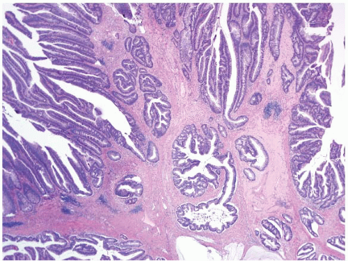
FIGURE 4.22 Pseudoinvasion in a tubular adenoma. Neoplastic glands are seen here hernitated into the submucosa. Note the rounded contours and similar cytoarchitectural features as the adenoma within the mucosa.


FIGURE 4.23 Pseudoinvasion in a tubular adenoma. This higher magnification better shows the presence of accompanying lamina propria in the same case as Figure. 4.22.


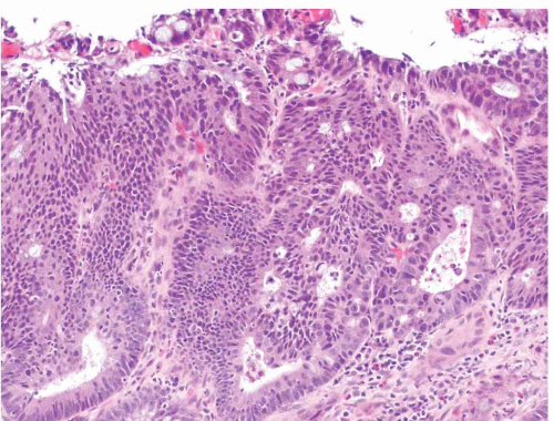
FIGURE 4.24 Adenoma with high-grade dysplasia. Note complex architecture with areas of cribriforming. An area suspicious for lamina propria invasion is seen in the lower right side of the field where cells show more abundant eosinophilic cytoplasm.


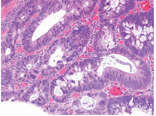
FIGURE 4.25 Adenoma with high-grade dysplasia. This high magnification image shows an area of cribriforming with bizarre cells and loss of nuclear polarity.

As for when to regard an adenoma as tubular versus tubulovillous versus villous, we do not know the answer. Adenomas with “villous features” are supposed to prompt closer surveillance than those without but there are no real criteria for when an adenoma has “villous features”—some colleagues are averse to reporting either high-grade dysplasia or “villous features” as there are no universal criteria for diagnosing them and they correlate with lesional size regardless, a piece of information the pathologist is usually unware of at the time of interpretation of mucosal samples (76). In daily practice, it is perhaps convenient to set a threshold in one’s own practice that correlates with findings in polyps in which the size is known than to frequently argue with clinical colleagues about why reporting villi and highgrade dysplasia may not be meaningful. We generally report lesions as tubulovillous when villi are well formed (Fig. 4.26) rather than hunt carefully for a focus that could be interpreted as a villous area. When most of the lesion is villous, we report this as well (Fig. 4.27) but we do not spend much time on this since this interpretation is, regardless, subjective.


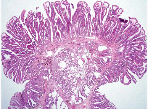
FIGURE 4.26 Tubulovillous adenoma. These polyps show a mixture of both tubular and villous components. Note prominent pseudoinvasion in this example.


PATHOLOGIC EVALUATION AND DIAGNOSIS OF EARLY COLORECTAL CANCERS TREATABLE BY ENDOSCOPIC POLYPECTOMY

Carcinomas are believed to develop in about one in 25 adenomas left in situ. When these are found in endoscopically removed polyps, a management decision must be made, and the role of the pathologist is important.
To justify colectomy, the risk of the patient having a metastasis must be higher than the risk of the patient undergoing surgery to remove a segment of the colon. Criteria to make this decision have appeared in the literature since the 1980s and have largely stood the test of time (77,78). Although early studies made a point that there were levels of invasion akin to those in a melanoma (78), other protocols have proved more reliable and more readily assessed in biopsies (79).


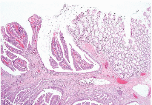
FIGURE 4.27 Villous adenoma. These polyps are composed predominantly of villous architecture characterized by slender, well-formed villi.

The diagnosis and treatment of colorectal cancers by endoscopic polypectomy have become commonplace. “Malignant polyps” are adenomas that contain any amount of invasive carcinoma, which is defined as a tumor that has gone through the muscularis mucosae into the submucosa. They also include polypoid carcinomas, in which the entire polyp head is replaced by carcinoma. By definition, malignant polyps exclude adenomas containing intraepithelial carcinoma or intramucosal carcinoma because these polyps lack biologic potential for metastasis. Polyps containing invasive carcinoma comprise about 5% of all adenomas. The chance that any given adenoma contains invasive carcinoma increases with polyp size, and the incidence of invasive carcinoma in adenomas >2 cm ranges from 35% to 53%. Therefore, any polyp >2 cm in diameter should be approached with the suspicion that it might harbor an invasive cancer. When technically possible, it is recommended that these polyps be removed intact, rather than piecemeal, with as great a margin as possible at the base or stalk. Identification of the resection margin is necessary for determining both the adequacy of the excision and the closest approach of the tumor, a parameter that predicts the risk of tumor recurrence.

Malignant polyps often constitute a form of early carcinoma (pathologic T category pT1) curable by endoscopic polypectomy alone (79) (Fig. 4.28, e-Figs. 4.105-4.113). However, the incidence of an unfavorable outcome (i.e., lymph node metastasis or local recurrence from residual malignancy) for malignant polyps treated by polypectomy alone varies from 0% to about 20% in the literature. Pathologic evaluation is critical in defining polyps with an increased risk of residual or recurrent disease, and the subsequent clinical management of the patient may be based, in part, on the findings. The histopathologic parameters that are known to be associated with a significantly increased risk of adverse outcome are listed below (79).


  • 1. A high tumor grade including poorly differentiated adenocarcinoma, signet ring cell carcinoma, small cell carcinoma, or undifferentiated carcinoma (e-Figs. 4.106, 4.107 and 4.114). It remains unclear in the literature whether poorly differentiated carcinomas—that are apparently confined to the lamina propria (e-Fig. 4.115)—have the biologic potential to metastasize. In our study, which included a limited number of patients, polypectomy alone was adequate management for such patients but there are reports of adverse outcomes for patients with intramucosal poorly differentiated colorectal carcinoma (80,81).


  • 2. A tumor ≤1 mm from the resection margin (some authors advise ≤2 mm). We assess this by the not-so-high-tech method of making two small dots (one at the leading edge of the tumor and the other at the nearest cauterized margin) and measuring the distance between them with a ruler. Remember that cauterized tissue contracts, a process that can pull the normal tissue margins together and gives a false impression of positive margins (see e-Fig. 4.112).


  • 3. Involvement of a small (thin-walled) vessel, presumably lymphatic, by the tumor (e-Figs. 4.108-4.110, 4.112, and 4.113).

In the presence of one or more of these features, the risk of an adverse outcome following polypectomy is estimated to be about 10% to 25% (79). Therefore, if one or more of these high-risk features is found on pathologic examination, further therapy may be indicated. Optimal management is decided on an individual case basis, but segmental resection of the involved colonic segment, local excision (e.g., transanal disk excision for a low rectal lesion), or radiation therapy may be considered. In the absence of high-risk features, the chance of adverse outcome is extremely small, and polypectomy alone is considered curative.

In the pathologic evaluation of malignant polyps, assessment of small vessel invasion is hampered by interobserver variability (79,82). In fact, small vessel invasion may be impossible to diagnose definitively in some cases and, ultimately, may be judged as being indeterminate. An absolute diagnosis of vessel invasion is dependent upon finding carcinoma cells
within an endothelial-lined space. Contraction artifact in the tissue, tumorinduced stromal sclerosis, and extracellular pools of mucin secreted by tumor cells may all complicate the evaluation of vessel invasion. The dilemma may or may not be resolved by the examination of additional tissue levels of the specimen, review by a second observer, and/or immunohistochemical staining for endothelial markers. In published cases in which the malignant polyps have lacked definitive evidence of high-risk features—but the patients have died of their disease—lymphatic invasion had been judged (on blinded review) as indeterminate because of a lack of interobserver agreement (79,82). This suggests that even the suspicion of small vessel invasion on pathologic examination should be considered as potentially important. When there were no adverse features at all, there were no adverse events in this study.


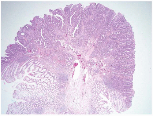
FIGURE 4.28 Invasive adenocarcinoma arising in a tubular adenoma. Malignant polyps are characterized by invasion of malignant glands into the submucosa. Note the adenocarcinoma at the level of large submucosal glands with associated desmoplasia.


Colorectal Adenocarcinoma

Most colorectal carcinomas are easy to diagnose on colon mucosal biopsies but sometimes diagnosing them requires correlation with the clinical findings. Additionally, there is some room for error. Invasive carcinoma is characterized by angulated glands and single cells set in a desmoplastic stroma (Figs. 4.29 and 4.30). Most examples are moderately-differentiated with well-developed glands that frequently contain necrotic debris in which the individual necrotic cells show an apoptotic nuclear pattern as well as neutrophils. The main problem that arises is that mucosal biopsies are just that—biopsies of the mucosa. Since colorectal carcinoma is not regarded as truly invasive (T1) unless there is invasion of the submucosa,
sometimes there can be doubt in samples that do not contain submucosa for evaluation.


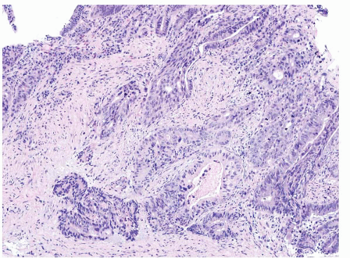
FIGURE 4.29 Colon adenocarcinoma. This biopsy of a colon mass shows angulated glands with central necrosis in a desmoplastic stroma.


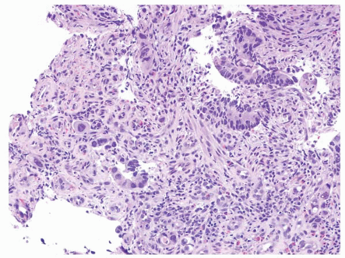
FIGURE 4.30 Rectal adenocarcinoma. This biopsy of a rectal mass shows individual cells and small collections of cells with abundant eosinophilic cytoplasm and prominent nucleoli.

Ideally, to diagnose colorectal cancer in a biopsy, we would like to see submucosa that is invaded by carcinoma. However, this is not always realistic since background adenomas can result in thick lesions such that it is difficult for our colleagues performing the biopsies to obtain samples that contain submucosa. However, if there is well-developed desmoplasia in the lamina propria associated with invasion into this structure (Fig. 4.31), there is almost invariably an underlying invasive carcinoma (into at least submucosa). In this situation, we correlate with the endoscopic impression and if there is a mass lesion, we report adenocarcinoma and do not qualify that we see only intramucosal invasion. If we truly believe we see invasion restricted to the lamina propria on a fragmented biopsy, we report it as such (e.g., “at least intramucosal carcinoma/invasion into the lamina propria/Tis arising in association with a tubulovillous adenoma…”). However, in some cases, the endoscopist sees a mass and biopsies show only an in situ component (adenoma with or without high-grade dysplasia). In this case, we report the adenoma and any high-grade dysplasia. If such a sample is from the right colon, it is reasonable to offer the patient a right colectomy based on the technical difficulty in removing large polyps from the right colon as well as the likelihood that a large polyp may contain occult carcinoma regardless of biopsy findings.

When invasive carcinoma is diagnosed, it is good practice to report whether there is an associated adenoma component as this ensures that
the lesion is primary (associated with a precursor) and essentially excludes the possibility of a metastasis to the colon.


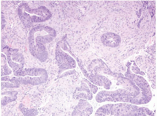
FIGURE 4.31 Rectal adenocarcinoma. This biopsy shows malignant glands associated with well-established desmoplasia.

Immunolabeling is generally not required to diagnose colorectal carcinoma on biopsies but it can be part of a molecular evaluation as below. There are, of course some cases in which the issue of a metastasis arises and immunolabeling can be important in such instances. Colorectal carcinomas are typically CK20+, CK7-, and CDX2+ but some examples with MMR defects have altered CK7/20 profiles (83), so correlation with imaging studies can be required to arrive at a confident diagnosis. As another caveat, remember that reactive nonneoplastic colonic mucosa expresses CK7 (e-Fig. 4.116).

Extremely rarely, colorectal adenomas can be complicated by pseudomyxoma peritonei presumably based on cells that are released into the peritoneum during an operation to remove a large adenoma (84), but this phenomenon is so rare that we have seen only one such case (e-Figs. 4.117-4.124).


MOLECULAR TESTING IN COLORECTAL CANCER

Although molecular testing of colorectal cancers is principally an issue in resected tumors, it can also be performed on biopsies. Since stage often directs testing, we often wait until staging information is available, although sometimes both a liver metastasis and a mucosal biopsy might provide sufficient information to direct testing. Table 4.3 shows the protocol that we follow at Johns Hopkins Hospital but other institutions might prefer a different protocol depending on the practices of local oncology colleagues.


TABLE 4.3 Suggested Reflex Testing of Colorectal Cancers


Stage I: (T1, N0, M0 or T2, N0, M0):


Order MSI testing if patient is under age of 50 (need both tumor and normal)


Stage II: (T3, N0, M0 or T4, N0,M0):


Order MSI testing for all cases


Stage III: (any T, N1 or N2, M0):


Order MSI testing if patient is under age 50


Order KRAS mutational testing and BRAF mutational testing (only need tumor)


Stage IV: Any T, any N, M1:


Order MSI testing if patient is under age of 50


Order KRAS mutational testing and BRAF mutational testing


Microsatellite Analysis and Mismatch Repair Immunohistochemistry

Microsatellites, also known as short tandem repeats (STRs), are repetitive DNA elements in which the repeating unit is 1 to 6 bases long, and the units are repeated 10 to 60 times. The repetitive nature of microsatellites creates inherent instability during replication. However, normal cells possess a system of DNA mismatch repair (MMR) that rapidly corrects replication errors to maintain microsatellite length. Proteins encoded by the mutL homolog 1 (hMLH1), postmeiotic segregation increased 2 (hPMS2), mutS homolog 2 (hMSH2), and mutS homolog 6 (hMSH6) genes correct these errors. Microsatellite instability (MSI) is defined as “a change of any length due to either insertion or deletion of repeating units, in a microsatellite within a tumor when compared to normal tissue.”

Histopathologic features suggesting MSI in colorectal tumors include intense lymphocytic infiltrate (Figs. 4.32, 4.33 and 4.34), mucinous or signet ring cell features (e-Fig. 4.114), Crohn-like reaction, and/or a medullary growth pattern (e-Figs. 4.125-4.127). These features can sometimes be encountered on biopsies. Such tumors are often in the right colon in patients younger than 50 years. While these features are incorporated into the revised Bethesda criteria (Table 4.4) (85), there is no single histologic feature that accurately predicts MSI, but a combination of them can predict MSI with great accuracy (86). Adenomatous colorectal polyps are less frequently affected even in Lynch syndrome patients, although they may be useful for testing in the context of a significant family history (we typically do not test them). In the diagnostic setting, reflex testing of colorectal tumors with the above stated features for MSI evaluation is becoming increasingly common. Variant forms of Lynch syndrome are Muir Torre syndrome in which patients manifest sebaceous neoplasms and colorectal cancer (e-Figs. 4.128-4.131) and Turcot syndrome (brain tumors and colorectal cancers).


FIGURE 4.32 Colon carcinoma associated with microsatellite instability (MSI). Note the associated lymphocytic infiltrate.

MSI evaluation typically involves microdissection (usually manual) of tumor and normal tissue from submitted formalin fixed paraffin embedded tissue sections followed by DNA isolation, a polymerase chain reaction (PCR) using primers directed at a number of microsatellite markers and analysis of the PCR products by capillary electrophoresis for patterns
of MSI. As such, when colorectal cancers are biopsied in a young patient (<50 years), we encourage our clinical colleagues to also sample benign flat mucosa to facilitate this testing, but of course the testing can always be performed later on resected material in which there is abundant normal tissue to use as a control. Electropherograms from both tumor and normal tissues are usually compared when conducting this analysis. The two most commonly used panels are a Promega Microsatellite Analysis System and a reference panel recommended by an NCI-sponsored consensus committee. The Promega system consists of five mononucleotide markers, while the NCI-sponsored reference panel consists of three dinucleotides and two mononucleotides. However, mononucleotide markers show the greatest sensitivity for MSI. MSI is diagnosed when microsatellite lengths are
shifted from the germline pattern. Three possible data interpretations exist: MSI-High (MSI-H), MSI-Low (MSI-L), and Microsatellite Stable (MSS). When five markers are used, a tumor that shows MSI in greater than or equal to two loci is considered MSI-H, one that shows MSI in one locus is termed MSI-L, and one that shows no MSI at any locus is diagnosed MSS.


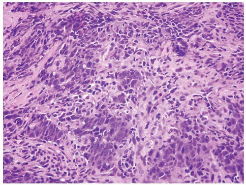
FIGURE 4.33 Colon carcinoma associated with microsatellite instability (MSI). Note the associated lymphocytic infiltrate in this higher magnification of Figure 4.32.


TABLE 4.4 The Revised Bethesda Guidelines for Testing Colorectal Tumors for MSI


Criterion


Commentary


Colorectal cancer diagnosed in a patient who is <50 years of age.


Presence of synchronous, metachronous colorectal, or other hereditary nonpolyposis colorectal cancer (HNPCC)-associated tumors, regardless of age.


HNPCC-related tumors include colorectal, endometrial, stomach, ovarian, pancreas, ureter and renal pelvis, biliary tract, and brain tumors (usually glioblastoma— Turcot syndrome), sebaceous gland adenomas and keratoacanthomas (Muir-Torre syndrome), and carcinoma of the small bowel.


Colorectal cancer with the MSI-H† histology diagnosed in a patient who is <60 years of age.


Key histologic features: Presence of tumor infiltrating lymphocytes, Crohn’s-like lymphocytic reaction, mucinous/signet-ring differentiation, or medullary growth pattern.


Colorectal cancer diagnosed in one or more first-degree relatives with an HNPCC-related tumor, with one of the cancers being diagnosed under age 50.


Colorectal cancer diagnosed in two or more first-or second-degree relatives with HNPCC-related tumors, regardless of age.


Adapted from Table 14.2, Iacobuzio-Donahue C, Montgomery E. Gastrointestinal and Liver Pathology. 2nd ed. Philadelphia, PA: Elsevier; 2012:446.


† MSI-H, microsatellite instability-high in tumors refers to changes in two or more of the five National Cancer Institute-recommended panels of microsatellite markers.


Immunohistochemical studies are an alternative approach to MMR defect screening. Loss of nuclear IHC staining of MMR proteins (Fig. 4.34) demonstrates a loss of function, can be used as a surrogate for the presence of a truncating MMR mutation, and generally correlates well with the presence of MSI. MMR IHC is also of value as it allows the clinician to order DNA sequencing on the gene most likely to be defective. However, positive IHC staining confirms MMR protein expression, but it does not measure function and does not preclude the possibility of a missense mutation causing loss of function (seen as MSI by microsatellite analysis) while retaining antigenicity.

HNPCC represents approximately 2% to 5% of all colorectal cancers and has an autosomal dominant mode of transmission with approximately 80% to 90% penetrance. Of known MMR genes, MLH1 and MSH2 most commonly harbor germline mutations followed by a much smaller mutation incidence in MSH6 and PMS2. When the MMR mutation is identified in a proband, the patient is said to have Lynch Syndrome. If such a patient develops colorectal cancer, the tumor will most likely have lost the second allele of that gene (the so-called second-hit) and manifest MSI. Identification of specific MMR gene mutation can have significant implications on entire families, prompting genetic screening and leading to increased disease surveillance in individuals carrying the mutated allele. NCI recommendations
for families with a proband that has MSI-H positive tumor, yet who has a negative genetic mutation screen, include counseling the proband and at-risk members as if Lynch Syndrome had been confirmed and beginning a high-risk surveillance program. Patients with MSI-H stage II colorectal cancer have an improved survival relative to other patients. However, it is important to recall that most patients whose tumors have loss of MLH1 protein do not have Lynch syndrome but instead have inactivation of the gene by promoter methylation. However, essentially all patients who show loss of MSH2 by immunostaining (Fig. 4.34) have germline mutations of MSH2. For this reason, we do not perform immunohistochemistry as our first test to evaluate for MMR but instead start with MSI testing. MSI positive tumors can be a result of either germline mutations or sporadic promoter methylation and thus MSI testing does not prove whether a patient has Lynch syndrome. If a patient’s tumor is MSI-H, a discussion can be had after correlation with history as to whether genetic counseling and sequencing of MMR genes is needed. In contrast, if immunolabeling is done reflexively and the patient has loss of MSH2 and the patient has not yet had genetic counseling or given permission for genetic testing, this can be a difficult situation to resolve. Individual institutions need to develop their own protocols for this issue and this testing.


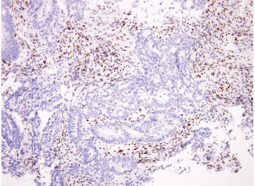
FIGURE 4.34 Colon carcinoma associated with microsatellite instability (MSI). This tumor shows loss of MSH2 by immunohistochemistry.

MSI testing may also be useful for prognosis and response to chemotherapy. Patients with an MSI-H tumor typically do not benefit from 5-fluorouracil (5-FU) chemotherapy and may actually have shorter diseasefree survival when treated with 5-FU alone. Recent studies raise the possibility that MSI-H status predicts a better response to adjuvant therapy when irinotecan is administered.

As above, MSI is observed in approximately 15% of sporadic colorectal cancers. Of colorectal tumors that are MSI-H, most are sporadic in origin and present in older patients while a minority are derived from germline mutations and manifest as Lynch Syndrome. Hence, the recommendation was made to perform MSI testing generally in patients <50 years of age. Sporadic MSI-H tumors typically demonstrate MLH1 gene promoter methylation which silences the MMR gene and results in loss of expression by IHC and the presence of MSI.

In the genetic screening setting, differentiating germline-origin from sporadic-origin MSI is necessary to identify Lynch syndrome kindreds. BRAF testing and MMR gene promoter methylation analysis may be performed to differentiate MLH1 promoter methylation from an MMR germline defect. Of those patients who are found to be BRAF mutant negative, immunohistochemistry to detect expression of MMR proteins can then be performed to further identify the appropriate MMR gene sequencing target. MSI-H tumors harboring the BRAF

Only gold members can continue reading. Log In or Register to continue

Stay updated, free articles. Join our Telegram channel

Jun 18, 2016 | Posted by in GASTROENTEROLOGY | Comments Off on Colon

Full access? Get Clinical Tree

Get Clinical Tree app for offline access