Diverticulosis
Asymptomatic
Diverticulitis
Noninflammatory
Symptoms without inflammation
Acute
Symptoms with inflammation
Simple
Localized
Complicated
With perforation
Chronic
Persistent, low grade
Atypical
Symptoms without systemic signs
Recurring, persistent
Symptoms with systemic signs (may be intermittent)
Complex
With fistula, stricture, obstruction
Malignant
Severe, fibrosing
Diverticulosis refers to the presence of diverticula with no related symptoms. This applies to the vast majority (80–90 %) of patients with diverticular disease.
Diverticulitis can be subclassified into noninflammatory, acute (simple or complicated), chronic (atypical or recurring/persistent), or complex disease.
Noninflammatory Diverticular Disease
Noninflammatory diverticular disease describes those patients with symptoms of diverticulitis but without associated inflammation. The diagnosis is made at the time of elective operation when no inflammatory changes are found in the specimen. This has been reported in 15–35 % of resections.
The term “atypical” has been applied to patients with chronic symptoms who never develop the necessary clinical and laboratory criteria to be judged as having acute diverticulitis.
Acute Diverticulitis
Acute diverticulitis is represented by signs and symptoms of acute inflammation and may be simple (limited to the colonic wall and adjacent tissues) or complicated (with perforation or fistula). Simple acute disease is usually accompanied by systemic signs of fever and leukocytosis, while complicated acute disease may have the added signs of tachycardia and hypotension.
Complicated acute diverticulitis can be classified according to the extent of spread of the inflammatory process. A common classification for diverticulitis with perforation is the Hinchey classification (Table 22.2 ).
Table 22.2
The Hinchey classification (proposed by Hinchey et al. in 1978)
Hinchey Stage I
Localized abscess (para-colonic)
Hinchey Stage II
Pelvic abscess
Hinchey Stage III
Purulent peritonitis (the presence of pus in the abdominal cavity)
Hinchey Stage IV
Feculent peritonitis
Chronic Diverticulitis
Patients with chronic diverticulitis remain symptomatic (left lower quadrant pain) despite standard treatment. It is considered atypical if systemic signs never develop. With systemic signs, chronic disease may manifest as recurring, intermittent episodes of acute disease or as persistent, symptomatic low-grade disease. This is frequently associated with the presence of a phlegmon. If resection is performed, there will be evidence of inflammatory changes within the specimen.
Complex Diverticular Disease
Complex diverticulitis refers to disease in those patients who manifest sequelae of chronic inflammation including fistula, stricture, and obstruction.
Presenting Symptoms
Patients with acute diverticulitis typically complain of left lower quadrant abdominal pain. The pain is generally constant in nature, not colicky. Radiation may occur to the back, ipsilateral flank, groin, and even the leg. The pain may be preceded or accompanied by episodes of constipation or diarrhea. It commonly is progressive in nature if appropriate treatment is not instituted.
Nausea and vomiting are unusual in the absence of obstruction. Bleeding is not an associated finding. Symptoms of dysuria or urgency suggest possible bladder involvement due to an adjacent inflammatory mass or a colovesical fistula. Pneumaturia, fecaluria, or passage of gas and stool through the vagina suggest a colovesical or colovaginal fistula, respectively. Fever is common and proportional to the amount of inflammatory response present. A high fever suggests a perforation with abscess or peritonitis.
Physical Findings
Patients presenting with acute diverticulitis will be tender to palpation in the left lower quadrant and left iliac region. There may be limited rigidity or localized guarding to deeper palpation. With resolution of the acute phase, palpation may reveal a mass in the left lower quadrant. Classically, there is no prodromal epigastric pain with diverticulitis as one might expect to see with appendicitis.
In the event of a perforation with development of fecal or purulent peritonitis, the area of pain will spread throughout the abdomen. Guarding will become prominent and the abdominal wall will become rigid.
Diagnostic Evaluation
Abdominal X-Rays
The primary value of abdominal X-rays is to rule out pneumoperitoneum or to assess for a possible obstruction; therefore plain films of the abdomen should include supine and upright or left lateral decubitus views. Computed tomography (CT) scan is often the evaluation of choice in the face of acute abdominal pain, so in many centers, the plain abdominal film is rarely used.
Contrast Studies
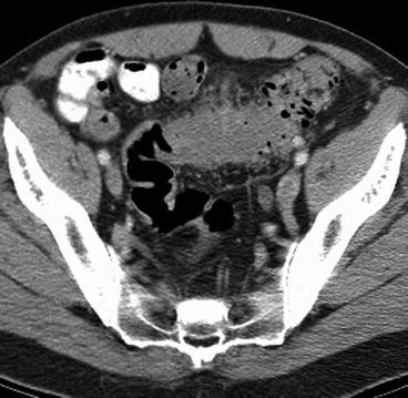
Barium or water-soluble contrast studies have their proponents for utilization; however, CT scans provide a significantly more thorough evaluation, making it the preferred imaging study in many centers (Fig. 22.1 ). Nonetheless, due to costs, some clinicians will utilize CT scan only if there is clinical suspicion of an abscess or other complicating feature for which an alternative to standard bowel rest and antibiotics might be applied. A water-soluble contrast study can evaluate the lumen of the bowel if there is concern about distal bowel obstruction. It may be an important part of the assessment for the possible use of a colonic stent if malignant disease is suspected.

Fig. 22.1
CT scan reveals uncomplicated diverticulitis with bowel wall thickening and streaky fat in the mesentery
In most centers, contrast studies, if used at all, are used in a limited fashion to evaluate the anatomy of the colon prior to an operation.
CT Scan
An important advantage of a CT scan is the ability to document diverticulitis, even if uncomplicated, when the diagnosis is in doubt.
It has been demonstrated that CT can recognize and stratify patients according to the severity of their disease.
It can distinguish uncomplicated disease with a predictably short length of hospital stay from complicated disease as defined by abscess, fistula, peritonitis, or obstruction and a predictably long length of stay.
It also provides information about extracolonic pathology and anatomic variation, which is useful for surgical planning.
Early CT-guided drainage of abscesses allows down-staging of complicated diverticulitis, converting an otherwise urgent or emergent operation with its attendant increases in morbidity and mortality to the safety of an elective operation. In some selected cases there may be no need for elective resection.
Colonography
Preliminary studies utilizing magnetic resonance imaging (MRI) colonography have shown a high correlation with CT findings in patients with diverticular disease without exposure to ionizing radiation. Three-dimensional rendered models and virtual colonoscopy can be performed only in the nonacute setting. These comprehensive 3-D models, rather than barium enema, may have a role in presurgical planning with concurrent assessment of the residual colon.
Ultrasonography
Transrectal ultrasound (TRUS) has been utilized in the evaluation of diverticular disease in conjunction with transabdominal ultrasound (TAUS). TRUS may prove to be a useful adjunct in selected cases of rectosigmoid diverticulitis and perirectal involvement by diverticular disease in centers where CT scanning is not readily available.
Endoscopy
Endoscopy in the face of acute diverticulitis must be undertaken with extreme caution due to risk of perforation and decreased chance of successful cecal intubation. Generally, in the absence of an urgent indication, colonoscopy should be delayed until resolution of the acute episode is complete.
In the case of elective colonoscopy, the unexpected finding of acute diverticulitis (manifested as erythema, edema, pus, or granulation tissue at a diverticular opening) is distinctly unusual, occurring in just 0.8 % of patients. Treatment with antibiotic therapy for such findings is generally unnecessary as follow-up has shown that symptoms of diverticulitis do not develop following the colonoscopy.
Differential Diagnosis
The differential diagnosis for diverticular disease includes IBS, carcinoma, inflammatory bowel disease (IBD), appendicitis, bowel obstruction, ischemic colitis, gynecologic disease, and urologic disease. Of these, IBS is perhaps the most difficult to differentiate in many patients.
Irritable Bowel Syndrome
In many ways, the distinction between chronic diverticulitis and noninflammatory diverticular disease relies upon the pathologist, while the distinction between noninflammatory diverticular disease and IBS relies on the diagnostic acumen of the clinician and the long-term outcomes of resection. Due to the prevalence of diverticular disease, many patients with IBS will have concomitant diverticular disease. However, due to the fact that diverticular disease is most commonly asymptomatic, the presence of diverticulosis in these patients will often not be the source of their symptoms but rather just a source of confusion in the differential. It is helpful to be familiar with the Rome II criteria (Table 22.3 ) for the diagnosis of IBS in order to sort through this differential.
Table 22.3
The Rome II criteria for irritable bowel syndrome
Irritable bowel syndrome can be diagnosed based on at least 12 weeks (which need not be consecutive), in the preceding 12 months, of abdominal discomfort or pain that has two of three of these features:
1. Relieved with defecation
2. Onset associated with a change in frequency of stool
3. Onset associated with a change in form (appearance) of stool
Symptoms that cumulatively support the diagnosis of IBS:
1. Abnormal stool frequency (>3 stools per day or <3 stools per week)
2. Abnormal stool form (lumpy/hard or loose/watery stool)
3. Abnormal stool passage (straining, urgency, or feeling of incomplete evacuation)
4. Passage of mucus
5. Bloating or feeling of abdominal distension
Red flag symptoms which are NOT typical of IBS:
Pain that often awakens/interferes with sleep
Diarrhea that often awakens/interferes with sleep
Blood in stool (visible or occult)
Weight loss
Fever
Abnormal physical examination
Colonic Neoplasia
Distinguishing diverticular disease from cancer can be difficult and occasionally a resection is necessary. Findings on barium enema (BE) that supports a diagnosis of diverticular disease include preservation of the mucosa, long strictures, and the presence of diverticula. A BE can also assess the extent of the diverticulosis prior to resection. Colonoscopy can frequently resolve this issue, but it is not always possible due to acute angulations or narrowing of the lumen. CT evaluates the entire abdomen, which can identify concurrent disease and may give clues to underlying colonic pathology.
The increasing incidence of colonic neoplasia with increasing age parallels that of diverticular disease. Polyps and cancer must be considered whenever a diagnostic workup for diverticular disease is begun.
Inflammatory Bowel Disease
Crohn’s disease can be a particularly difficult diagnosis to make. Both Crohn’s and diverticular disease may present with similar complications including fistulas, phlegmons, and abscesses. Rectal involvement, anal disease, extracolonic signs, and bleeding suggest Crohn’s disease. Recurrent “diverticulitis” requiring a repeat resection should always raise the question of possible Crohn’s disease.
Other Colitides, Appendicitis, and Gynecologic and Urologic Disease
Endoscopy can be an important adjunct in differentiating IBD, ischemic colitis, and other forms of colitis although caution must be used in the acute setting. A major advantage of the CT scan is the ability to evaluate for many of the other potential differentials including appendicitis and gynecologic and urologic disease.
Special Considerations
Diverticulitis in Young Patients
There continues to be some debate as to the issue of recurrence in patients younger than 50 years old. It does appear that there is an increased incidence in younger patients presenting with diverticulitis.
In a recent study by Etzioni et al. evaluating the nationwide inpatient sample data for changing patterns of diverticular disease and treatment, a 73 % increase in the rate of admission for patients aged 18–44 years with diverticulitis was found between 1998 and 2005. While an increase was also found in patients aged 45–74 year, the increase was only 29 %.
Historically, diverticular disease in patients less than age 50 has been described as more virulent and with more serious complications.
Despite the increased number of younger patients with the disease, its virulence does not appear to be any different compared to older counterparts. It is now doubtful that age itself should be a primary consideration in the decision to operate.
Young patients with diverticular disease are more commonly male and obese and have a higher incidence of right-sided diverticulitis.
Young patients undergoing operation were frequently misdiagnosed preoperatively with appendicitis being the most common misdiagnosis.
Many recommend that patients less than age 50 have an elective resection after a single episode of acute disease.
Recent evidence is mixed and supports a more conservative trend to recommending resection for uncomplicated diverticulitis.
Some have recommended elective resections at a younger age to avoid the increased morbidity and mortality associated with urgent or emergent surgery in the elderly (0 % vs. 34.9 %). Some recommendations for elective resection in the young patient are based on cost savings related to definitive surgical management vs. the higher costs of ongoing medical treatment for recurring disease. These types of recommendations assume a high risk of recurrent disease.
There is evidence that diverticular disease in young patients is changing. It is not as rare as in the past and continues to become more prevalent. And recent evidence suggests there is no increased risk of complications from diverticular disease in the young. Based on these findings, resection following a single episode of diverticulitis is not recommended.
Current recommendations for resection are based on the predicted risk of developing a serious complication that would lead to emergency surgery with increased morbidity and mortality. To improve management, we must become better at predicting who is at risk for recurrent disease. Age alone does not appear to be a reliable factor. The use of CT to identify “severe” or “complex” diverticular disease seems most promising.
Giant Colonic Diverticulum
Giant diverticula of the colon are rare entities associated with sigmoid diverticular disease. They are generally pseudodiverticula with inflammatory rather than colonic mucosal walls. They usually arise off of the antimesenteric border of the sigmoid colon.
Stay updated, free articles. Join our Telegram channel

Full access? Get Clinical Tree


