Hyponatremia
- Hyponatremia develops due to an excess of total body water in relation to total body sodium.
- Determination of extracellular volume status and urinary indices aids in the classification of hyponatremia.
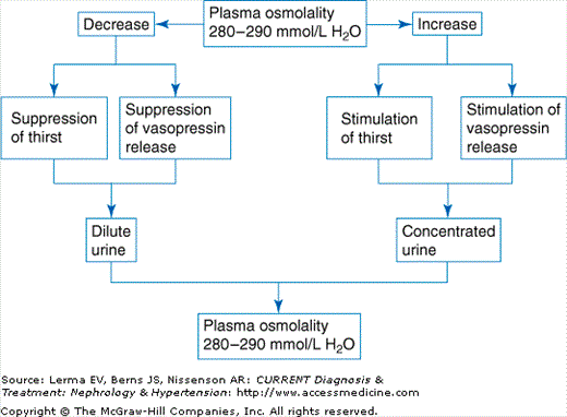
Hyponatremia is present when the serum sodium concentration falls below 135 mEq/L. In healthy subjects, the sodium concentration is closely regulated to remain between 138 and 142 mEq/L despite wide variations in water intake (Figure 3–1). When excess water is ingested, the normal kidney dilutes the urine, excretes excess water, and prevents the development of hyponatremia. Hyponatremia develops when the intake of water exceeds the ability to excrete it leading to dilution of total body sodium.
Sodium concentration is the major determinant of plasma osmolality, therefore hyponatremia usually indicates a low plasma osmolality. Plasma osmolality can be estimated by the following equation:
Low plasma osmolality rather than hyponatremia, per se, is the primary cause of the symptoms of hyponatremia. Hyponatremia not accompanied by hypoosmolality does not cause signs or symptoms and does not require specific treatment.
The limitation in the kidney’s ability to excrete water in hyponatremic states is, in most cases, due to the persistent action of antidiuretic hormone (ADH, vasopressin). ADH acts at the distal nephron to decrease the renal excretion of water. The action of ADH is, therefore, to concentrate the urine and, as a result, dilute the serum. Under normal circumstances, ADH release is stimulated primarily by hyperosmolality. However, under conditions of severe intravascular volume depletion or hypotension, ADH may be released even in the presence of serum hypoosmolality. Disease states characterized by a low cardiac output or systemic vasodilation result in “effective” intravascular volume depletion and may also stimulate ADH release.
Importantly, ADH alone is not sufficient to cause hyponatremia. Only when the intake of water exceeds its excretory capacity can hyponatremia result. In some cases, massive water ingestion or a defective urinary concentrating mechanism can cause hyponatremia despite the complete absence of circulating ADH.
The symptoms and signs of hyponatremia most likely result from cellular and cerebral edema. Headache, lethargy, confusion, weakness, psychosis, ataxia, seizures, and coma can all occur. Although no consistent correlation between the degree of hyponatremia and neurologic manifestations exists, patients with seizures and altered sensorium generally have serum sodium concentrations less than 120 mEq/L.
Understanding the physiology of water movement is essential to understand the symptomatology and proper treatment of the disorders of water balance. In hyponatremia, the fall in plasma osmolality causes osmotic movement of water from the hypotonic extracellular compartment into relatively hypertonic cells. When the movement of water into cells occurs rapidly and exceeds the ability of the cells to compensate, cellular edema occurs and the symptoms of hyponatremia can result. Over a period of days the cells of the brain are able to adapt to a decreased extracellular tonicity by losing osmolytes, thereby decreasing intracellular tonicity. Once cells have adapted, rapid correction of hyponatremia can leave the extracellular space relatively hypertonic. This causes water to move out of cells into the hypertonic extracellular space causing cellular dehydration. In the brain, this can lead to central pontine myelinolysis (CPM).
Consequently, acute hyponatremia developing over less than 48 hours is more likely present with typical symptoms due to the lack of complete cerebral adaptation. In contrast, hyponatremia developing over more than 48 hours, even when severe, may be entirely asymptomatic due to the adaptive capacity of the brain.
Typical clinical findings of hypovolemia include diminished skin turgor, flattened neck veins, dry mucous membranes and axillae, orthostatic hypotension, and tachycardia. In mild cases, hypovolemia may not be clinically apparent but usually results in renal sodium avidity and a urinary sodium concentration of less than 10 mEq/L. Vomiting is sometimes accompanied by metabolic alkalosis that obligates urinary bicarbonate and sodium losses, increasing the urinary sodium concentration to greater than 20 mEq/L. In this situation, hypovolemia can be confirmed by measuring the urinary chloride concentration, which is typically less than 10 mEq/L in this setting. Renal volume losses generally occur due to the administration of diuretics and result in a urinary sodium concentration greater than 10 mEq/L.
An increased extracellular volume may be evidenced by distended neck veins, pulmonary edema, ascites, or lower extremity edema. Hypervolemic hyponatremia is generally due to volume-retaining states such as congestive heart failure, cirrhosis, nephrotic syndrome, or advanced renal failure. In the absence of diuretic administration, hypervolemic hyponatremia is usually accompanied by “effective” hypovolemia and, consequently, a urinary sodium concentration less than 10 mEq/L.
Clinical determination of euvolemia is confirmed by the absence of signs of hypovolemia or hypervolemia. The urinary sodium concentration is greater than 20 mEq/L.
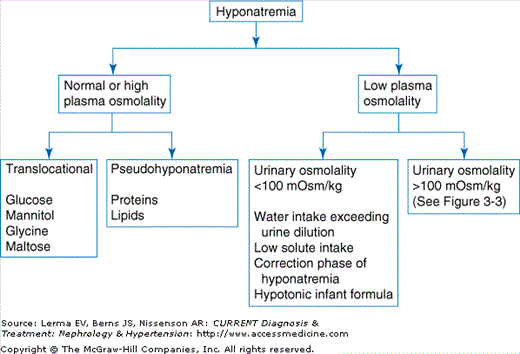
Hyponatremia should be approached systematically using an algorithm. The first step involves determining whether the observed hyponatremia is associated with a decreased, normal, or even elevated plasma osmolality (Figure 3–2).
Hyponatremia can be present in the absence of hypoosmolality when one of two situations is present. In the most common situation, osmotically active substances unable to enter the cell, such as glucose (in the absence of insulin), mannitol, or glycine (employed in hysteroscopy, laparoscopy, and transurethral resection of the prostate), cause water to move from the intracellular to the extracellular space. This water movement dilutes the extracellular sodium resulting in hyponatremia but, importantly, not hypoosmolality. Since serum tonicity does not change, symptoms of hyponatremia do not develop. When hyperglycemia is present, the underlying sodium concentration can be estimated by adding 1.6–2.4 mEq/L to the reported sodium concentration for every 100 mg/dL increase in the plasma glucose.
When present, severe hyperlipidemia or hyperproteinemia alter the usual ratio of serum water to solute. Since most clinical laboratories assume a constant ratio and perform a dilution of the serum prior to measuring sodium concentration, the serum water content is overestimated resulting in the reporting of incorrect or “pseudohyponatremia.” This error can be avoided by measuring the sodium concentration in undiluted serum using a sodium-selective electrode.
Most often, hyponatremia is associated with a low plasma osmolality. In these cases, determination of the urinary osmolality allows differentiation between hyponatremia resulting from a functioning urinary diluting system that is overwhelmed by hypotonic fluid administration (urine osmolarity <100 mOsm/kg) and hyponatremia resulting from an inability to appropriately dilute the urine (urine osmolarity >100 mOsm/kg)(Figure 3–2).
Hyponatremia that develops despite normal urinary dilution is caused by excessive fluid ingestion or inadequate solute intake, or develops during the correction phase of hyponatremia.
Primary polydipsia is a common problem in psychiatric patients, particularly in those with schizophrenia. In contrast to the syndrome of inappropriate antidiuretic hormone (SIADH), primary polydipsia develops despite maximally suppressed ADH secretion and a maximally dilute urine. Though psychogenic stress may cause defects in urinary dilution, the primary cause of the hyponatremia is the ingestion of massive quantities of water that overwhelm the renal excretory capacity. Urine output in these patients is typically very high.
Subjects that ingest very little dietary solute may develop an ADH independent impairment in renal water excretion. Modest increases in water intake may lead to hyponatremia despite maximal urinary dilution. Poorly nourished, chronic beer drinkers classically develop this condition but other malnourished patients may be similarly affected.
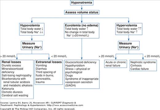
When hyponatremia is associated with low plasma and elevated urinary osmolality it is useful to classify patients based on extracellular volume status (Figure 3–3). Since total body sodium content is the primary determinant of extracellular volume, patients with low, normal, or high extracellular volumes have low, normal, or high total body sodium contents, respectively. Hyponatremia occurs due to an increase in total body water relative to total body sodium.
In hypovolemic hyponatremia nonosmotic release of ADH occurs in response to hypovolemia. Despite serum hypoosmolality, circulating ADH causes urinary concentration, water retention, and hyponatremia.
A patient with hypovolemia has a deficit of total body sodium resulting from either extrarenal or renal sodium losses. Extrarenal sodium loss can occur from the gastrointestinal tract in the form of vomiting or diarrhea, skin, or through third-space fluid sequestration. Common causes of renal sodium loss occur following diuretic administration or osmotic diuresis. Rarer causes of renal sodium loss occur due to cerebral salt wasting, salt-losing nephropathy, or mineralocorticoid deficiency.
Euvolemic hyponatremia is the most common form of hyponatremia in hospitalized patients. Normally, euvolemic hyponatremia develops due to inadequate urinary dilution evidenced by an inappropriately elevated urine osmolality (urine osmolality > 100 mOsm/kg H2O).
SIADH is the commonest cause of euvolemic hyponatremia but remains a diagnosis of exclusion (Table 3–1).
Decreased extracellular fluid effective osmolality (<270 mOsm/kg H2O) |
Inappropriate urinary concentration (>100 mOsm/kg H2O) Clinical euvolemia |
Elevated urinary Na+ concentration (>20 mEq/L) under conditions of a normal salt and water intake |
Absence of adrenal, thyroid, pituitary, or renal insufficiency or diuretic use |
Under normal circumstances, in the setting of hypoosmolality and euvolemia, ADH is maximally suppressed and urine is maximally dilute. In SIADH, however, ADH is inappropriately released and the urine, consequently, is concentrated. Despite abnormal water handling, sodium regulatory mechanisms remain intact and patients do not become hypervolemic. Hypouricemia is commonly seen in SIADH due to both dilution and increased uric acid elimination.
SIADH is most commonly associated with medication administration (Table 3–2). With widespread use, selective serotonin reuptake inhibitor (SSRI) antidepressants deserve mention as frequent causative agents, particularly among the elderly. Malignancy, pulmonary or central nervous system (CNS) disease, infection, and trauma are responsible for the remainder of cases. SIADH is also been frequently described in association with human immunodeficiency virus (HIV) infection.
Vasopressin analogs |
Desmopressin (DDAVP) |
Oxytocin |
Drugs that potentiate renal vasopressin |
Chlorpropamide |
Cyclophosphamide |
Nonsteroidal anti-inflammatory agents |
Acetaminophen |
Drugs that enhance vasopressin release |
Chlorpropamide |
Clofibrate |
Carbamazepine–oxycarbazepine |
Vincristine |
Nicotine |
Narcotics |
Antipsychotics/antidepressants |
Ifosfamide |


