Fig. 4.1
CT enterography performed in a patient with multifocal Crohn’s ileitis. Images reveal a penetrating ulcer (arrow) and a complex enteroenteric fistula (arrow head)
Technique
CTE is the culmination of targeted modifications designed to enhance intestinal mural assessments. This includes the use of large volume (approximately 1,350–1,500 ml) of neutral oral contrast to distend the small intestine.[5] This maneuver is of great benefit for analyzing wall thickness, enhancement, and stricture detection. Various oral products have been utilized including water, mannitol, low contrast barium solution (Volumen), or polyethylene glycol.[6] Iodinated intravenous contrast is provided with image acquisition typically in the enteric phase (45–50 s after contrast injection) when peak small bowel enhancement occurs.[7] High resolution images are constructed in multiple planes with a slice thickness of ≤ 3 mm. Unlike magnetic resonance enterography (MRE), antispasmodic agents are not required for high quality CTE imaging. Exams should extend through the perineum to detect perianal disease. This process for CTE imaging maximizes the detection of enhancing small bowel lesions, inflammation, penetrating disease, and strictures. CTE has become preferred over CT enteroclysis due to similar diagnostic accuracy (80 % and 88 % respectively), but greater patient tolerance with CTE.[8]
Performance Characteristics
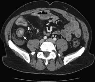
Various CTE parameters have been evaluated for their ability to detect active small bowel inflammation. Candidate variables have included mural hyperenhancement, bowel wall stratification, wall thickening, increased mesenteric fat density (fatty proliferation), and dilated vase recta (comb sign) (Fig. 4.2). Using ileoscopy as the gold standard, mural hyperenhancement and increased wall thickness appear to be sensitive features for active intestinal inflammation.[9, 10] While not all studies have demonstrated a correction between elevated serum C-reactive protein (CRP) levels and abnormal small bowel imaging (small bowel follow-through or CTE) [11], a large retrospective study (n = 143) has reported a relationship between elevated CRP concentrations and increased mesenteric fat density.[12] While this remains an area of debate and active research, the ideal predictive model for active small bowel inflammation may include both mural hyperenhancement and dilated vasa recta, having a receiver operating characteristic (ROC) curve with an area under the curve (AUC) of 0.75. This model was not improved with the addition of any additional clinical or laboratory variables.[13]


Fig. 4.2
Crohn’s disease patient with active ileocolonic Crohn’s disease. CTE demonstrates intestinal regions with mural hyperenhancement and thickening (arrows)
CTE has been widely assessed in comparison to other small bowel imaging modalities. A prospective 4-way comparison trial was performed utilizing ileoscopy, CTE, capsule endoscopy (CE), and small bowel follow-through in 41 patients with established or suspected Crohn’s disease.[14] CTE and CE had similar sensitivity for detecting active small bowel inflammation (82 % and 83 % respectively), but CTE had a significantly higher specificity (89 % versus 53 %). MRE appears to have a similar performance profile (sensitivity and specificity), but CTE demonstrates higher image quality and greater interobserver agreement.[15] Additional advantages of CTE over MRE include its lower cost, wider availability, and shorter image acquisition time. Limited prospective data is available comparing CTE to small bowel ultrasound,[16] and it is unclear whether ultrasound will be able to accurately and consistently detect strictures and penetrating disease as is noted with CTE.
Indications/Applications
The indications for CTE continue to expand in IBD cases. In patients with suspected Crohn’s disease, it can be used to further establish the diagnosis, assess luminal extent, and determine severity of disease. It can also be used to help determine direction (antegrade versus retrograde) when balloon-assisted endoscopy (BAE) is needed for histologic confirmation of the diagnosis. For individuals with established Crohn’s disease, CTE can provide an objective measure of response to treatment[17], detect penetrating complications and strictures, and note extra-intestinal disease manifestations. These applications have earned CTE a prominent role in IBD diagnostic and management algorithms.
Future Innovations
Additional modifications and new applications are on the horizon for CTE. A key concept remains ionizing radiation dose reduction. This focus is driven by the desire to minimize potential patient risks, acknowledging that the data behind this risk assumption is limited.[18, 19] It is an area of great debate that will likely become less of an issue as low-dose CTE becomes standard practice.[20]
Stay updated, free articles. Join our Telegram channel

Full access? Get Clinical Tree


