Age of diagnosis
A1 <16 years
A2 17–40 years
A3 >40 years
Location
L1 ileal
L2 colonic
L3 ileocolic
L4 isolated upper
Behavior
B1 nonstricturing nonpenetrating
B2 stricturing
B3 penetrating
P perianal disease
While only 15 % of patients experience an alteration in anatomic location, nearly 80 % of individuals with inflammatory disease ultimately demonstrate stricturing or penetrating behavior.
Symptoms and Signs
Chronic diarrhea is the most common presenting symptom and is defined as a decrease in fecal consistency for more than 6 weeks to adequately differentiate this from self-limited infectious diarrhea.
Abdominal pain and weight loss are seen in about 70 and 60 % of patients before diagnosis, respectively.
Blood or mucus in the stool can be seen in 40–50 % of patients with Crohn’s disease of the colon but is unusual in patients with ileal or isolated upper gastrointestinal disease.
The most common of the recognized extraintestinal manifestations are abnormalities involving the axial and peripheral joints of the musculoskeletal system, which are most frequently seen when Crohn’s disease affects the colon.
Table 30.2 shows the Perianal Crohn’s Activity Index.
Table 30.2
Perianal Crohn’s disease activity index
Feature
Score
Abscess
None or 0
First occurrence, single abscess or
1
First occurrence, multiple abscesses or
3
First recurrence, single or multiple abscesses or
4
Multiple recurrence, single or multiple abscesses
5
Maximum abscess score 8
Fistula
None
0
Short-term (<30 days) fistula or
1
Long-term (>30 days) fistula or
2
Persistent postsurgery fistula or
3
Recurrent fistula
3
Multiple fistulas
3
Rectovaginal/rectourethral fistula or
4
Recurrent rectovaginal/rectourethral fistula
6
Maximum fistula score
14
Ulcer and fissure
None
0
Short-term (<30 days) ulcer/fissure or
1
Long-term (>30 days) ulcer/fissure or
2
Single ulcer/fissure or
1
Multiple ulcers/fissures
2
Maximum ulcer/fissure score
4
Stenosis
None
0
Short-term (<30 days) stenosis or
1
Long-term (>30 days) stenosis
2
Recurrent stenosis
4
Maximum stenosis score
6
Incontinence score
No incontinence or
0
Incontinence score of 1–6 or
1
Incontinence score of 7–14 or
3
Incontinence score >14
5
Maximum incontinence score
5
Concomitant disease a
None or
0, 0, 0
Moderate or
3, 2, 1
Severe
4, 3, 2
Active fistula
4, 3, 2
Maximum concomitant disease score
18
Diagnosis
The initial diagnosis of Crohn’s disease is based on an amalgamation of clinical, laboratory, imaging, endoscopic, and histologic findings.
No single diagnostic test provides an unequivocal verdict.
Anemia and thrombocytosis represent the most common changes in the complete blood count.
The C-reactive protein (CRP) and erythrocyte sedimentation rate (ESR) are standard laboratory surrogates of the acute-phase response to inflammation.
CRP broadly correlates with disease severity.
The ESR less accurately measures intestinal inflammation because it reflects changes in both plasma protein concentration and packed cell volume.
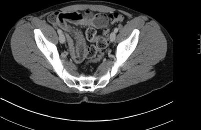
Computed tomographic (CT) enterography and enteroclysis differ from standard CT imaging by using intraluminal bowel distention with neutral enteric contrast, multidetector CT with narrow slice thickness and reconstruction interval, and intravenous contrast administration followed by delayed scans that optimize bowel wall enhancement.
CT enterography has largely supplanted barium examinations because the CT study is more sensitive and allows improved visualization of small bowel loops within the pelvis.
Contrast-enhanced magnetic resonance imaging (MRI) enterography (Fig. 30.1 ) and enteroclysis appear to provide results comparable to those seen with CT studies without the risk of exposure to ionizing radiation.

Fig. 30.1
CT enterography
Natural History
Patients’ initial presentations are equally distributed among ileitis, colitis, and ileocolitis.
Disease location remains relatively stable over time.
The majority of patients have nonstricturing, nonpenetrating disease at the time of diagnosis but tend to evolve into a stricturing or penetrating phenotype over their lifetime.
It is too early to accurately understand how immunomodulators and biologic agents will impact long-term disease activity and relapse rates.
The cumulative risk for surgery within 10 years of diagnosis is 40–55 %, and the risk of a second operation has been estimated to be 16, 28, and 35 % at 5, 10, and 15 years following the initial procedure, respectively.
Overall mortality was slightly but significantly higher than that seen in the general population.
Regarding the cause-specific mortality, there is a significantly increased risk of cancer death.
Chronic obstructive pulmonary disease, gastrointestinal diseases, and genitourinary diseases are more commonly implicated as a cause of death.
Operative Indications
The indications for operative management of Crohn’s disease include acute disease complications, chronic disease complications, and failed medical therapy. The acute complications are hemorrhage, perforation, and severe colitis with or without associated megacolon.
Chronic disease complications include extraintestinal manifestations, growth retardation, and neoplasia.
Hemorrhage
Crohn’s disease may infrequently cause life-threatening lower gastrointestinal hemorrhage and even exsanguination.
Localization of the bleeding site is essential.
In a stable patient with colonic disease, endoscopic evaluation is preferred.
A patient who requires ongoing resuscitation to maintain hemodynamic stability or in whom a small bowel source of active bleeding is suspected should undergo emergent mesenteric angiography to localize the source of hemorrhage and arrest ongoing bleeding through superselective angiographic embolization.
If the hemorrhage is localized but cannot be controlled, the catheter is left in position and intraoperative angiography is performed to accurately identify the bleeding site and guide a limited bowel resection.
An operation is warranted if the patient’s hemodynamic state cannot be sustained, bleeding persists despite 6 units of transfused blood, hemorrhage recurs, or another indication for surgery exists.
Resection with or without anastomosis is usually required for ongoing hemorrhage, whereas intraoperative enteroscopy with endoscopic therapy might be employed in less emergent settings.
Perforation
Free perforation of the small bowel is unusual and typically occurs at or immediately proximal to a stricture site.
Stay updated, free articles. Join our Telegram channel

Full access? Get Clinical Tree


