Fig. 19.1
Variation in biliary anatomy and aggressive hilar dissection in patients with large tumors close to hilum are risk factors for biliary injuries. a Variation in biliary anatomy of the right biliary system is more common than that in left side. Normal anatomy where the right anterior ( RA) and posterior ( RP) sectoral combine to form right hepatic duct which then joins the left hepatic duct to form common hepatic duct ( CHD) is seen only in 60 % of the patients. b In about 12 % of patients, right anterior and posterior sectoral ducts do not join to form right hepatic duct but in fact join left hepatic duct at the same point to form a trifurcation. c In a fifth of all patients, the right posterior sectoral duct crosses the midline to join the left-sided duct. The right anterior sectoral duct joins the left hepatic duct to form the CHD. d In a minority of patients, the right anterior instead of posterior sectoral duct crosses midline to drain into left hepatic duct. e MRI shows a large tumor encroaching on the hilum. Such large tumors place the contralateral outflow at risk
Difficult Surgical Resection and Reoperation
Reoperative surgery is a perfect setup for biliary injury as scarred hilum as well as extensive adhesions make identification of segmental anatomy difficult. In addition, excessive blood loss, which is considered a risk factor for biliary injury [2, 7], can make the procedure even more challenging by impairing visualization and by making proper identification of hilar structures difficult.
Type of Liver Resection
Left-sided resection is typically associated with higher risk of biliary complications including large duct injuries [1, 2, 8]. There are two potential explanations for this phenomenon. Firstly, as mentioned, there is increased incidence of variant anatomy in the right ductal system. Secondly, the outflow of the right liver has a short extrahepatic course and in an attempt to get margin on the left duct, the operator may encroach on the remnant right ductal system.
Aggressive Dissection and Devascularization of Bile Ducts
Another situation where the contralateral outflow is in peril is when the tumor is encroaching upon the hilum (Fig. 19.1e). In such patients in an effort to get enough clearance from the tumor, the operator can encroach and injure the contralateral duct. The contralateral duct is at even increased risk when a stapler is used to divide the bile duct in the presence of large hilar tumor as a bulky stapler can impair vision and needs more clearance from tumor as compared to division and suture closure. Also, preoperative endoscopic and interventional procedures including stent placement leads to intense inflammation and can make the hilar dissection difficult. In such cases, aggressive circumferential dissection of the hilar ducts in an attempt to get control can lead to devascularization and predispose to postoperative bile leak or late stricture formation.
Initial Investigations and Management
Initial Investigations
Patients with injury to the biliary outflow of the remnant will typically present with biliary leak, biliary obstruction, or a combination of the two. Postoperative fever and rising white count may point to the presence of a collection necessitating a computed tomography (CT) scan (Fig. 19.2a). CT scan with contrast, besides demonstrating a collection, may demonstrate biliary dilatation suggesting a component of distal obstruction (Fig. 19.2a). Rising bilirubin, which does not plateau or which settles at much higher level than expected for the size of remnant, suggests a possibility of biliary injury and obstruction and may prompt an ultrasound or CT scan with contrast. Derangement of alanine transferase/aspartate transferase (ALT/AST) is very common after hepatectomy and is generally not helpful. Alkaline phosphatase will also be elevated in case of biliary obstruction.

Fig. 19.2
Presentation and management of contralateral bile duct injury during hepatectomy. a Abdominal CT scan of a patient who suffered injury to left hepatic duct following right hepatectomy. CT scan demonstrates a large fluid collection ( asterisk) which turned out to be a bilioma when drained percutaneously. CT scan also demonstrates left intrahepatic biliary system dilatation ( arrowhead). b Contrast study through percutaneously placed drain opacifies the left biliary system and shows an abrupt cutoff at the hilum. c Schematic depicting the management of contralateral outflow injury
Stabilization and Operative Planning
In patients with biliary injury leading to leak or obstruction, the stabilization of the patient by control of sepsis and relief of any obstruction is the priority (Fig. 19.2b). When an intraabdominal collection is observed on CT scan in setting of fever and rising white count, percutaneous drainage should be performed under ultrasound or CT guidance (Fig. 19.2b). Most bilomas are due to leak of bile from the cut surface of the remnant rather than from major bile duct injury. The ongoing output from percutaneously placed drain suggests the presence of biliary fistula. Any evidence of biliary obstruction (intrahepatic biliary radical dilatation on imaging or elevated bilirubin which does not show a trend toward normalization), should prompt decompression of biliary system. The decompression is best achieved by percutaneous transhepatic route, though it may rarely be possible through endoscopic retrograde cholangiopancreatography (ERCP; see below).
Once the patient is stabilized and the collection is adequately drained, more information is needed to guide further management. The site of injury to the biliary system, severity of injury, adequacy of drainage, and the presence or absence of distal obstruction need to be ascertained. A good quality follow-up liver protocol CT scan can suggest the presence of any undrained collections and information about associated vascular injury. Contrast study through the drain can suggest the site of large bile duct injury (if it shows communication with biliary system) and also evaluate for presence of distal obstruction (if contrast does not drain into the intestines) (Fig. 19.2b). Further management depends on the presence or absence of distal obstruction.
No Evidence of Distal Obstruction with Fistula
If no distal obstruction is suspected (contrast study through the percutaneous drain drains freely into the biliary system and then into the duodenum and there is no intrahepatic biliary radical (IHBR) dilatation), then an ERCP and sphincterotomy can decompress the biliary system and provide radiologic evaluation of distal biliary system and site of leak (Fig. 19.2b). In such cases, prolonged conservative management with nutrition, correction of electrolyte and fluid deficits due to fistula losses, replacement of fat soluble vitamins, and treatments of infection is in order. Many fistulas with no distal obstruction will heal with conservative management and endoscopic stenting.
Evidence of Distal Obstruction with Fistula
If distal obstruction is suspected on the drain study or on CT scan then ERCP is rarely of utility. In such circumstances, the goal is to adequately drain the biliary system to prevent adverse consequences of biliary obstruction (inadequate remnant hypertrophy, cirrhosis, and portal hypertension). The biliary system may already be adequately decompressed through the fistula. However, if any suggestion of inadequate decompression is present, e.g., dilated IHBR on CT scan or ultrasound (US) or elevated bilirubin, then adequate drainage of the biliary system with percutaneous transhepatic approach is in order (Fig. 19.2b). Once all the collections are drained and the biliary system is adequately decompressed, conservative management should be instituted and the surgeon should wait for 4–6 weeks to let the inflammation settle down before attempting operative correction.
Evidence of Distal Obstruction but no Fistula
In patients who present with stricture without any fistula, the foremost priority is to decompress the biliary system. A liver protocol CT scan or US done to evaluate for the etiology of elevated bilirubin will demonstrate dilated intrahepatic biliary radicals. Drainage in these patients is best achieved through percutaneous transhepatic method (Fig. 19.2b). Drainage catheter also helps in identification of ductal structures intraoperatively at the time of operative repair, by palpation.
Detailed information about the ductal anatomy is critical in planning operative repair of the biliary stricture. This detail can be provided by cholangiography performed through the percutaneously placed drainage tube (Fig. 19.2c) or through magnetic resonance cholangiopancreatography (MRCP). MRCP not only provides striking images and detailed anatomic information but can also help in evaluation of liver parenchyma as well as relationship of ducts with vascular structures (Fig. 19.2b).
Definitive Management
Anatomy Relevant to Operative Repair of Biliary Outflow of Remnant
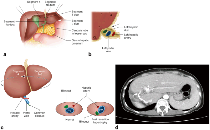
In contrast to the biliary anatomy of the right hemiliver, the left biliary system anatomy is relatively consistent. Also, fortuitously, the left hepatic duct has a long extrahepatic course as it runs along the undersurface of segment IVb. In this transverse course, the left hepatic duct is accompanied by left portal vein and this portal diad (notice the absence of left hepatic artery, normal or variant) is ensheathed in the peritoneal reflection of the gastrohepatic ligament (Fig. 19.3a). Thus, the left duct can be exposed at the base of segment IVb by dividing the reflection of Glisson’s capsule onto the gastrohepatic ligament, a maneuver called “lowering the hilar plate” (Fig. 19.3b). Left hepatic artery joins the “portal diad” at the base of the umbilical fissure. As the left hepatic duct runs in the umbilical fissure, it receives biliary ducts from segments II, III, and IV. Often, the left duct in the umbilical fissure is covered by a bridge of hepatic tissue that crosses from the left lateral section to the base of segment IV, and these need to be divided to gain access to the biliary and vascular structures in the umbilical fissure.
 Management of Airway, Hoarseness, and Vocal Cord Dysfunction After Esophagectomy
 Jejunal Feeding Tube Complications
 Bile Reflux and Gastroparesis
 Chyle Leak After Esophageal Surgery
 Complications After TEM (Transanal Endoscopic Microsurgery) and TAMIS (Transanal Minimally Invasive Surgery)
 Management of Rectovaginal Fistula


Fig. 19.3
Essentials of anatomy and anatomical alterations for biliary reconstruction. a Normal anatomy of biliary system. The right hepatic duct has a short extrahepatic course. On the other hand, the left hepatic duct has a long extrahepatic course and runs transversely at the base of segment IV before entering the umbilical fissure. In the umbilical fissure, the left hepatic duct gives rise to segment IVa and IVb ducts on the right and segment II and III ducts on the left. b Schematic demonstrating the lowering of hilar plate. Left portal pedicle runs transversely between quadrate lobe ( Q) and caudate lobe ( CL). Division of the reflection of Glisson’s capsule onto the gastrohepatic ligament in the plane shown by the arrow lowers the hilar plate and exposes the left hepatic duct which is situated deeper to the portal vein. c Schematic demonstrating how hypertrophy of the left liver after right hepatectomy displaces the hilum posteriorly and laterally and also changes the normal orientation of structures in the hilum. d CT scan in a patient postright hepatectomy depicting how posterolateral displacement of the hepatic hilum due to left liver hypertrophy may lead to difficulty in access to the portal structure and may require the use of thoracoabdominal incision
< div class='tao-gold-member'>
Only gold members can continue reading. Log In or Register to continue
Related posts:
Management of Airway, Hoarseness, and Vocal Cord Dysfunction After Esophagectomy
Jejunal Feeding Tube Complications
Bile Reflux and Gastroparesis
Chyle Leak After Esophageal Surgery
Complications After TEM (Transanal Endoscopic Microsurgery) and TAMIS (Transanal Minimally Invasive Surgery)
Management of Rectovaginal Fistula
Stay updated, free articles. Join our Telegram channel
Full access? Get Clinical Tree
