Figure 11.1 Endoscopic appearance of a pale ulcer in the duodenal cap with mild surrounding erythema, without evidence of recent bleeding and no visible vessel. This case presented with abdominal pain and malaena. There was associated Helicobacter pylori infection and antral gastritis.
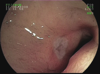
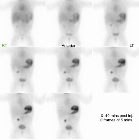
Figure 11.2 Gamma-emitting 99Tc scan appearances of a Meckel’s diverticulum, with uptake in the right iliac fossa, suggesting ectopic gastric mucosa. This infant presented with dark followed by bright red blood passed per rectum.
The colour of the blood in stool is indicative of the site of bleeding:
- Black: upper GI tract, e.g. varices
- Claret: midgut, e.g. Meckel’s
- Red: lower bowel, e.g. fissure, polyp, colitis
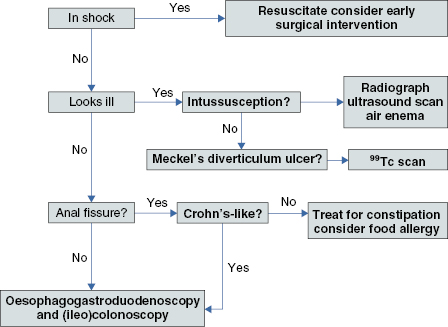
Investigations (see Algorithm 11.1)
- FBC: anaemia or thrombocytopoenia
- Coagulation screen:
- Prolonged PT suggests liver disease or vitamin K deficiency
- Prolonged APTT suggests factor deficiency or other coagulopathy: seek Haematology advice
- Prolonged PT suggests liver disease or vitamin K deficiency
- Liver function tests
- Consider sepsis, especially infants
- Apt’s test for maternal haemogloblin in neonates or breast-fed infants
- Meckel’s scan: if abdominal pain and/or claret-coloured stool
- Endoscopy/colonoscopy: seek specialist advice
Algorithm 11.1 Investigation of lower gastrointestinal bleeding
Related posts:


Stay updated, free articles. Join our Telegram channel

Full access? Get Clinical Tree


