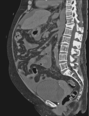
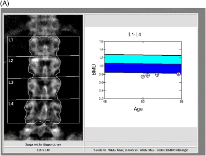
Chapter 25 Jane Collier John Radcliffe Hospital, Oxford, UK Osteomalacia, osteoporosis, and osteonecrosis are bone diseases seen in patients with liver disease. Osteomalacia is surprisingly uncommon in adults with cirrhosis, despite the high prevalence of vitamin D deficiency in this population. Osteonecrosis, usually of the femoral neck, is a rare condition seen predominately post-liver transplantation, partly related to immunosuppressive therapy [1]. This leaves osteoporosis as the most common bone disease affecting patients with cirrhosis. In a study of 360 patients with primary sclerosing cholangitis (PSC) and primary biliary cirrhosis (PBC) requiring liver transplantation, only 1.4% had a history of osteonecrosis [1]. This was often bilateral and usually at the femoral neck. Not all patients were on steroids which is the major risk factor. Although rare, the condition should be considered in patients on steroids who develop hip, shoulder, or knee pain, particularly on movement, as it is easy to miss. The classic description of osteomalacia is of hypocalcemia, hypophosphotemia, increased alkaline phosphatase and parathyroid hormone, although serum calcium and phosphate levels are often normal. Hepatic osteomalacia as defined histologically by bone biopsy is now rare in adults with cirrhosis and cholestatic liver disease. In a study from the Mayo Clinic of 33 patients with end-stage PBC and PSC undergoing liver transplantation, no cases of osteomalacia were identified on bone biopsy and none were seen in 60 patients undergoing liver transplantation at another center in the UK [2,3]. Case reports of osteomalacia complicating cirrhosis in the literature are confined to patients with malabsorption such as in celiac disease and cystic fibrosis. It may also occur in untreated chronic cholestatic disease of childhood. In contrast to osteomalacia, vitamin D deficiency is common in cirrhotic patients [4]. Vitamin D deficiency has been defined as 25-hydroxyvitamin D levels <20 ng/mL (50 nmol/L) with sufficient levels as >30 ng/mL (72 nmol/L) [5]. Of patients awaiting liver transplantation, 90–96% have low vitamin D levels [1,4]. In a study of 43 hepatitis C cirrhotic patients, 78% were vitamin deficient [6]. The severity of vitamin D deficiency has been associated with liver dysfunction rather than etiology [7]. Vitamin D is obtained from endogenous skin synthesis following exposure to sunlight leading to production of cholecalciferol (D3), both ergocalciferol (D2) and vitamin D3 are also acquired through food (reviewed in [5]). The liver is involved in bile salt production, absorption of vitamin D, and subsequent 25-hydroxylation of vitamin D. Intestinal absorption of cholecalciferol and 25-hydroxycholecalciferol is only affected in the presence of severe cholestasis where patients are jaundiced. Subsequent hepatic 25-hydroxylation of vitamin D3 has not been studied in humans but in cirrhotic rats this process is not impaired. Although low vitamin D does not lead to osteomalacia it is a associated with reduced bone mineral density (BMD), high bone turnover, and increased risk of hip fracture in the elderly. The precise contribution of low vitamin D levels to osteoporosis in patients with cirrhosis is unclear [8]. Osteoporosis is a major risk factor for fragility fractures, which impact on morbidity including quality of life. Osteoporotic fractures of the vertebral spine tend to occur about a decade before those in the femoral neck. Corticosteroids tend to cause vertebral rather than hip fractures. The former are often initially asymptomatic and are often detected incidentally in cirrhotic patients on cross-sectional imaging (e.g., CT scans looking for hepatocellular carcinoma) (Figure 25.1). Figure 25.1 Thoracic spine osteoporotic fracture seen on a computed tomography (CT) scan in a 64-year-old woman with diuretic-resistant ascites due to alcoholic cirrhosis. She presented with a myopathy leading to the diagnosis of cirrhosis. The fracture was identified on a CT scan showing ascites and a small liver prior to a transjugular intrahepatic portosystemic shunt (TIPS). She was treated with alendronate once-weekly based on the presence of an osteoporotic fracture without bone mineral density (BMD) assessment. She had normal vitamin D levels of 52.9 nmol/L and no other risk factors of osteoporosis apart from cirrhosis and alcohol excess. BMD is not needed to start treatment but may be useful as a baseline to assess if therapy needs to be continued after 5 years, ideally with ascites drained. BMD also falls in the first 3 months following liver transplantation leading to an increase in fractures in the first 2 years post-transplant. Although the rates of post-transplant fracture have fallen with the reduction in post-transplant steroid dosage, it is important to detect in the small proportion of cirrhotic patients suitable for liver transplantation. Osteoporosis is characterized by loss of bone mass and strength. Peak bone mass is reached in the third decade and then declines in both sexes but more rapidly in women. The risk of fracture depends on bone density but also on trabecular architecture and bone turnover as well as the tendency to fall. The latter often occurs in cirrhotic patients, because of low body mass index (BMI) and loss of muscle mass, as well as in alcoholics. Bone density is measured by dual-energy X-ray absorptiometry (DEXA) scan but is only part of the assessment of fracture risk (Figure 25.2). Figure 25.2 Bone mineral density in a patient with cirrhosis: (A) lumbar spine; (B) femoral neck. This 57-year-old man with a biliary cirrhosis due to combined variable immunodeficiency has a T score of –2.7 at vertebral spine and –3.0 at the femoral neck. His other risk factors for fracture include malabsorbtion due to chronic cryptosporidia and a budesonide-treated enteropathy. He has a low vitamin D level of 42 nmol/L. According to the FRAX index his fracture risk is 15% at 10 years and that of a femoral neck fracture 8.4%. The FRAX index underestimates the risk on steroids and its reliability in the presence of cirrhosis is unknown. He was treated with a single dose of zoledronic acid intravenously because of malabsorption – ibandronic acid would have been an alternative – and his vitamin D levels corrected. Risk factors for osteoporosis are frequently found in patients with cirrhosis: poor nutrition, excess alcohol, corticosteroid use, and hypogonadism (Table 25.1). Patients with end-stage PBC are also usually postmenopausal women. Corticosteroid use, the equivalent of 5 mg prednisolone taken for more than 3 months, is a particularly important independent risk factor for fractures (reviewed in [9]). Previous fracture is a further major clinical risk factor for further fractures independently of BMD. Once a vertebral fracture has occurred, the risk of a further vertebral fracture increases 10-fold and that of a hip fracture two- to threefold. The 5–10 year risk of fracture in an individual with low BMD can be calculated using several algorithms such as the World Health Organization’s fracture risk assessment tool (FRAX index) [10,11]. Although cirrhosis has been incorporated into the FRAX index (as secondary osteoporosis), the relative increased contribution of cirrhosis to fracture risk in an individual is not clear because of the relatively small numbers of liver disease patients in prospective studies. However, FRAX is useful tool in showing how age, BMI, and so on contribute to fracture risk. It is less reliable in patients on corticosteroids as it uses femoral neck BMD rather than that of the lumbar spine. Table 25.1 Risk factors for osteoporosis independent of cirrhosis. Various studies over the last two decades have shown that the prevalence of osteoporosis in patients with cirrhosis is between 12% and 55% (Table 25.2) [12–14]. The difference between studies is probably a reflection of differences in age, etiology of liver disease, nutritional state, hypogonadism, and severity of liver disease. In a study of 58 patients with viral cirrhosis, the risk of osteoporosis was shown to be associated with the severity of cirrhosis, with Child–Pugh A patients having a higher BMD than those with Child–Pugh C disease [15]. In a study of 243 patients with mixed end-stage liver disease requiring transplantation, the only independent risk factors for osteoporosis were lower BMI in women and increasing age [16]. Osteoporosis in hemochromatosis has been associated with the degree of iron overload [17]. Table 25.2 Prevalence of osteoporosis and fractures in cirrhosis and chronic biliary disease [12].
Bone Disorders
Introduction
Osteonecrosis
Osteomalacia
Vitamin D Deficiency
Osteoporosis
Background
Risk Factors
Age
Previous fragility fracture
Oral glucocorticoid therapy (>5 mg for 3 months)
Body mass index <19 kg/m2
Alcohol intake >3–4 units/day
Maternal/paternal history of hip fracture
Premature menopause <45 years
Prevalence
n
Type of cirrhosis
Osteoporosis (%)
Referred OLT
Related posts:
![]()
Stay updated, free articles. Join our Telegram channel

Full access? Get Clinical Tree

 Get Clinical Tree app for offline access


