Video capsule endoscopy (VCE) that was launched 10 years ago has become a first-line procedure for examining the small bowel, especially in the case of obscure gastrointestinal bleeding. Other major indications include Crohn disease (CD), celiac disease, and intestinal polyposis syndrome. In the case of small bowel diseases, the use of VCE must be integrated in a global diagnostic and therapeutic approach. More recently, wireless endoscopy has been adapted for examining the colon, opening up larger perspectives for colorectal cancer screening or colon examination. Technologic modifications of the second-generation colon capsule increase the sensitivity of this method for detecting polyps. Other new developments, including remote magnetic manipulation, power management, drug delivery capsule, microbiopsy capsule, and adaptation of technologies such as chromoendoscopy, are sure to enhance the capabilities of wireless endoscopy in gastrointestinal disorders.
The introduction of wireless or video capsule endoscopy (VCE) has revolutionized imaging modalities in gastroenterology. In the last 10 years, the number of scientific publications on VCE has continuously increased, promoting a new interest for small bowel (SB) diseases. Initially, VCE was used to examine the SB but was rapidly considered as a first-line procedure for several intestinal disorders. Now, the procedure is largely used in clinical practice globally. Subsequently, VCE was adapted for examining the esophagus and, more recently, the colon. A new era of research and development is open. Indeed, many people believe that wireless endoscopy is still an incompletely developed technology. In this article, the authors focus on the use of VCE for SB and colon diseases and describe the new and potential capabilities of VCE.
Small bowel video capsule endoscopy
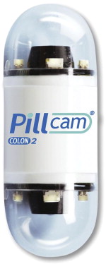
The VCE system comprises a capsule containing the video camera; a sensing system comprising an array of sensor pads, a data recorder, and a battery pack; and a workstation. A portable external viewer is also available for directly monitoring the images during the procedure. VCE was initially developed by Given Imaging (Yoqneam, Israel) for examining the SB (PillCam SB capsule). Subsequently, this company also developed the PillCam ESO capsule and the PillCam colon capsule for examining the esophagus and the colon, respectively. The small bowel video capsule endoscope (SBVCE) is 11 to 24–27.9 mm long, weighs approximately 3.4 to –6 g, and obtains 2 to 3 images per second. There are 5 capsule endoscopy systems available: PillCam (Given Imaging), EndoCapsule (Olympus, Center Valley, PA, USA), MiroCam (IntroMedic, Seoul, Korea), OMOM capsule (Jinshan Science and Technology, Chongqing, China), and Sayaka (RF SYSTEM lab, Nagano, Japan); however, only PillCam and EndoCapsule are currently approved by the Food and Drug Administration (FDA) for use in the United States. PillCam uses a complementary metal-oxide semiconductor (CMOS) chip, and the others use a charge-coupled device (CCD) chip for imaging ( Figs. 1–6 ).


PillCam and EndoCapsule use a radiofrequency (RF)-based communication technology, whereas MiroCam is currently undergoing FDA approval trials in the United States and uses a CMOS chip for imaging and the human body communication technology for transmission of images.
Most of the available literature on SBVCE is confined to PillCam. There are very little data on the EndoCapsule, MiroCam, OMOM, and Sayaka in the literature. Technical characteristics of the capsules are described in Table 1 .
| PillCam Colon | PillCam ESO | PillCam SB2 | EndoCapsule | MiroCam | OMOM | Sayaka | |
|---|---|---|---|---|---|---|---|
| Length (mm) | 26 | 26 | 26 | 26 | 24 | 27.9 | NA |
| Weight (g) | 3.4 | 3.4 | 3.4 | 3.8 | 3.4 | 6 | NA |
| Number of cameras | 2 | 2 | 1 | 1 | 1 | 1 | NA |
| Frames (per second) | 4–35 | 18 | 2 | 2 | 3 | 2 | NA |
| Image sensor | CMOS | CMOS | CMOS | CCD | CCD | CCD | NA |
| Battery life (h) | 8 | 8 | 8 | 9 | 11 | 8 | NA |
| Antennas | 8 | 3 | 8 | 8 | 9 | 14 | NA |
| Sleeping mode | Yes | No | No | No | No | No | NA |
Agile Patency Capsule
SBVCE is contraindicated for patients with known or suspected small-bowel strictures because of the risk of capsule retention and impaction that may necessitate removal either endoscopically or surgically. Screening fluoroscopic procedures, such as small bowel enteroclysis and small bowel follow-through, are associated with high doses of radiation and false-negative results.
The Agile Patency Capsule (Given Imaging) is a disintegrating, time-controlled capsule developed to identify patients with strictures that may cause retention of the video capsule. This capsule was approved by the FDA in 2006. It consists of a disintegrating capsule with an RF identification tag (RFID) and an RFID scanner.
The Agile Patency Capsule is of the same size as the SBVCE. It has cellophane walls that are filled with lactose (mixed with barium) and surround an RFID. When retained in a fluid-filled environment, the core of the patency capsule dissolves after approximately 40 hours, allowing the insoluble outer membrane to collapse and pass. The physician determines the presence of the patency capsule in the body of the patient using the scanner. The Agile Patency Capsule is expected to eliminate the risk of capsule retention in patients with known intestinal strictures who undergo capsule endoscopy.
SB preparation before SBVCE
The optimal SB preparation for SBVCE is an area of debate in the literature. Manufacturers of capsule endoscopy systems recommend only a clear liquid diet and an 8-hour fast. Several investigators have studied the effect of bowel preparation using polyethylene glycol (PEG) or sodium phosphate on SB visualization. However, the results of these studies have been contradictory. Some studies reported that bowel preparation resulted in an improvement in visualization, whereas others found no differences in SB visualization or diagnostic yield. A recent meta-analysis has shown that small bowel purgative preparation (PEG solution or sodium phosphate) improves the diagnostic yield of the examination. Another meta-analysis and a systematic review (presented as an abstract) that examined the effectiveness of bowel preparation for VCE also included studies using prokinetics and simethicone (in contrast to the previous study) and showed that bowel preparation had no effect on VCE diagnostic yield. Although adverse events and patient intolerance might be associated with the use of bowel purge for VCE, as inferred from colonoscopy studies, these have not yet been reported.
A new cleansing score system for SB preparation assessing 2 visual parameters was recently published: (1) proportion of visualized mucosa (4-step scale ranging from 0 to 3: score 3, >75%; score 2, 50%–75%; score 1, 25%–50%; score 0, <25%) and (2) degree of obscuration (4-step scale ranging from 0 to 3: score 3, <5% [no obscuration]; score 2, 5%–25% [mild obscuration]; score 1, 25%–50% [moderate obscuration]; score 0, >50% [severe obscuration]).
There is no consensus about the necessity of intestinal preparation for capsule endoscopy, and it should be interesting to develop adequate guidelines to improve its efficacy and tolerability.
Indications for SBVCE
A recent systematic review of 227 original English-language articles involving 22,840 procedures showed that obscure gastrointestinal bleeding (OGIB) is the most common indication (66%) for SBVCE, followed by “clinical symptoms” (10%), definite or suspected Crohn disease (CD) (10%), and other indications (34%).
OGIB
An OGIB is considered in patients with a digestive bleeding when the source of the bleeding remains unexplained after routine endoscopic procedures, including upper gastrointestinal (GI) endoscopy and colonoscopy and more frequently SB imaging modalities. OGIB represents about 5% of all GI bleeding. An overt OGIB with melena or hematochezia must be distinguished from occult digestive bleeding that is characterized by iron-deficient anemia with positive fecal occult blood test (FOBT) result. Digestive bleeding is now classified as upper GI bleeding, mid-GI bleeding, and lower GI bleeding.
The most common causes of OGIB are located in the SB and are considered as mid-GI bleeding. Nevertheless, 10% to 15% of the so-called OGIB is because of missed lesions located either in the upper GI tract or in the colon. Arteriovenous malformations or angioectasias account for most of the lesions, followed by SB tumors, drug-related lesions, and CD.
Several factors have been identified for selecting patients with OGIB in whom the risk of detecting a lesion is high: a serum hemoglobin level less than 10 g/dL, an ongoing overt bleeding (within 15 days), the presence of anemia or recurrent bleeding for more than 6 months, the occurrence of more than 1 episode of bleeding, the coexistence of renal insufficiency, and an occult bleeding with continuous positive FOBT results.
Should all patients with an OGIB be investigated? The American Gastroenterology Association Institute stated that “some patients with OGIB are managed clinically with intermittent blood transfusion along with other non specific supportive measures such as avoidance of anticoagulants as well as oral iron supplementation.” In clinical practice, the evaluation of OGIB is a function of the extent of the bleeding and the age of the patient.
Since the last 10 years, several endoscopic methods are available for examining the SB; these methods are either noninvasive, such as the SBVCE, or invasive, such as the assisted-device enteroscopy. Several comparative studies that have been compiled in the meta-analysis have demonstrated that SBVCE is superior to push enteroscopy for detecting small-bowel lesions in the case of OGIB. Some studies suggest that the double-balloon method has a diagnostic yield similar to that of SBVCE but is obviously more invasive. The diagnostic accuracy of SBVCE by obtaining a final diagnosis at 1 year has a negative predictive value close to 100%.
Besides the diagnostic accuracy of SBVCE in OGIB, it is mandatory to determine the therapeutic effect of this investigation. Several studies reported a therapeutic effect in 33% to 66% of the patients. In a well-designed protocol comparing push enteroscopy with SBVCE as the first-line strategy, De Leusse and colleagues showed that SBVCE has a therapeutic effect (43%) superior to that of push enteroscopy (34%), with less additional procedures.
Is there a role for a second-look SBVCE or other modalities after a nondiagnostic SBVCE first test? The rebleeding rate after a normal SBVCE is very low. However, if the rebleeding presentation changes from occult to overt or if the hemoglobin value decreases more than 4 g/dL, there is a need for further investigation. It has been reported that gross abnormalities, including tumors, may be missed by SBVCE, especially in the proximal jejunum.
Long-term follow-up in patients with OGIB showed an 11% to 33% total recurrent bleeding. In the case of positive results in SBVCE and subsequent therapeutic enteroscopy, there was 80% no rebleeding. However, Laine and colleagues recently reported a prospective study that enrolled 135 patients with OGIB and a negative workup, including upper GI endoscopy, colonoscopy, and push enteroscopy. These patients were randomized to undergo either SBVCE or SB contrast radiography with a follow-up of 12 months. The diagnostic yields for SBVCE and the SB radiography were 30% and 5%, respectively. However, there was 30% further bleeding in the SBVCE group and 24% in the SB radiography group (no statistical difference).
In conclusion, SBVCE has an excellent diagnostic yield in OGIB, reaching 50% to 60%; it is well admitted that SBVCE should be the first-line procedure in OGIB. If the SBVCE result is negative, the risk of rebleeding is low, but the patient’s characteristics and bleeding features should be further investigated.
CD
CD is an inflammatory bowel disease (IBD) that can affect the entire GI tract, with the SB being the most commonly affected site. It is the only site involved in 30% of the cases and, therefore, is difficult to diagnose. Symptoms of CD are heterogeneous but include diarrhea for more than 6 months, abdominal pain, or weight loss. Systemic symptoms of malaise, anorexia, or fever are common. A single gold standard for the diagnosis of CD is not available. The diagnosis is confirmed by clinical evaluation and a combination of endoscopic, histologic, radiologic, or biochemical investigations.
For suspected CD, ileocolonoscopy and biopsies from the terminal ileum and each colonic segment to look for microscopic evidence are first line to establish the diagnosis. The advent of VCE, along with device-assisted enteroscopy (push, single and double balloon, and spiral), has revolutionized SB imaging and has major implications for diagnosis, classification, and therapeutic decision making. A position paper reached by a group of experts in endoscopy and IBD, endorsed by the European Crohn Colitis Organisation and the World Organisation of Digestive Endoscopy, has recently been published. The working parties performed a systematic literature search of their topic with the appropriate keywords using Medline/PubMed/EMBASE and the Cochrane database. The evidence level was graded according to the system of the Oxford Center for Evidence-Based Medicine.
In patients with suspected CD, ileocolonoscopy must be performed before SBVCE. Moreover, SB cross-sectional imaging should generally precede SBVCE. SBVCE is likely to identify mucosal lesions that are compatible with CD in some patients in whom conventional endoscopic and small bowel radiographic imaging modalities have been nondiagnostic, but the diagnosis of CD should not be based on the appearance at the capsule endoscopy alone. Indeed, there is a lack of validated diagnostic criteria for SBVCE for the diagnosis of CD. Endoscopic differentiation of small bowel CD from drug-induced lesions or other diseases is unreliable.
However, SBVCE is better than small bowel follow-through enteroclysis, computed tomography (CT)/magnetic resonance enterography, or enteroclysis for detecting mucosal lesions related to CD. A normal SBVCE result has a very high negative predictive value, essentially ruling out SB CD. However, the use of SBVCE is limited by a lack of specificity when SB CD is suspected. Indeed, more than 10% of healthy subjects demonstrate mucosal breaks and erosions in their SB.
In patients with established CD, the role of SBVCE is to focus on the unexplained symptoms when other investigations are inconclusive, if it will alter management. In these patients, the risk of SBVCE retention is increased, particularly in those with known intestinal stenosis. The Agile Patency Capsule reduces the risk of retention. For the assessment of postoperative recurrence of CD, SBVCE should be considered only if ileocolonoscopy is contraindicated or unsuccessful.
In patients with indefinite IBD, SBVCE is helpful in identifying mucosal lesions related to CD. A negative SBVCE result does not exclude a future diagnosis of CD. In a recent report, it has been confirmed that preoperative wireless capsule endoscopy does not predict the outcome after ileal pouch anastomosis.
SBVCE can be safely used in selected children who are even younger than 8 years.
In conclusion, SBVCE should be reserved for patients in whom the clinical suspicion for CD remains high despite negative results with ileocolonoscopy and radiologic examinations.
Other Indications for SBVCE
SB tumors
In patients with SB tumors, the most common indication for SBVCE is an obscure bleeding; in patients who underwent SBVCE for various reasons, the incidence of SB tumors ranged from 2% to 9%. SBVCE has good sensitivity in diagnosing malignant tumors as adenocarcinomas, lymphomas, carcinoids, sarcomas, and hamartomas. SBVCE also plays a role in identifying benign tumors as GI stromal tumors ( Fig. 7 ). Tumors are located in the jejunum (40%–60%), the ileum (25%–40%), and less frequently in the duodenum (15%–20%).

Celiac diseases
Although SBVCE has good sensitivity and specificity for detecting mucosal atrophy, the procedure has limited application in patients with suspected celiac disease because a biopsy specimen is needed to confirm the diagnosis; SBVCE is probably more useful in selected cases with refractory or complicated celiac disease.
Hereditary polyposis syndromes
It has been shown that SBVCE is better for diagnosis and surveillance of SB polyps than for that of SB enteroclysis ; however, magnetic resonance imaging provides a better estimation of the site and size of the polyps. Although SBVCE is recommended for searching for SB polyps in patients with familial adenomatous polyposis who have duodenal lesions, endoscopy, mainly with a side-view duodenoscope, is preferable to examine the duodenum, especially the ampullary region. SBVCE should be considered as the first-line screening modality for surveillance in patients with Peutz-Jeghers syndrome.
Colon capsule endoscopy
Colorectal cancer (CRC) is the second leading cause of cancer-related deaths in the Western world. Most of the cases of CRC are believed to arise from adenomatous polyps that progress over the course of many years to invasive adenocarcinoma. Current evidence-based guidelines recommend the screening of average-risk adults, because the early detection and removal of adenomatous polyps has been shown to reduce the incidence of CRC and CRC-related mortality. Such screening can reduce CRC incidence and mortality, but effectiveness depends on its quality, ease of use, and patient adherence.
Optical colonoscopy (OC) is currently considered to be the gold standard procedure for CRC screening despite some limitations such as the suboptimal performance of colonoscopy and the poor quality of bowel preparation. Moreover, limited endoscopy resources may limit access to large, population-based screening programs, and many individuals are reluctant to undergo OC because of its perceived inconvenience and discomfort. Compliance to CRC screening is only about 50% in the United States and typically 20% or lower across Europe.
The main objective of a CRC screening program is to lower the population mortality rate related to this tumor. Besides the sensitivity of any method for detecting a lesion, the development of well-accepted, noninvasive, and, if possible, cheaper methods is mandatory to reach this goal.
Based on the success of the SB capsule and the technical adaptations of the ESO capsule with a camera on both sides, it seems challenging to develop a PillCam colon capsule endoscope (PCCE) that could be an alternative method for CRC screening.
The development of a wireless endoscope for examining the colon raised several challenging questions: (1) the duration of the colon transit time, requiring to save batteries, (2) the cleanliness of the colon, requiring an excellent colon preparation, (3) the caliber of the colon lumen, taking into account the impossibility of inflating the colon as during a colonoscopy.
Related posts:
 Endomicroscopy of Intestinal Metaplasia and Gastric Cancer
 Wide View and Retroview During Colonoscopy
 High-Definition Endoscopy and Magnifying Endoscopy Combined with Narrow Band Imaging in Gastric Cancer
 New Options of Cholangioscopy
 High-Definition and Filter-Aided Colonoscopy
 Molecular Imaging of Gastroenteropancreatic Neuroendocrine Tumors
Stay updated, free articles. Join our Telegram channel

Full access? Get Clinical Tree


