Vaginal Repair of Urethrovaginal and Vesicovaginal Fistulae

14 Vaginal Repair of Urethrovaginal and Vesicovaginal Fistulae


Worldwide, genitourinary fistulae are most often a devastating consequence of prolonged or obstructed labor. In the industrialized world, however, this is rarely the situation, and fistulae generally occur as a result of gynecological surgery, most commonly hysterectomy, or radiation therapy to the pelvis. Vesicovaginal fistula (VVF) is much more common than urethrovaginal fistula. The latter more commonly occurs after urethral surgery, such as a midurethral sling procedure in which synthetic material was placed or eroded into the lumen of the urethra. Repair of both types of fistulae can almost always be addressed via a vaginal approach. The following important principles for a successful repair apply to both types of fistulae:


The vaginal approach is an ideal approach that allows exposure of the fistula and preparation of the tissue layers and a well-vascularized flap for interposition. This chapter reviews and demonstrates the technical steps required to successfully correct urethrovaginal and vesicovaginal fistulae via a vaginal approach.


Preoperative Evaluation


History


In industrialized countries, women with VVF usually have varying degrees of incontinence that began after recent hysterectomy or pelvic surgery. Typically, the incontinence is described as continuous, both day and night, and unrelated to activity or an urge to void. In some cases, however, the incontinence may be somewhat positional or gravity dependent. If the fistula is quite sizable, the patient rarely voids volitionally, because the bladder never fills to any significant volume. In other cases in which the fistula is small (i.e., several millimeters in diameter), incontinence may be intermittent and more difficult to characterize. An important symptom in most patients with VVF is incontinence that occurs in the supine position, even during sleep. If the patient had an indwelling urethral catheter following pelvic or gynecological surgery, she will often report sudden onset of incontinence when the catheter was removed postoperatively. The patient may also have experienced gross hematuria immediately following the surgery, such as after hysterectomy.


Less commonly, a fistulous tract can be present between the urethra and vagina causing total incontinence, sporadic incontinence, or vaginal voiding without incontinence depending on its location. A fistula in the proximal portion of the urethra, proximal to the external sphincter complex, may present with continuous incontinence (similar to a VVF), especially if the bladder neck is incompetent. If the fistula is located in the middle to distal urethra, distal to the sphincter complex, the incontinence can be sporadic and may present as postvoid dribbling. In addition, recurrent urinary tract infections are common in women with urethrovaginal fistula because of migration of vaginal bacteria through the fistulous tract. Today, urethrovaginal fistula may commonly occur following midurethral synthetic sling surgery because of unrecognized perforation of the urethra during dissection or sling passage, inadvertent dissection into the periurethral fascia resulting in sling placement in the urethral wall, unrecognized placement of a sling through the urethra, or late urethral erosion. In such cases fistula repair often involves excision of the eroded foreign material in addition to fistula closure. Urethrovaginal fistula may also result from other surgeries on the urethra, such as urethral diverticulectomy, or from urethral trauma.


It is also important to consider the timing of the presentation of incontinence relative to the event that may have caused a fistula, particularly for a VVF. When there is an unrecognized or inadequately repaired cystotomy at the time of pelvic surgery, urine leakage usually occurs immediately after surgery. After cautery or devascularization injuries (e.g., from suture placement), leakage usually becomes evident 7 to 10 days after surgery. However, in some cases a VVF may present weeks or months after pelvic surgery, particularly in a patient who has undergone pelvic irradiation.


Physical Examination


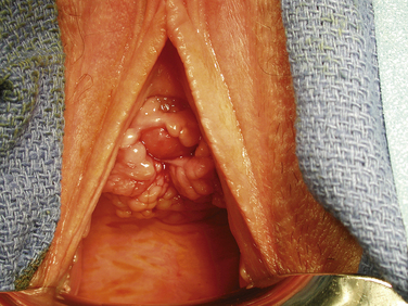
Vaginal speculum examination should be performed to visualize the fistula. For a VVF, when a full speculum is placed, the first observation is usually that of a urine-filled vaginal vault. Depending on the patient’s anatomy, the fistula may best be seen by using a full speculum to retract both the anterior and posterior vaginal wall. This is particularly true when the fistula is located at the vaginal cuff, the most common site for a VVF occurring as a result of hysterectomy. When the fistula is more distal, it is helpful to use the bottom blade of a Graves speculum to facilitate careful visualization of the anterior vaginal wall. The half-speculum is placed along the posterior vaginal wall with mild traction downward to expose the aforementioned compartments. The fistula is often surrounded by granulation tissue. Palpation of the vaginal cuff and anterior vaginal wall may reveal the opening as well as suture material at the vaginal cuff near a presumed fistula. In addition, a urethral catheter can be placed and the bladder filled with a solution of either indigo carmine or methylene blue diluted in sterile saline. This maneuver can help both to confirm and to identify fistula location. If extravasation of blue-tinged fluid is not seen during speculum examination, a tampon or gauze packing can be placed vaginally and the patient can ambulate or sit upright for 10 to 15 minutes. Subsequent inspection of the gauze revealing blue staining is indicative of a lower urinary tract fistula. If there is no blue staining of the vaginal gauze but the gauze appears saturated with yellow urine, a ureterovaginal fistula must be suspected. This possibility can be further investigated with computed tomographic (CT) urography (see later section on imaging).


To identify a urethrovaginal fistula, it may be necessary to place a urethral catheter to elongate the urethra and allow inspection of the entire vaginal epithelium overlying the urethra. If any portion of the catheter becomes visible on vaginal examination, a sizable fistula is present. The same thing can be accomplished by placing a lubricated cotton-tipped applicator (Q-tip) into the urethra.


Imaging


Imaging is not always mandatory to diagnose and treat a VVF, though it is important to rule out a ureterovaginal fistula in cases where such a finding is possible (e.g., after pelvic surgery such as hysterectomy). Standard cystography, CT cystography, or pelvic magnetic resonance imaging can allow identification and further characterization of a VVF. In cases in which the fistula is not obvious on physical examination or endoscopy, we prefer standard cystography as the simplest and most reliable initial method of diagnosis. A Foley catheter is placed via urethra and the bladder is filled under gravity drainage with a contrast solution. Standard radiographic or fluoroscopic images are obtained in both the anteroposterior and lateral orientations. A VVF is typically most readily identifiable in the lateral orientation as a wisp of contrast beyond the border of the bladder wall, with filling or pooling of the vaginal canal just beneath the bladder visible on the radiograph. The same applies to a urethrovaginal fistula located proximal to the external sphincter.


In any case of VVF following pelvic surgery, the possibility of a ureterovaginal fistula or even ureteral obstruction should be evaluated preoperatively with upper urinary tract imaging using CT or magnetic resonance urography. The radiologist should be given sufficient clinical information so that the distal ureters are properly opacified and evaluated on delayed images during the study, typically by administration of a diuretic following contrast-enhanced imaging. This is critical in evaluating for ureterovaginal fistula. Alternatively, and particularly in cases in which use of intravenous contrast is contraindicated, retrograde ureteropyelography can be done at the time of fistula repair.


Patient Selection and Surgical Planning


A transvaginal surgical route is the best approach in any patient with a urethrovaginal fistula. In patients in whom the fistula occurred secondary to synthetic sling placement it is critical to the procedure to completely excise the foreign sling material in and around the urethra to allow proper healing of the urethral mucosa and periurethral fascia. Accurate preoperative assessment of stress incontinence is often difficult, so patients are counseled regarding the possible need for a delayed, second-stage procedure to treat stress incontinence, such as injection of a urethral bulking agent, or even repeat sling placement, which is usually done with an autologous fascial sling in cases of prior synthetic sling complications. When the urethrovaginal fistula is proximal and stress incontinence can be assessed preoperatively, placement of a pubovaginal sling of autologous fascia can be performed at the time of fistula repair. The sling can provide both a backboard of support for the urethra and an additional tissue layer for the repair. We believe that concurrent placement of a synthetic sling is contraindicated in the setting of urethrovaginal fistula repair and/or urethral reconstruction.


The timing of VVF repair depends primarily on the surgeon’s experience and preference. Our practice is to repair VVF upon presentation in most cases, but it is also reasonable to provide time for healing and wait for the tissues appear healthy and demonstrate a well-formed fistulous tract. Most patients come for treatment several weeks after the gynecological surgery and are therefore candidates for repair at the time of presentation. There is no objective evidence to indicate that waiting 3 to 6 months, as suggested by older published series, is truly beneficial.


May 30, 2016 | Posted by in GASTOINESTINAL SURGERY | Comments Off on Vaginal Repair of Urethrovaginal and Vesicovaginal Fistulae

Full access? Get Clinical Tree

Get Clinical Tree app for offline access