Transtar
David Jayne
Antonio Longo
Introduction
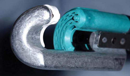
This chapter on Contour30® Transtar (Ethicon Endosurgery Inc., Cincinnati, OH, USA) should be read in conjunction with that on Rectocele: STARR. Both STARR and Transtar are procedures for the correction of obstructed defecation associated with intussusception with or without rectocele. In essence, both procedures aim to produce the same surgical outcome, namely resection of the distal rectal redundancy with restoration of normal anorectal anatomy. STARR was the procedure first described, prior to the introduction of the specially designed Contour30® stapling device, and used two PPH-01 staplers. The PPH-01 stapler had been designed for the treatment of prolapsing hemorrhoids and its application to internal prolapse and rectocele was not without potential drawbacks. It was for this reason that a specific stapler, the Contour30®, was designed for Transtar. The Contour30® consists of a curved stapling device that holds a reloadable cartridge (Fig. 27.1). When deployed, the stapler simultaneously fires three staple lines and cuts the contained tissue. The potential benefits of the Contour30® Transtar over the PPH-01 STARR include:
An ability to resect a greater volume of prolapse
An ability to tailor the extent of the prolapse resection to the individual patient
Improved visibility of the resection during the procedure
A true full-thickness, circumferential resection
In this chapter, the term Transtar will be used to denote transanal circumferential rectal resection with the Contour30® stapler.
The indications and contraindications for Transtar are identical to those outlined for STARR. In summary, the patient should have symptoms consistent with obstructed defecation syndrome (ODS) and distal rectal redundancy in the form of intussusception with or without significant rectocele demonstrable on dynamic pelvic floor imaging. Ideally, the patient should have reasonable anal sphincter function. Transtar is contraindicated in the presence of other concomitant pathology that might predispose to poor anastomotic healing or the development of septic complications. Successful outcome is dependent on correct patient selection, which in turn demands thorough preoperative investigation.
Like many operations for functional disorders, successful outcome is dependent on correct patient assessment and selection, which in turn demands thorough preoperative investigation. As a minimum, this should include quantification of symptoms using an appropriate scoring system (1,2), exclusion of coexistent pathology by appropriate colorectal imaging, assessment of pelvic floor anatomy by defecating proctogram or dynamic magnetic resonance imaging, and anorectal physiology and endoanal ultrasound. Once this information has been obtained, an informed decision can be made regarding the suitability of the patient for Transtar. No additional preoperative workup is required for Transtar as compared to PPH-STARR.
Transtar can be performed under either spinal or general anesthesia. The lower bowel should be prepared by administration of a phosphate enema to ensure that the anorectum is empty. The patient is placed on the operating table in the supine position. The legs are supported in stirrups with the hips flexed to at least 90 degrees and the table tilted to 30 degree head-down for maximal exposure of the perineum. A single dose of broad-spectrum perioperative antibiotics is administered. An examination under anesthesia is performed to confirm the presence of internal prolapse with or without rectocele and to exclude coexistent pathology and other pelvic organ prolapse.
The following describes the steps involved in the Transtar procedure:
The anal canal is gently dilated and the circular anal dilator is inserted and secured with 1/0 silk sutures to the anal verge.
The extent and apex of the internal rectal prolapse are assessed by insertion and withdrawal of a dry swab.
Prolene traction sutures (2/0) are placed circumferentially around the apex of the prolapse in the 2, 12, 10, 8, and 5 o’clock positions. Each suture is loosely tied and held by an artery forceps (Fig. 27.2).
A marking suture of 1/0 Vicryl is inserted to the depth of the prolapse to be resected at the 3 o’clock position. This suture is tied tightly and a loop is created toward the free end, through which the Transtar stapler can be passed.
The resection starts at the 3 o’clock marking suture with a radial firing of the stapler, which has the effect of “opening up” the prolapse (Fig. 27.3). The resection then
Stay updated, free articles. Join our Telegram channel

Full access? Get Clinical Tree


