Fig. 9.1
Advantage of transanal minimally invasive surgery (TAMIS) for rectal cancer. Compared with the conventional laparoscopic approach, the TAMIS approach offers an in-line vantage point to the distal rectum, and this facilitates perianal dissection, especially in patients with a narrow pelvis or a bulky tumor
9.2 Patient Position and Operative Setup
The patient is placed in a lithotomy position with slight head-down tilting. The perineal region is prepped and draped. The location of the monitor and surgical devices is shown in Fig. 9.2. As an operative platform, we usually use GelPOINT mini or GelPOINT path device (Fig. 9.3); the latter is the only device that was designed specifically for transanal use. Three trocars are placed in the Gelseal cap: one for the camera and the other two for the surgeon to use. A rigid endoscope (0° or 30°) with a high-definition camera is used for the operation. Electrocautery or ultrasonic scissors can be used as the energy device, though we prefer electrocautery. Appropriate selection of a CO2 insufflator is very important for this procedure because the operative space in this approach is small, and smoke evacuation and instability of the operative field due to loss of pneumoperirectum are major problems. A real-time pressure sensor (AirSeal, SurgiQuest, or PneumoSure, Stryker) is helpful in maintaining a constant CO2 pressure and stable operative field (Fig. 9.4). In particular, the AirSeal device constantly evacuates the smoke through automatic circulation of CO2.
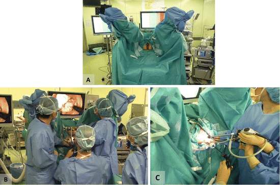
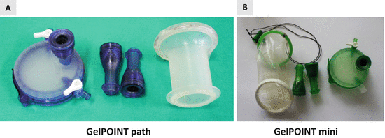
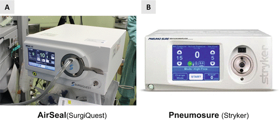

Fig. 9.2
Operative setup. (a) Patient position. (b) Location of the surgeons and operative monitor. (c) Closer view of the setup

Fig. 9.3
Platform for transanal access. (a) GelPOINT path. (b) GelPOINT mini (Applied Medical)

Fig. 9.4
CO2 insufflator. (a) AirSeal (SurgiQuest). (b) PneumoSure (Stryker)
9.3 Operative Procedure
Using the TAMIS approach, several types of rectal surgery can be performed, including local excision, TME, intersphincteric resection (ISR), and abdominoperineal resection (APR) (Fig. 9.5). In the following section, the technical details of TAMIS-TME/ISR and APR will be discussed.
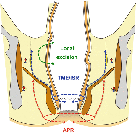

Fig. 9.5
Schema of operative procedures using the TAMIS approach
TAMIS-TME/ISR
The operative schema of TAMIS-TME/ISR is shown in Fig. 9.6.
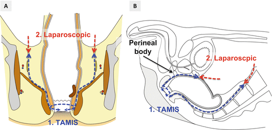

Fig. 9.6
Operative schema of TAMIS-intersphincteric resection (ISR)/total mesorectal excision (TME). (a) Coronal view. (b) Sagittal view
9.3.1 Distal Transection of the Rectum
The anus is gently dilated and the location of the tumor is confirmed. The intersphincteric groove is identified by palpation and the external anal sphincter is dilated with six threads. Use of a Lone Star Retractor is also beneficial. Determination of the distal margin is an important part of this procedure, and it depends on the location of the tumor and how the distal rectum (or anal canal) is transected. When the tumor is very low lying near the anal verge, full-thickness circumferential incision of the anal canal under direct vision is usually made with a sufficient distal margin. Subsequently, intersphincteric dissection is performed under direct vision to some extent (2–3 cm) to fix the sleeve portion of the GelPOINT device to the anal canal (Fig. 9.7). When the tumor is located more proximally, the GelPOINT path is inserted into the anal canal, and rectal occlusion and distal incision of the rectal wall are performed under endoscopic vision (Fig. 9.8). Secure closure of the distal stump and sufficient irrigation of the surgical field are important to prevent spillage of tumor cells and bacterial contamination.
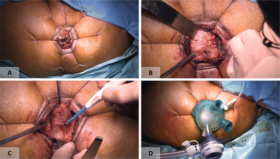
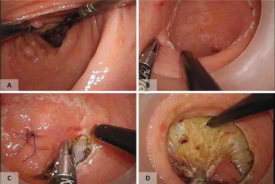

Fig. 9.7
(a) Full-thickness incision of the anal canal is made following the assessment of tumor location. (b) Intersphincteric dissection. (c) Posterior dissection. (d) Placement of GelPOINT mini

Fig. 9.8
Distal incision of the rectum through the TAMIS approach (optional). (a, b) The location of the tumor is examined and circumferential marking is made with an appropriate distal margin. (c, d) The rectum is tightly closed and a full-thickness rectal incision is made
9.3.2 TAMIS Mesorectal Dissection
Pneumoperirectum is maintained at 8–12 mmHg. Posterior presacral dissection is usually performed first because the anatomical dissection plane is easier to identify posteriorly (Fig. 9.9). It should be noted that posterior dissection tends to migrate into the mesorectum because of the sharp angle of the dissection line due to the pull of the puborectal sling at the anorectal flexure (Fig. 9.10) [7]. Once the appropriate dissection plane is found, posterior dissection is performed along the curve of the sacrum until the sacral promontory is reached. Because the dissection goes easily behind the layer of hypogastric nerves, care should be taken not to injure them. After sufficient mobilization of the posterior side, identification and preservation of pelvic autonomic nerves are crucial at the posterior-lateral side (Figs. 9.11a and 9.12a). The dissection between the mesorectum and pelvic autonomic nerves progresses laterally, and it will help in the identification of the neurovascular bundle running alongside the prostate and vagina as well as facilitate the dissection of the anterior side (Figs. 9.11b and 9.12b).

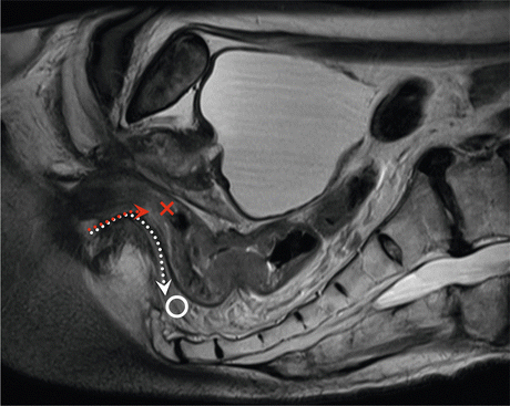
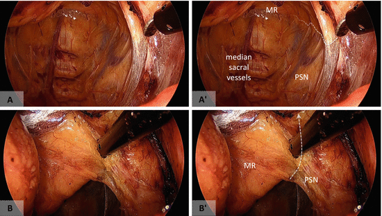
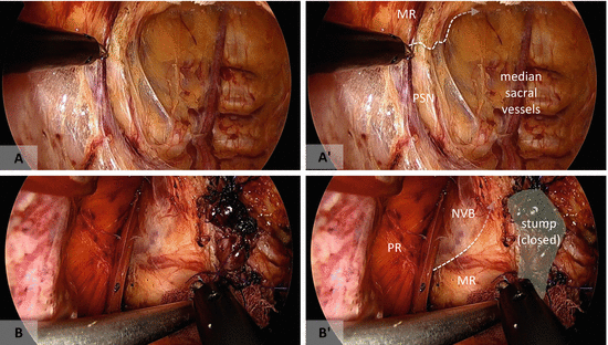

Fig. 9.9
Posterior dissection (TAMIS-ISR). (a) The rectal dissection is usually initiated at the posterior side. (b) The mesorectal plane is easy to identify at the posterior side. (c) Dissection tends to go behind the hypogastric nerves (HGN). MR mesorectum, PR puborectal muscle

Fig. 9.10
Median sagittal view of the anorectal region on magnetic resonance imaging. As indicated by the red dotted line, posterior dissection tends to migrate into the mesorectum (or posterior rectal wall) because of the sharp angle of the dissection line made by the puborectal sling. The white dotted line should be followed

Fig. 9.11
(a, b) Identification and preservation of the left pelvic autonomic nerves. The pelvic splanchnic nerves (PSN) can be identified at the posterolateral side and dissected from the mesorectum (MR). White broken lines indicate mesorectal dissection line

Fig. 9.12
(a, b) Identification and preservation of the right pelvic autonomic nerves. The pelvic splanchnic nerves (PSN) are identified as was done on the left side. When dissection is preceded anteriorly, the lateral aspect of the neurovascular bundle (NVB) is identified. Imaginary dissection line is indicated in white broken lines
When the tumor is low lying, rectal dissection starts from the intersphincteric plane, and the dissection layer of the anterior side is usually obscure because of the perineal body (rectourethral) muscle (Figs. 9.13 and 9.14). On the other hand, when the level of the distal rectal incision is above the perineal body, it is relatively easy to identify the proper anterior dissection plane (Fig. 9.15). Once the proper dissection plane is established between the anterior side of the rectum and the prostate or vagina, maintaining this plane is relatively easy under good exposure and close endoscopic view (Fig. 9.16). Finally, lateral mesorectal dissection is performed by connecting the dissection layer of the anterior and posterior sides. It should be kept in mind that there is an easily dissectible layer outside of the autonomic nervous system (Fig. 9.17). The appearance of the operative field after TAMIS dissection is shown in Figs. 9.18 and 9.19. Anteriorly, the peritoneum at the cul-de-sac may be opened to enter into the peritoneal cavity (Fig. 9.20). However, premature entry into the peritoneal cavity might disturb exposure of the operative field.
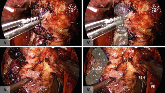
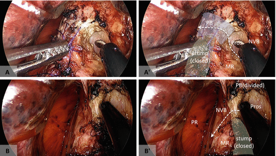
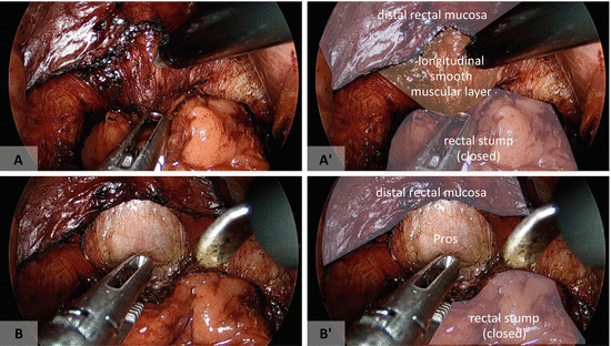
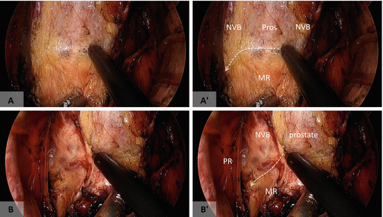

Fig. 9.13
Anterior dissection in TAMIS-ISR. (a) The anterior dissection plane is usually obscure because of the perineal body (PB). (b) Dissection from the lateral to anterior aspects while identifying the pelvic autonomic nervous system (white broken line) might be useful for identifying the appropriate anterior dissection plane. MR mesorectum, PSN pelvic splanchnic nerve, NVB neurovascular bundle, PR puborectal muscle

Fig. 9.14
Anterior dissection in TAMIS-ISR. (a) Identification of the prostate (Pros) from the left side facilitates division of the perineal body (PB; white broken line) (b). Division of the perineal body is followed by dissection between the mesorectum (MR) and right NVB (white broken line)

Fig. 9.15
Anterior dissection (optional). (a, b) When the distal incision is made above the level of the perineal body, an appropriate anterior dissection plane behind the prostate (Pros) is easy to identify

Fig. 9.16


Anterior dissection. (a) Once the appropriate dissection plane is identified, it is easy to dissect the mesorectum (MR) from the prostate (Pros) and neurovascular bundle (NVB) bilaterally (b). The small vessels branching from the right NVB are divided. Imaginary dissection lines are indicated in white broken lines
Stay updated, free articles. Join our Telegram channel

Full access? Get Clinical Tree


