Fig. 5.1
Positioning and instruments. The patient is placed in a modified lithotomy position using a levitator, with both arms fixed to the body trunk. Shoulder pads are used to prevent the patient from slipping downward (a). The set of instruments used for laparoscopic colectomy (b). A detachable intestinal clip is used to occlude the rectal lumen before irrigating and transecting the rectum (c)
5.4 Instruments
A 12-mm flexible endoscope or a 30° rigid scope, a spatula-shaped electric scalpel, three 5-mm double-action intestinal forceps, a 5-mm single-action intestinal forceps, and a 5-mm Babcock forceps are used (Fig. 5.1b). Transection of the rectum is performed using a detachable intestinal clip (Fig. 5.1c), a 60-mm linear stapler, and a 29-mm circular stapler; anastomosis is performed using the double-stapling technique (DST). In cases of anal stenosis and long DST, the use of a smaller 25-mm circular stapler allows it to be more easily inserted into the rectum.
5.5 Port Placement
A vertical intraumbilical incision is made and a 12-mm camera port is installed via the open method (Figs. 5.2, 5.3, and 5.4). A 12-mm port for the surgeon’s right hand is placed in the right lower abdomen at the lateral border of the right rectus abdominis muscle and approximately 2 cm cranial to the right inferior epigastric vessels. A 5-mm port for the surgeon’s left hand is placed approximately 4 fingerbreadths cranial to the 12-mm port placed in the right lower abdomen. Two 5-mm ports for the assistant are placed symmetrically opposite the two ports for the surgeon in the left lower abdomen. The surgery is performed via these five ports.
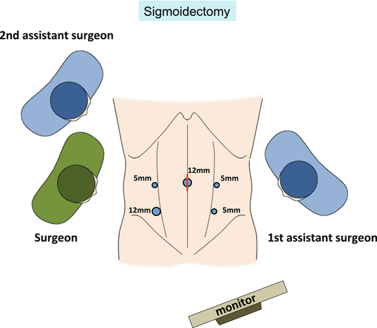
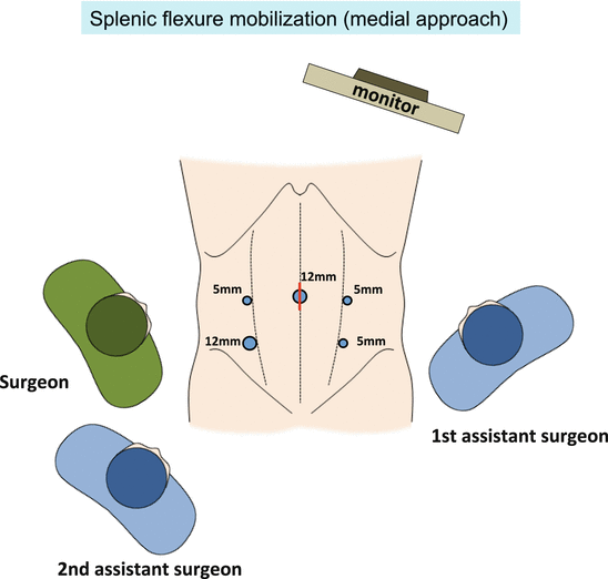
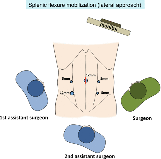
Fig. 5.2
Port placement and the arrangement of surgeons in laparoscopic sigmoidectomy
Fig. 5.3
Port placement and the arrangement of surgeons in laparoscopic splenic flexure mobilization via a medial approach
Fig. 5.4
Port placement and the arrangement of surgeons in laparoscopic splenic flexure mobilization via a lateral approach
5.6 Surgical Anatomy
Loose connective tissue lies between the mesorectum/mesocolon and pelvic wall/retroperitoneal organs. By applying and maintaining adequate tension between tissues, the loose connective tissue extends into a layer of foamy appearance and thus can be identified as the dissectable layer [2] (Figs. 5.5 and 5.6).
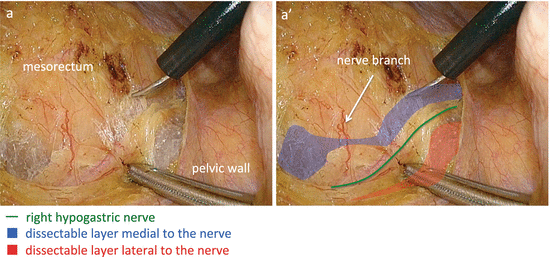
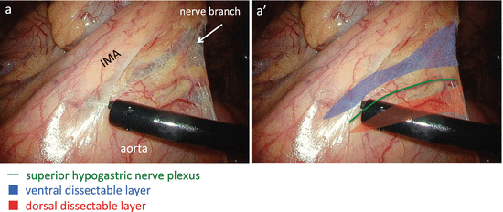
Fig. 5.5
The dissectable layer around the right hypogastric nerve. The right hypogastric nerve lies within the foamy loose connective tissue (i.e., the dissectable layer). The dissectable layer lies both medial and lateral to the nerve
Fig. 5.6
The dissectable layer around the superior hypogastric nerve plexus. The superior hypogastric nerve plexus runs within the foamy loose connective tissue identified as the dissectable layer. The dissectable layer lies both ventral and dorsal to the nerve
The autonomic nervous system—consisting of the lumbar splanchnic nerve, superior hypogastric nerve plexus, hypogastric nerve, pelvic splanchnic nerve, pelvic nerve plexus, and neurovascular bundle—and the left ureter and left gonadal vessels lie within the dissectable layer (Figs. 5.7 and 5.8). In particular, note that the dissectable layer exists on both the ventral and dorsal sides of the autonomic nerves, ureter, and gonadal vessels. Care must be taken to avoid straying into the dorsal side of the structures (Figs. 5.5, 5.6, 5.7, and 5.8).
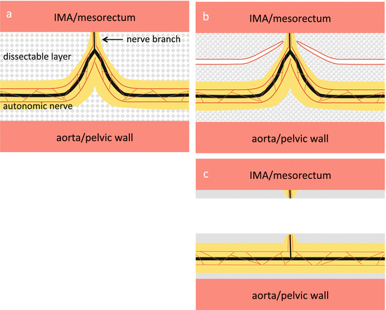
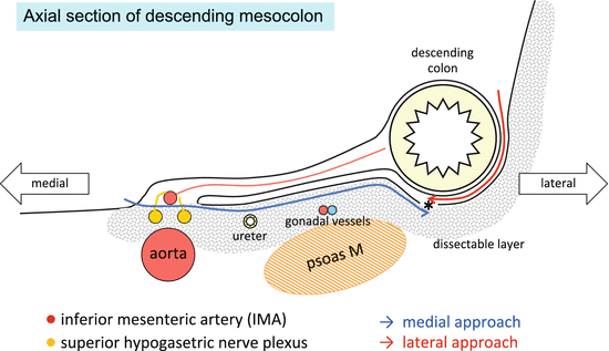
Fig. 5.7
The relationship between the autonomic nerve and the dissectable layer. The autonomic nerve runs within the dissectable layer accompanied by adipose tissue (yellow paint) and thin vessels (red line) and is pulled toward the IMA and mesorectum by the nerve branch (a). After cutting the nerve branch at the peak of pulled autonomic nerve, the dissectable layer contracts and the autonomic nerve is preserved (b, c). IMA inferior mesenteric artery
Fig. 5.8
Axial section of the descending mesocolon. The autonomic nerve, the left ureter, and the left gonadal vessels lie within the dissectable layer. The descending mesocolon is mobilized by a medial approach. The Toldt’s white line (*) can be incised also by a lateral approach
The autonomic nerve runs longitudinally through the dissectable layer accompanied by adipose tissue and thin vessels (Fig. 5.7a). This autonomic nerve is pulled toward the rectum and inferior mesenteric artery (IMA) by the nerve branch that penetrates the dissectable layer and runs toward the mesorectum and IMA (Fig. 5.7a). The peak that is caused by this pulling action—that is, the nerve branch—is cut and the autonomic nerve is preserved (Fig. 5.7b, c). Once the dissectable layer is resected, the “foamy” loose connective tissue, which has now lost the tension maintained by its connection to the structures on both sides, now contracts and resembles a membrane, and the structures on both sides—the mesorectum and mesocolon on one side and the pelvic wall and retroperitoneal organs on the other side—are now covered in this membranous tissue (Fig. 5.7c). This membranous tissue, derived from the contracted foamy loose connective tissue, is variously referred to as the proper rectal fascia, retroperitoneum, Gerota’s fascia, pre-hypogastric nerve fascia, Denonvilliers’ fascia, and so forth, depending on the location. These membrane-like structures can be created artificially by dissecting the foamy loose connective tissue (i.e., the dissectable layer).
By dissecting the space between the descending mesocolon and retroperitoneal area in the cranial direction, the dissection proceeds behind the pancreas, because the inferior mesenteric vein (IMV) is contained within the descending mesocolon and runs behind the pancreas (Figs. 5.9 and 5.37b, c). At the inferior margin of the pancreas, the dissection plane is intentionally shifted so that it is between the root of the transverse mesocolon and pancreas, and then fenestration of the anterior lobe of the transverse mesocolon will allow access to the omental bursa (Figs. 5.9 and 5.38).
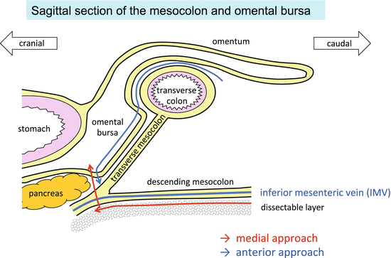
Fig. 5.9
Sagittal section of the mesocolon and omental bursa. The descending mesocolon is mobilized and the omental bursa is opened by a medial approach. The omental bursa can be opened also by an anterior approach
5.7 Procedure
5.7.1 Sigmoidectomy
5.7.1.1 Creation of the Operative Field
The patient’s head is tilted downward by approximately 15° until the small intestines move out of the pelvis, with the right side slanted approximately 5° lower. After moving the omentum and transverse colon to the epigastric region, the mesentery and small intestines are moved to the right abdominal area as if turning a page of a book, enabling visualization of the duodenum, root of the IMA, left abdominal area, and pelvic cavity (Fig. 5.10).
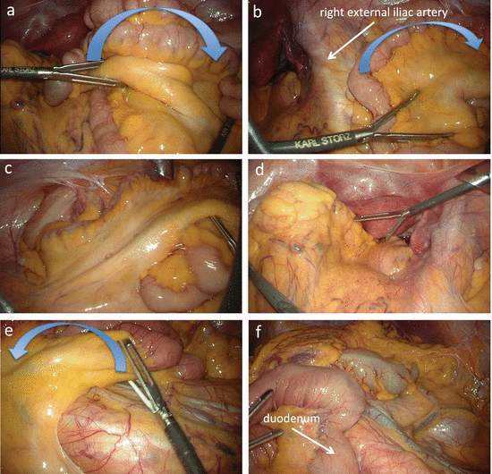
Fig. 5.10
Creation of the wide operative field. The distal small intestines are moved to the right abdominal area to empty the pelvic cavity (a–d). The proximal small intestines are moved to the right upper abdomen to observe the duodenum, IMA, and descending mesocolon in the same field of view (e, f)
5.7.1.2 Mobilization of the Upper Rectum
Before further preparations, the sigmoid colon is drawn out of the pelvis using intestinal forceps, with the surgeon using both hands to straighten the rectum. Using the intestinal forceps, the surgeon grasps the fatty appendage on the right side of the sigmoid colon and lifts it in the left ventrocranial direction. The assistant then uses the intestinal forceps in the right hand to press the right side of the sigmoid mesocolon in the lateral direction, allowing identification of the vascular pedicle of the sigmoidal vessels (Fig. 5.11a). Using the Babcock forceps in the left hand, the assistant grasps the vascular pedicle of the sigmoidal vessels and draws it in the left ventrocranial direction and then uses the intestinal forceps in the right hand to grasp the fatty appendage on the right side of the upper rectum and elevate it in the left ventrocaudal direction (Fig. 5.11b, c). Then, by drawing the tips of the forceps in both hands away from each other, the assistant can stretch out the right side of the mesorectum (Fig. 5.11d).
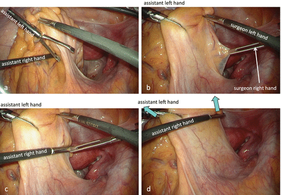
Fig. 5.11
Stretching out the right side of mesorectum. With the Babcock forceps in the assistant’s left hand, the vascular pedicle of the sigmoid vessels is pulled in the left ventrocranial direction (a, b). With the intestinal forceps in the assistant’s right hand, the fatty appendage on the right side of the rectum is elevated in the left ventrocaudal direction (c, d). By drawing the tips of the forceps in both hands away from each other, the assistant can stretch out the right side of the mesorectum (d)
The traction applied by the assistant is slightly relaxed to confirm the curvature of the right pelvic peritoneum and the right surface of the mesorectum, that is, the imaginary incisional line of the peritoneum (Fig. 5.12a). Then the right surface of the upper mesorectum is once again stretched and the imaginary incisional line is verified again (Fig. 5.12b). With the intestinal forceps in the left hand, the surgeon pulls the right pelvic peritoneum in the right direction using friction force (Fig. 5.12b). Once the peritoneum on the right side of the upper rectum is incised slightly caudal to the promontory, carbon dioxide gas permeates beneath the peritoneum and the dissectable layer is clearly revealed (Fig. 5.12c, d).
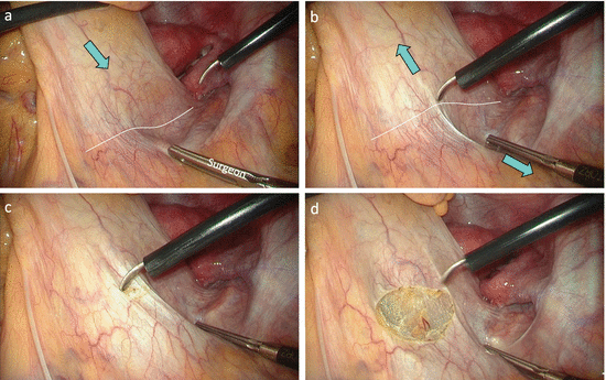
Fig. 5.12
The imaginary incisional line on the right side of the mesorectum. The retraction of the mesorectum is slightly relaxed (a) and the right surface of the mesorectum is stretched out once again (b) to verify the imaginary incisional line (white dotted line). After cutting the imaginary incisional line, the dissectable layer is clearly revealed (c, d)
The right hypogastric nerve, which is surrounded by adipose tissue through which thin vessels run longitudinally, is identified first (Figs. 5.5, 5.7, and 5.13). The nerve branch extending toward the mesorectum is dissected to preserve the right hypogastric nerve. By elevating the mesorectum ventrally using the opened intestinal forceps in the left hand or by pushing down the right hypogastric nerve in the lateral–dorsal direction, the foamy loose connective tissue posterior to the rectum (i.e., the dissectable layer) is clearly revealed and is dissected caudally from the posterior to posterolateral direction in the manner of a swinging pendulum. Then the left hypogastric nerve covered in adipose tissue is identified and preserved (Figs. 5.13, 5.14, and 5.15). After resecting this loose connective tissue, it contracts toward the mesorectum and pelvic wall, where it appears like a membrane. The loose connective tissue contracted against the pelvic wall is identified as the so-called pre-hypogastric nerve fascia (Fig. 5.15b, b′).
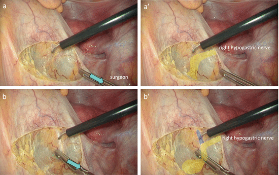
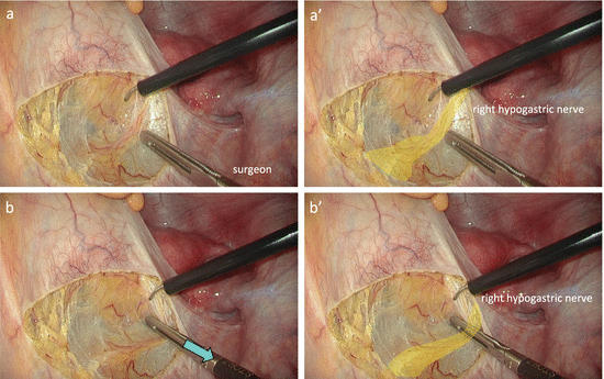
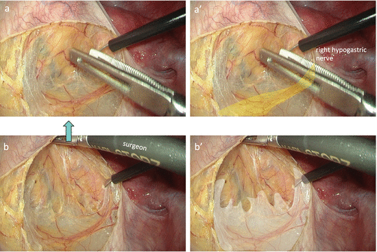
Fig. 5.13
Identification of the right hypogastric nerve. The right hypogastric nerve is surrounded by adipose tissue through which thin vessels run longitudinally (a, a′). The nerve branch extending toward the mesorectum (blue paint) is dissected to preserve the nerve (b, b′). Care must be taken not to stray into the dissectable layer behind the nerve
Fig. 5.14
The dissectable layer between the mesorectum and right hypogastric nerve. By pushing down the right hypogastric nerve in the lateral–dorsal direction, the foamy loose connective tissue between the mesorectum and the right hypogastric nerve is clearly revealed
Fig. 5.15
The dissectable layer behind the mesorectum. By elevating the mesorectum ventrally with the opened intestinal forceps, the foamy loose connective tissue behind the mesorectum is clearly revealed (a, b). The loose connective tissue contracted against the pelvic wall is identified as the so-called pre-hypogastric nerve fascia (white paint) (b, b′)
When performing sigmoidectomy, it is enough to dissect posterior to the rectum up to the level of the midpoint of the promontory and peritoneal reflection.
5.7.1.3 Mobilization of the Mesocolon
The assistant elevates the vascular pedicle of the sigmoidal vessels, grasped with the Babcock forceps in the left hand, in the lateral–ventral direction, and the surgeon grasps the cut edge of the parietal peritoneum and pulls it in the medial–ventral direction to apply tension to the root of mesocolon (Fig. 5.16a, b). After verifying the location of the duodenum, the peritoneum is incised in the cranial direction approximately 5 mm right to the IMA (Fig. 5.16).
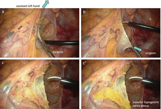
Fig. 5.16
The dissectable layer between the IMA and superior hypogastric nerve plexus. With the Babcock forceps in the assistant’s left hand, the vascular pedicle of the sigmoidal vessels is elevated in the lateral–ventral direction (a), and the surgeon grasps the cut edge of the parietal peritoneum and pulls it in the medial–ventral direction (b) to apply tension to the root of mesocolon. The dissectable layer between the IMA and superior hypogastric nerve plexus is revealed (c, c′)
After incising the peritoneum, the loose connective tissue between the IMA and aorta takes on a foamy appearance (Fig. 5.16c, c′). Using the intestinal forceps in the left hand, the surgeon grasps the peritoneal cut edge near the IMA and rotates it in the ventral–lateral direction. Alternatively, using the dorsal blade of the opened intestinal forceps in the left hand (Fig. 5.17), the surgeon elevates the IMA in the ventral–lateral direction, which allows the superior hypogastric nerve plexus to be drawn toward the IMA. The loose connective tissue ventral to the nerve and the nerve branch extending toward the dorsal side of the IMA are resected, and the superior hypogastric nerve plexus is then separated from the IMA and is preserved dorsally (Figs. 5.16 and 5.17).
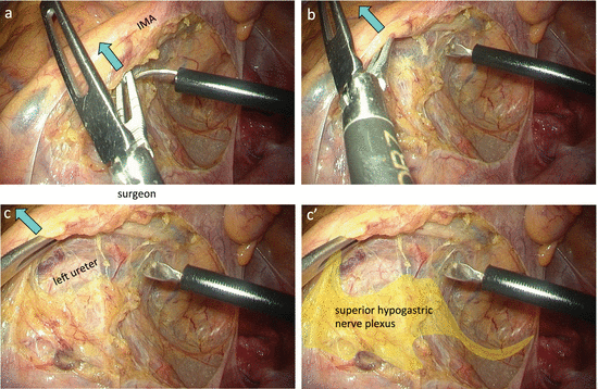
Fig. 5.17
The superior hypogastric nerve plexus drawn toward the IMA. Using the dorsal blade of the opened intestinal forceps, the surgeon elevates the IMA in a ventral–lateral direction (a, b). The nerve branch extending toward the dorsal side of the IMA is resected, and the superior hypogastric nerve plexus is preserved dorsally (c, c′)
After separating the IMA and superior hypogastric nerve plexus, with the Babcock forceps in the left hand, the assistant grasps and draws the IMA in the ventral–lateral direction (Fig. 5.18). This allows better tension to be maintained between the mesocolon and retroperitoneal tissue. By elevating the descending mesocolon ventrally with the opened intestinal forceps in the surgeon’s left hand, the dissectable layer is clearly visualized. Dissection proceeds along the dorsal surface of the descending mesocolon, and the left ureter is identified and preserved. When the left psoas muscle is exposed, the dissection plane strays into the dorsal side; therefore, it should be carefully corrected to return near the descending mesocolon. The left gonadal vessels are identified and preserved prior to resecting the left colic artery (LCA) and IMV.
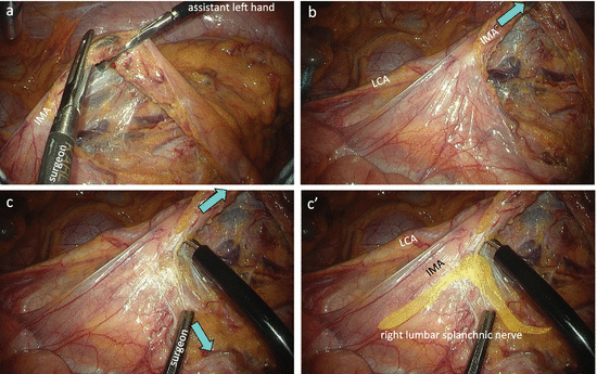
Fig. 5.18
Identification of the right lumbar splanchnic nerve. With the Babcock forceps in the assistant’s left hand, the IMA is drawn in the ventral–lateral direction (a, b). The right lumbar splanchnic nerve is pulled toward the nerve sheath around the IMA (c, c′)
As the mobilization of the mesocolon proceeds, the assistant moves the Babcock forceps in the left hand, grasping the IMA ventrally so as to position the IMA in an obtuse angle to the aorta (Fig. 5.18c). This applies satisfactory tension to the space between the IMA and lumbar splanchnic nerve, allowing the surgeon to confirm that the right lumbar splanchnic nerve is being pulled toward the nerve sheath around the IMA (Fig. 5.18c, c′). The nerve branch extending toward the IMA from the right lumbar splanchnic nerve is resected, and the right lumbar splanchnic nerve is preserved (Figs. 5.18c, c′ and 5.19c, c′). At the site immediately cranial to the root of the IMA, the direction of the peritoneal incision is changed toward the patient’s left side (Fig. 5.19a, b). The dissection between the descending mesocolon and retroperitoneum proceeds cranially, and then the left lumbar splanchnic nerve drawn toward the lateral–dorsal side of the IMA is identified (Fig. 5.19c, c′).
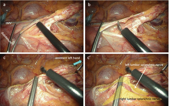
Fig. 5.19
Proximal lymph node dissection. The adipose tissue surrounding the lymph nodes is transected at the level of the root of the IMA (a, b). After dissecting the nerve branch extending toward the IMA from the right lumbar splanchnic nerve, the left lumbar splanchnic nerve drawn toward the lateral–dorsal side of the IMA is identified (c, c′)
5.7.1.4 Proximal Lymph Node Dissection and Transection of the IMA
The assistant moves the tip of the Babcock forceps in the left hand, grasping the IMA dorsally and caudally, and lays the IMA down (Fig. 5.19c). This provides a good view of the ventral side of the IMA. When possible, the assistant grasps the descending mesocolon near the LCA with the forceps in the right hand and draws it in the ventral–lateral direction. This stretches the descending mesocolon, and the site where the LCA branches off the IMA is thus identified.
Related posts:
 Evidence of Laparoscopic Surgery for Colorectal Cancer
 Laparoscopic Transverse Colectomy
 Laparoscopic Total Mesorectal Excision (TME) for Rectal Cancer
 Robotic Total Mesorectal Excision
 Laparoscopic Right Lateral Pelvic Lymph Node Dissection (LPLND) with Pelvic Autonomic Nerve Preservation
 Transanal Minimally Invasive Surgery for Rectal Cancer
Stay updated, free articles. Join our Telegram channel

Full access? Get Clinical Tree


