and Hubert Lepidi1
(1)
UER Médecine, Aix-Marseille Université, Marseille, France
Abstract
This vascularisation plays an extremely important double role. It ensures not only the normal nutritional physiological circulation of this organ but also the functional circulation of the erectile bodies, which allows the intumescence and clitoral erection phenomena during sexual arousal in women.
10.1 General
This vascularisation plays an extremely important double role. It ensures not only the normal nutritional physiological circulation of this organ but also the functional circulation of the erectile bodies, which allows the intumescence and clitoral erection phenomena during sexual arousal in women.
10.1.1 The Arteries
The arteries mostly originate from the branches of the internal pudendal artery (internal shameful artery according to previous authors), which is, itself, one of the branches from the division of the hypogastric artery. This pudendal artery follows the same pathway as the pudendal nerve. With the pudendal veins, it is routed with the nerve, on the lateral wall of the true pelvis, against the inferior insertions of the obturator internus muscle, inside a fibrous canal: the pudendal canal of Alcock. It is then divided into the perineal artery and the dorsal artery of the clitoris. The bulbar artery originates from the deep branch of the perineal artery, at the level of the posterior edge of the anterior perineum. The arteries of the bulbo-clitoral organ are thus the bulbar artery and the dorsal artery of the clitoris.
10.1.1.1 The Bulbar Artery
It detaches from the deep perineal artery (of which it often forms the terminal branch) at the level of the ischio-bulbar triangle (Figs. 12.1 and 13.2). It is routed obliquely towards the posterolateral part of the bulb and thus irrigates the greater vestibular gland. It is then divided into several rami, which approach the convex part of the bulb, via its lateral surface. The deep perineal artery also emits a bulbo-urethral collateral vessel, whose bulbar ramus also contributes to the vascularisation of the bulb.
10.1.1.2 The Dorsal Artery of the Clitoris
This artery is the terminal branch of the internal pudendal artery. It has the same ischio-pubic trajectory as the homologous nerve (see Chap. 8). Once it has left the canal of Alcock, it appears at the level of the lateral part of the infra-pubic canal, under the arched ligament of Lauth, wrapped in a sort of fibrous cone, under a similar cone, from which emerges the homologous nerve. These fibrous structures, whose protective role seems obvious, are formed by splitting of the transverse ligament of the pelvis (ligament of Krause), which itself is formed by coalescence, in front of the urogenital diaphragm, of the superior and inferior fascias covering this diaphragm. It is necessary to recall, such as we have already observed, that the arterial cone is always located under the nerve cone and that the 2 cones are adjacent and separated by a common fibrous lamina, a genuine fibrous “spacer”, according to the expression employed by G. Paturet (Fig. 8.2). The artery then approaches the dorsal surface of the clitoris; extends along the homologous pillar, thus providing this pillar with the deep artery; then passes rapidly under the retro-crural fascia; and rises obliquely up to the angle of the clitoris. The artery then penetrates the base of the suspensory ligament (Fig. 12.3) and progressively passes on the dorsolateral part of the descending portion of the clitoral body. It is then routed towards the glans, inside the nerve, on either side of the superficial dorsal vein. Over this trajectory, it remains underneath the clitoral fascia, which separates it from the cell tissue of the prepuce. On the lateral walls of the descending portion, it provides branches perpendicular to its axis: circumflex branches. It then supplies the glans and its hood. Anastomoses exist between the branches of this artery and the distal rami of the superficial perineal artery (which vascularises the prepuce, the labia majora and labia minora). The vascularisation of the latter is particularly rich and thus explains the involvement of these formations in the turgescence phenomena and their extremely characteristic histological aspect.
10.1.1.3 The Cavernous Artery
English authors refer to this artery as the deep artery of the clitoris. It generally originates from the dorsal artery of the clitoris, shortly after the point where this vessel reaches the clitoral pillar. It can also originate from the pudendal artery itself, along the ischio-pubic trajectory of this vessel, which then seems to have 2 terminal branches, the dorsal artery of the clitoris and the cavernous artery. In the first case, it is very short. In the 2nd case, it is longer and can measure more than 2 cm. When it is a collateral vessel of the dorsal artery of the clitoris, it detaches from this vessel almost nearly at a right angle and penetrates the clitoral pillar via its lateral surface (Fig. 8.5). It rapidly divides inside the pillar into 2 rami, a proximal ramus, referred to as recurrent, which irrigates the pillar and the pre-angular ascending portion of the body, and a distal ramus, which follows the direction of the clitoral body, in the centre part of the corpus cavernosum. It can remain single or divide into 2 or 3 rami. In the corporeal corpora cavernosa, these cavernous arteries approach the median septum and remain in contact with it via a connection consisting of a fibrous crown, appendage of this septum.1 The branches originating from the cavernous artery, and which have penetrated the right pillar, remain on the right of the septum. The branches originating from the artery, and which have penetrated the left pillar, remain on the left. In his dissections, Kobelt observed, with great frequency, a retro-cavernous anastomotic arch joining together the 2 right and left cavernous arteries. From this arch originate “2 small rami, which deeply penetrate the corresponding corpus cavernosum and which would be the true deep arteries of the clitoris”. In any case, the cavernous arteries are of great importance for the operation of the corpora cavernosa, which is demonstrated not only by their size but also and especially by the very rich vegetative innervation of their wall (Fig. 10.1).
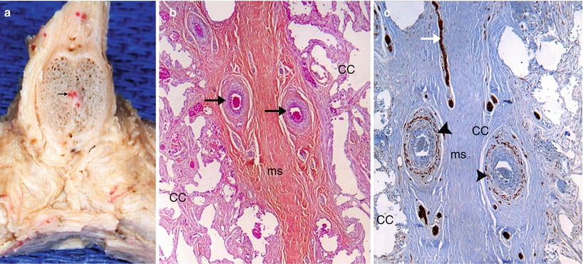
Fig. 10.1


The deep arteries of clitoris. (a) Situation of the deep arteries (black arrow) on a frontal section of the clitoral body, just after the joining of crura. (b) Microscopic aspect of the deep arteries, included in the fibrous tissue of the median septum (classical staining by HEAS). (c) Another aspect of the deep arteries: staining of the section by PS100. CC corpora cavernosa, ms median septum, right black arrows they show the deep arteries of clitoris, black arrowheads they show the vegetative nerve in the arteries’ wall, white right arrow it shows a nervous ramus oriented in the axis of the median septum and innervating this structure. Note: on (a) the position latero-septal of the deep arteries, their conjunctive wrapping connected with the median septum; on (b) the intra-septal position of the arteries; on (c) the rich vegetative innervation of the arteries
Related posts:


Stay updated, free articles. Join our Telegram channel
Full access? Get Clinical Tree
