Fig. 21.1
The serosa is incised, and the stump is exposed
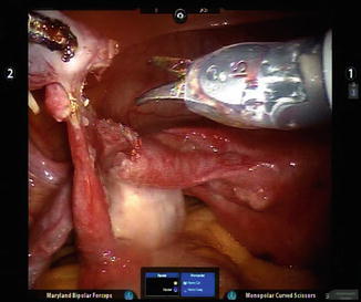
Fig. 21.2
Scissors are used without energy to reveal the tubal lumen
21.5.2 Reapproximation of Mesosalpinx
Often, the mesosalpinx separates widely after preparation of the tubal ends. To align the tubes and relieve tension on the anastomosis site, one or more 6-0 Vicryl (Ethicon, EndoSurgery Inc., Somerville, NJ) stitches are placed into the mesosalpinx to bring its edges closer together. Also at this time, a catheter is placed into the proximal and distal tubal ends to facilitate suturing of the lumina.
For a tubal stent we use the inner plastic cannula from the Novy Cornual Cannulation set (Cook Medical Inc., Bloomington, IN), cut to a 6- to 9- cm length. Other luminal stents and adaptations have been described, including simply using a piece of 0-Vicryl suture for this purpose.
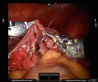
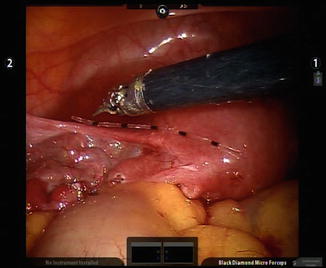

Fig. 21.3
Reapproximated mesosalpinx

Fig. 21.4
Plastic cannula as tubal stent
21.5.3 Tubal Reanastomosis
The tubal reanastomosis is performed using interrupted sutures of 8-0 Vicryl through the tubal muscularis and mucosal layers. In order, we place sutures at the 6, 3, 9, and 12 o’clock locations and tie them with intracorporeal knot-tying techniques. The suturing should position the knot outside of the tubal lumen. Precise placement of these sutures is another distinct advantage when using the surgical robot. We do not tie down the 3 and 9 o’clock knots until the 12 o’clock stitch is placed; otherwise, it may be difficult to identify the lumina and place the 12 o’clock stitch correctly. Great care must be taken to handle the tissue delicately while suturing and not to avulse the needle.
As with all robotic surgery, visual rather than tactile feedback can be used to provide the appropriate tissue tension. Transcervical injection of indigo carmine dye is again used to confirm tubal patency at the conclusion of this portion of the procedure. Revisions can occur if patency has not been established. Usually an additional suture placed at an area of dye leakage at the anastomosis site is all that is needed.
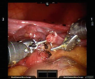
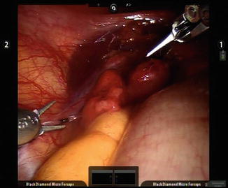

Fig. 21.5
Chromopertubation

Fig. 21.6
Serosal stitches
21.5.4 Serosal Repair
In a similar fashion to the above, the serosa is repaired with circumferential interrupted stitches of 8-0 Vicryl. Chromopertubation is again performed to ensure that the reanastomosis site has not been kinked or occluded by the placement of serosal sutures.
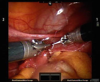

Fig. 21.7
Repair of serosa with circumferential stitches
21.6 Postoperative Care
In most cases, patients undergoing robotic tubal reanastomosis can be discharged home the same day with oral pain medications. A follow-up visit should be scheduled within 6 weeks. Patients can initiate attempts to conceive after two menstrual cycles. We recommend a hysterosalpingogram if the patient has not conceived within six cycles.
Stay updated, free articles. Join our Telegram channel

Full access? Get Clinical Tree


