Fig. 23.1
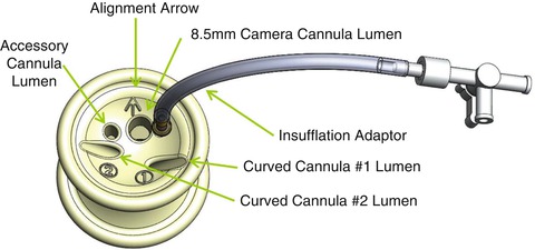
From left to right: two long curved laparoscope cannulae, 300 mm; long flexible obturator; two short curved cannulae, 250 mm; short flexible obturator; assistant cannula, 5 mm; assistant cannula, 10 mm

Fig. 23.2
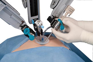
Single-site port

Fig. 23.3
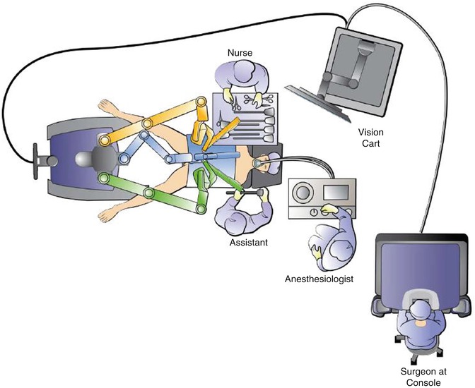
Extracorporeal environment with robotic side cart docked
23.3 Technique
23.3.1 Extracorporeal Environment
A 2.5-cm skin incision at the umbilicus is required using the Hasson technique for intraperitoneal access. Port placement is similar to the SILS port (Covidien; Mansfield, MA) with the exception that the material is much more delicate and will fracture easily. Therefore, the port should be “fed” incrementally into the incision using a curved clamp with care to slide rather than drag the port into place.
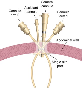
Insufflation can begin after port placement and gas egress is minimal even in the absence of trocars. The camera trocar is placed, and the robot center docked. Under direct visualization the short curved cannulae are placed, arm two before arm one, with the trocar concavity facing the midline. Within the port, the cannula channels cross (Fig. 23.4). Therefore, when placing a cannula, the tip will come into view on the contralateral side. While handedness is maintained at the console (i.e., the surgeon’s right hand controls the instrument tip field right), notice that arm two (patient left) holds the instrument in field right and arm one (patient right) holds the instrument in field left. Side docking is feasible; however, doing so limits the range of motion on the contralateral side. For most purposes, center docking is preferred (Fig. 23.5).


Fig. 23.4
Curved cannulas establish triangulation intracorporeally while achieving wide instrument separation extracorporeally


