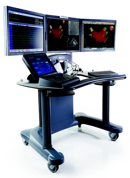
Fig. 38.1
Schematic representation of flexible robotic catheter control system (courtesy of Hansen Medical, Mountain View, CA. Used with permission)

Fig. 38.2
Setup of surgeon console
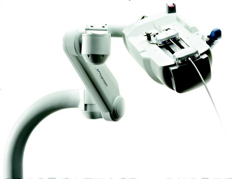
The functional remote element consists of the RCM, an arm that attaches to the operating table on which the steerable catheter sheath and guide catheter are attached. The steerable catheter system (Fig. 38.3) contains an outer catheter sheath (14 F/12 F), which is used to stabilize the catheter at the level of the ureteropelvic junction and an inner catheter guide (12 F/10 F), which is controlled remotely by the MID. A custom-designed flexible ureteroscope is fixed to and guided by the inner catheter. The space between the inner guide catheter and the flexible ureteroscope allows for inflow and efflux of irrigation fluid. The working channel of the ureteroscope provides additional drainage. Maximum deflection of the flexible catheter system is 270° and is not decreased by placement of accessory instruments through the working channel [2, 7].


Fig. 38.3
RCM with steerable guide catheter and sheath (courtesy of Hansen Medical, Mountain View, CA. Used with permission)
Preliminary Experience
The preliminary animal studies using the flexible robotic system were performed by Desai et al. in the swine model [2]. Flexible robotic ureteroscopy was performed in five swine kidneys bilaterally. In two animals (four kidneys) 4 mm human stones were placed in the collecting system and fragmented using a Ho:YAG laser. In one animal (two kidneys) one papilla from each caliceal group was laser fulgurated. Diagnostic ureteropyeloscopy was performed in the other two pigs (four kidneys).
Balloon dilation of the ureter was required in two of the ten kidneys (20%) to accommodate the robotic catheter system. 83 of 85 (98%) calices could be adequately inspected in the ten kidneys. Two lower pole minor calices could not be accessed due to small ostial size. The mean time to inspect the entire collecting system was 4.6 min (range 49 s to 15 min), but decreased considerably with experience.
A Visual analog scale (VAS; 1 = worst, 10 = best) was completed by the surgeon at the end of each procedure to evaluate the performance of the system. The flexible catheter was rated 10 out of 10 for ureteroscope tip stability, 10 out of 10 for reproducibility of access in each calyx, and 8 out of 10 for reproducibility of the auto-retract mechanism to retract the ureteroscope tip to the original position.
Intraoperatively, one complication of ureteral perforation was observed in one kidney unit (10%) at the ureteropelvic junction due to the surgeon retracting the robotic catheter in the flexed position. On histologic examination of the injured ureter, changes consistent with acute dilation were found, but no necrosis was observed.
On necropsy, significant irrigation extravasation was observed in the retroperitoneum and peritoneal cavity of all five animal subjects. Subsequently, the diameter of the prototype ureteroscope was reduced in size from 8.5 to 7.5 F to increase the space between the ureteroscope and the catheter guide and improve drainage of irrigant fluid. The changes improved drainage sufficiently to eliminate irrigant fluid extravasation on a mechanical and an ex vivo animal study.
Early Clinical Data
After successful application of the flexible robotic system to treat nephrolithiasis in the swine model, improvements were made based on findings from the preliminary animal studies. Applying lessons learned from the animal studies, the authors performed the first clinical study in urology using a flexible robotic system for ureteroscopy [7]. The prospective trial treated 18 patients with renal calculi using a modified Sensei robotic catheter system. All patients had a preexisting ureteral stent in place for approximately 2 weeks prior to the procedure. The robotic catheter system was manually inserted and advanced under fluoroscopic guidance to position the tip of the outer sheath, marked by a radiopaque marker, at the ureteropelvic junction. Stones were fragmented using a Ho:YAG laser with a 365 μm (micron) laser fiber. A ureteral stent was left in place for 2 weeks postoperatively.
Stay updated, free articles. Join our Telegram channel

Full access? Get Clinical Tree


