Fig. 8.1
The general scheme of surgical procedures for laparoscopic restorative proctocolectomy with mucosal resection. Maneuvers of mobilization and division are performed in order of indicated numbers
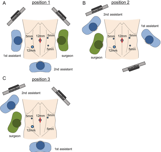
Fig. 8.2
The scheme of surgeon’s position, port placement, and monitor location. Each figure shows the setting for mobilization of (a) the right-side colon and the hepatic flexure, (b) the left-side colon and rectum, and (c) the splenic flexure
8.2.2 Mobilization of the Right-Side Colon and the Small Bowel
At first, the surgeon stands on the right side of the patient (Fig. 8.2a). In the Trendelenburg position, the small bowel is completely moved to the upper right quadrant of the abdomen. The small bowel mesentery is elevated and spread by the first assistant (Fig. 8.3a). This maneuver is a critical step in the “inferior approach,” because it enables the operator to thoroughly recognize the dissection line, that is, boundary between the root of the small bowel mesentery and the retroperitoneum. Subsequently, the small bowel mesentery is dissected all the way from the ligament of Treitz to the cecum. The ascending mesocolon is mobilized through the same dissection layer as far as possible toward the hepatic flexure (Fig. 8.3b, c). This dissection layer runs beneath the superior mesenteric artery and vein (SMA&V) and over the duodenum and the retroperitoneum, preserving the right ureter and the gonadal vessels (Fig. 8.3c, d). These maneuvers elongate the mesentery of the ileal pouch and reduce the tension on the IPAA. By retracting the cecum to the left, “the white line” at lateral side of the ascending colon is divided (Fig. 8.3e). During the mobilization of the right-side colon, it is essential not to injure the ileocecal vessels, because these will be the feeder of the ileal pouch.
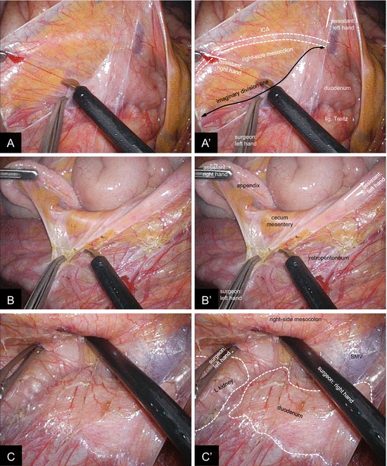
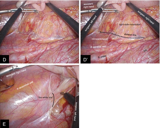


Fig. 8.3
Mobilization of the right-side colon by “inferior approach.” (a,a′) The small bowel mesentery is elevated and completely spread by the first assistant. The black arrow indicates the boundary between the small bowel mesentery and the retroperitoneum (i.e., imaginary division line). (b,b′) The cecum is mobilized from the retroperitoneum. (c,c′) The right-side mesocolon is further mobilized toward the hepatic flexure. The duodenum is swept dorsally. (d,d′) The right ureter and the gonadal vessels are carefully preserved. (e) Lateral attachment of the ascending colon is divided at “the white line.” ICA ileocolic artery. SMV superior mesenteric vein, IVC inferior vena cava
8.2.3 Mobilization of the Sigmoid Colon and the Rectum
Surgeons move as indicated in Fig. 8.2b. In the same patient’s position as previous step, the rectosigmoid mesocolon is dissected from its right side (Fig. 8.4a), and the hypogastric nerves are carefully preserved (Fig. 8.4b, c). The inferior mesenteric artery (IMA) is divided at distal side of the left colic artery (LCA) branching (Fig. 8.4d), and the inferior mesenteric vein (IMV) is divided at the same level. The descending mesocolon is mobilized first from medial side and then from lateral. The left ureter and the gonadal vessels are swept dorsally and preserved (Fig. 8.4e, f).
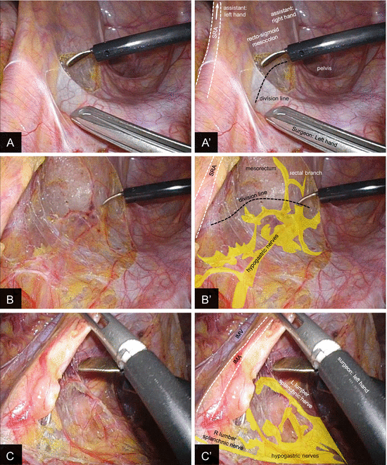
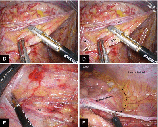


Fig. 8.4
Mobilization of the left-side colon. (a,a′) The rectosigmoid mesocolon is spread by the first assistant and is dissected from its right side. The loose connective tissue is divided. (b,b′) The hypogastric nerves are carefully preserved except the rectal branches. (c,c′) Bilateral splanchnic nerves are identified at the origin of IMA and preserved. (d,d′) The SRA is divided at the distal side of the branching of the LCA using vessel-sealing system. (e) The descending mesocolon is mobilized from the medial side. The left ureter and the gonadal vessels are preserved dorsally. (f) Lateral attachment of the descending colon is divided. SRA superior rectal artery, IMA inferior mesenteric artery, IMV inferior mesenteric vein, LCA left colic artery, S1 1st sigmoid colon branch of the superior rectal artery
The posterior side of the rectum is dissected on the mesorectal plane, identifying and preserving the hypogastric nerves and pelvic plexus (Fig. 8.5a, b) [14, 15]. In UC patients, we often experience difficulty in identifying the appropriate layer of dissection due to chronic inflammation. It is acceptable that dissection line penetrates into mesorectum except the case that rectal cancer is diagnosed. However, such dissection plane is time consuming by substantial bleeding. Thus, the layer of total mesorectal excision (TME) is more reasonable in this maneuver [16].
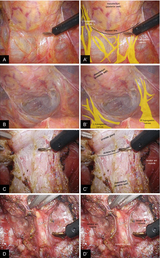

Fig. 8.5
Mobilization of the rectum. (a,a′) At the posterior side of the rectum, the loose connective tissue is divided close to the mesorectum preserving the hypogastric nerves. (b,b′) The posterior side of the mesorectum is dissected down to the pelvic floor. (c,c′) At the anterior aspect of the mesorectum, the area of glistening connective tissue is identified. In restorative proctocolectomy for ulcerative colitis patient, the connective tissue is divided on the line which preserves as much thickness as possible on the anterior pelvic wall. (d,d′) The hiatal ligament at the pelvic floor is divided under laparoscopic view
At the anterior aspect of the mesorectum, we encounter the area of glistening connective tissue by providing proper traction. We consider that it is recognized as “the Denonvilliers’ fascia” in cadavers by collapsing and condensing with formalin fixation [17]. The appropriate dissection line generally divides the connective tissue, and parts of the connective tissues remain at both sides to form the so-called fascia. In RPC for UC, the connective tissue is divided on the line which preserves as much thickness as possible on the anterior pelvic wall. By keeping this line, the seminal vesicles and the prostate gland are left covered by the connective tissue (Fig. 8.5c). This situation differs from that used for cancer resection, which proceeds more anterior in the connective tissue.
Subsequently, the rectum is circumferentially dissected down to the pelvic floor, and the hiatal ligament is divided under laparoscopic view (Fig. 8.5d).
8.2.4 Mobilization of the Splenic Flexure
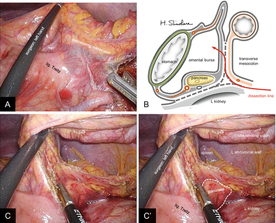
Surgeons stand at the same position as previous maneuver, using the monitor located on the left side of the patient’s head (position 3, Fig. 8.2c). The first assistant may stand between the patient’s legs depending on the situation. In the same table position, the descending mesocolon is further divided toward the upper left, by continuing the layer of the sigmoid mesocolon mobilization (Fig. 8.6a). The scheme of the subsequent dissection line is shown in Fig. 8.6b. The lower edge of the pancreas is identified, and the omental bursa is opened from this view by dividing the anterior layer of the transverse mesocolon (Fig. 8.6c). The transverse mesocolon is further divided toward the left carefully preserving the pancreas dorsally, and the remaining attachment of the descending colon (“the white line” at lateral side) is divided. In the case this approach is difficult to achieve, another option is to divide the omentum to enter the omental bursa from the ventral side and to mobilize the splenic flexure from the superior side after identifying the pancreas.
