Renal Cell Carcinoma: Common Types


Renal Cell Carcinoma: Common Types


Satish K. Tickoo


Besides the traditional radical nephrectomy, multiple management options for renal tumors have emerged in the recent years. With the options of cryo- or radio-frequency ablation, active surveillance, and therapies based on specific tumor type in metastatic settings, among others, needle core biopsies for renal tumors are becoming increasingly popular (1,2,3,4,5). Although the diagnostic accuracy of renal core biopsy was traditionally regarded as unsatisfactory, it has shown marked improvement in the more recent years, with the reported accuracy for tumor subtype in the range of 80% to greater than 95% (6,7,8,9,10,11,12,13,14,15,16,17,18,19,20). This likely is a result of the pathologists’ increased familiarity with renal needle core biopsies as well as the availability of more reliable ancillary methods, particularly immunohistochemistry, for the differentiation of renal cell tumors (20).


CLEAR CELL RENAL CELL CARCINOMA

Clear cell renal cell carcinoma, the most common subtype of renal cell carcinoma (RCC), constitutes approximately 55% to 70% of all diagnosed renal cell tumors according to the more recent published literature (21,22,23,24,25). This proportion in the contemporary literature has shown a downward trend from the much higher percentage described in the past. One of the likely reasons for the decrease in relative incidence has been the recent evolution in the classification of renal tumors with clear cell cytology. A number of tumors diagnosed as clear cell RCC in the past are now recognized as entities quite distinct from it (Table 3.1). Most studies published before the 1990s are particularly blemished, as a number of tumors then were classified as “granular cell” and “sarcomatoid” carcinomas, entities that are now regarded nonexistent. These entities are now known to represent morphologic variants within several tumor types. Thus, granular cell tumors of that era include clear cell RCC, chromophobe RCC, or even renal oncocytoma, among others. Similarly, sarcomatoid differentiation can be seen in clear cell, chromophobe, papillary, unclassified RCC, and collecting duct carcinoma. Additionally, a number of RCC subtypes (Table 3.2) have been extracted from
the category of clear cell RCC in more recent years and are reclassified now as clear cell papillary RCC (26,27,28,29,30), translocation-associated RCC (31,32,33,34,35,36,37,38), multilocular cystic RCC/neoplasm of low malignant potential (25,39,40,41,42,43), and unclassified RCC, among others. Recognition of these specific clinicopathologic entities with differing biologic behaviors makes it imperative that on interpretation of needle core biopsies, such tumors are not misclassified as clear cell RCC (Table 3.2).


TABLE 3.1 Diagnostic Features of the Three Most Common Subtypes of Renal Cell Carcinoma on Needle Core Biopsies


Clear cell renal cell carcinoma


  • Intricate, branching vascular septae enclosing cell nests, acini, and solid alveoli


  • Clear cytoplasm, often optically transparent; eosinophilic cytoplasm in varying proportions—focal to exclusive


  • Immunohistochemical positivity for pan-keratins, epithelial membrane antigen (EMA), and CD10 and carbonic anhydrase IX (CA-IX)—diffuse, box-like membranous pattern


Papillary renal cell carcinoma


  • Papillary, tubulopapillary, or glomeruloid or compressed—papillations architecture


  • Not extensively infiltrative growth pattern or prominent desmoplasia


  • Amphophilic and/or clear cell cytology with granular, flocculent cytoplasm (type 1), eosinophilic cytoplasm with nuclear pseudostratification (type 2), or eosinophilic cytoplasm with low-grade, nonstratified nuclei arranged away from basement membrane (oncocytic)


  • CK7 and AMACR positivity in type 1 (including oncocytic type)


Chromophobe renal cell carcinoma


  • Classical chromophobe RCC


    • Incomplete vascular septations


    • Clear cells with fibrillary cytoplasm; resinoid nuclear features with perinuclear halos (may be less prominent in biopsies compared to what is usual in resection specimens)


    • CA-IX, negative; CK7, positive; CD117, positive


  • Eosinophilic chromophobe RCC


    • Incomplete vascular septations with sheet-like architecture, or nests, tubules


    • Mostly round nuclei with at least some nuclei showing nuclear membrane irregularity with perinuclear halos (may be less prominent in biopsies compared to what is usual in resection specimens)


    • CA-IX, negative; CK7 mostly focal, rarely diffuse; CD117, positive


CK7, cytokeratin 7; AMACR, racemase; RCC, renal cell carcinoma.


TABLE 3.2 Common Differential Diagnostic Considerations for the Three Most Common Subtypes of Renal Cell Carcinoma on Needle Core Biopsies


Clear cell RCC


  • Classical chromophobe RCC


  • Papillary RCC with clear cytoplasm


  • Clear cell papillary RCC


  • Epithelioid angiomyolipoma with prominent clear cells


  • Translocation-associated RCC


  • Adrenocortical tumors


Papillary RCC


  • Collecting duct carcinoma


  • Clear cell RCC with pseudopapillations or true papillations


  • Clear cell papillary RCC


  • Translocation-associated RCC


  • Acquired cystic disease-associated RCC


  • Metanephric adenoma


  • Mucinous tubular and spindle cell carcinoma


Chromophobe RCC


  • Classic variant


  • Clear cell RCC


  • Eosinophilic variant


  • Renal oncocytoma


  • Low-grade oncocytic RCC, unclassified/low-grade oncocytic neoplasms with overlapping features


RCC, renal cell carcinoma.


Microscopic Features

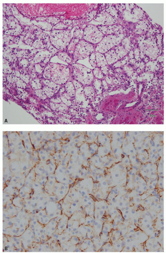
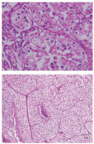
The most characteristic feature of clear cell RCC in both resection and biopsy specimens is the presence of delicate, intricately branching, richly vascular fibrous septae (Fig. 3.1A-D) that surround solid acini, alveoli, tubules, cysts, or papillary/pseudopapillary structures. Many tumors show a combination of these growth patterns in variable proportions (Fig. 3.2A-F, eFigs. 3.1 and 3.2). The rich capillary vascular network is retained in almost all but some high-grade, sheet-like, and sarcomatoid areas. Thus, this diagnostic hallmark of the characteristic vascularity should be the initial focus of attention in the diagnosis of clear cell RCC, and more so in needle core biopsies. In general, architectural pattern is loosely associated with grade (discussed later) because most low-grade lesions have

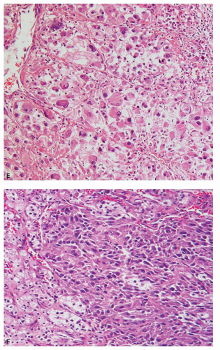
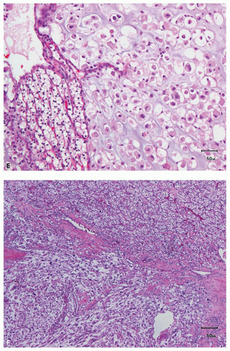
an acinar growth, whereas higher grade areas are more likely to exhibit solid, pseudopapillary, or sarcomatoid features (Fig. 3.2E,F). Tumors exhibiting a pseudopapillary growth pattern, which must not be confused with the true papillary growth pattern typically seen in papillary RCCs, are characterized by pseudopapillae lacking true fibrovascular cores. In our experience, pseudopapillations are particularly more common in core biopsies, especially at their edges, as these are the areas most prone to


lose the direct support and compression provided by the walls of biopsy needle on delivery from the needle. Such pseudopapillations occur more often in high-grade tumors, as they are less likely to have closely arranged fibrovascular septations and, therefore, abundant internal architectural support (Fig. 3.3). Occasionally, the biopsy may yield what at initial evaluation appears as fibrous tissue only. This often represents the biopsy needle
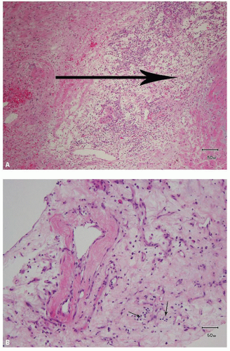
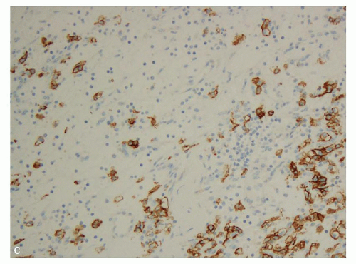
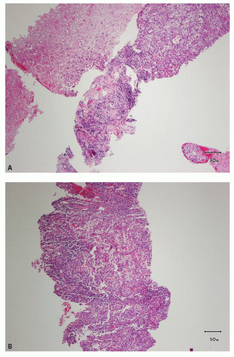
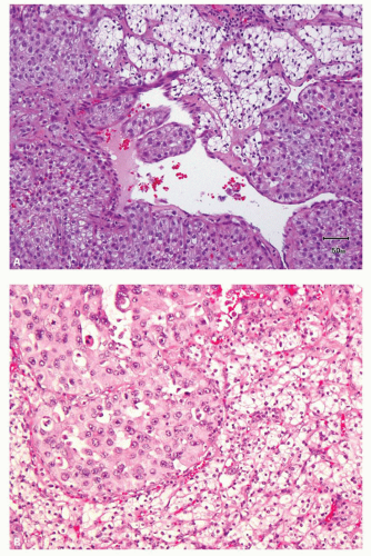
hitting a scar within the tumor. However, single or small clusters of cells— resembling small lymphocytes with scant clear cytoplasm—may often be noted dispersed within the fibrosis (Fig. 3.4A,B). These very frequently represent rare tumor cells that are still viable within scar tissue and must be investigated further, particularly with the help of immunohistochemical stains. The results on immunohistochemical staining in such material are frequently surprising as well as gratifying (Fig. 3.4C). Many of the tubules and acini show intraluminal hemorrhage—a characteristic but not specific finding in clear cell RCC (Fig. 3.5). This feature is common even at metastatic sites and should lead to the workup for a possible renal primary. Areas of necrosis, sometimes extensive (Fig. 3.6A), and hemorrhage are some other not uncommon histologic findings seen in core biopsies. Some tumors contain an inflammatory infiltrate, often predominantly lymphocytic in nature (Fig. 3.6B). Calcifications and osseous metaplasia, as well as intracytoplasmic hyaline globules or Paneth-like coarse granules (eFigs. 3.3 and 3.4) may also be seen in occasional biopsies.
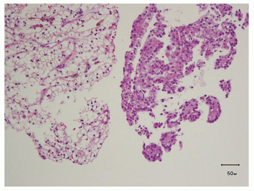


FIGURE 3.1 A: The most characteristic and diagnostically essential feature of clear cell RCC: intricate, arborizing vascular septations enclosing nests of cells. B: Arborizing vasculature in clear cell RCC, highlighted by CD31 immunohistochemical stain. C: Retained characteristic vasculature pattern in a high-grade clear cell RCC. D: The typical intricate vasculature maintained in a tumor with large solid alveolar growth pattern.


FIGURE 3.1 A: (Continued)


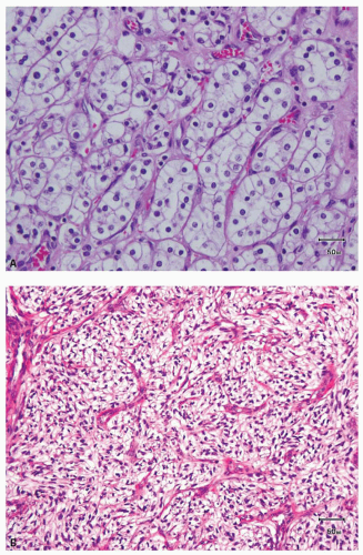
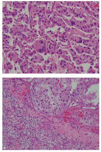
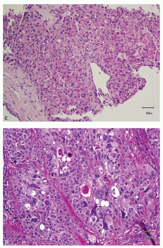
FIGURE 3.2 A: Optically transparent cytoplasm in a low-grade clear cell RCC. B: Optically transparent cytoplasm in a higher grade clear cell RCC, with partially solid architecture. C: Eosinophilic cells in clear cell RCC with high nuclear grade. D: Higher nuclear grade in eosinophilic cells compared to that in adjacent clear cells in a clear cell RCC—a typical occurrence. E: Higher nuclear grade in chondroid-appearing area compared to that in adjacent clear cells in a clear cell RCC. F: Higher nuclear grade in a sarcomatoid area with lower grade nuclei in better differentiated clear cell area.


FIGURE 3.2 A: (Continued)


FIGURE 3.2 A: (Continued)


FIGURE 3.3 Pseudopapillations in an area of higher nuclear grade in a needle core biopsy from a clear cell RCC.

Besides the pure clear cell cytology in many tumors, the majority of clear cell RCCs show a mixture of cytoplasmic features in various proportions, that is, combination of cells with clear and granular eosinophilic cytoplasm (Fig. 3.7A). The cytoplasmic features often correlate with tumor grade; low-grade (grade 1 or 2) lesions almost always contain clear cytoplasm, whereas the cytoplasm of higher grade lesions is more likely to be variably eosinophilic and granular (Fig. 3.7B). Some grades 3 and


4 lesions may altogether lack clear cytoplasm (Fig. 3.7C). Grade 4 lesions contain bizarre, pleomorphic and/or multilobulated, and multiple nuclei (Fig. 3.7D); cells with rhabdoid features (Fig. 3.7E); or spindle cells (sarcomatoid areas) (Fig. 3.7F).
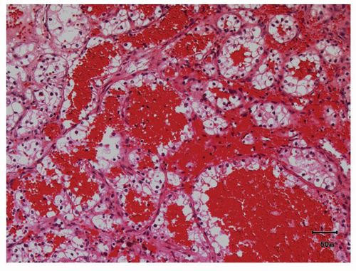


FIGURE 3.4 A: A surgical resection specimen of clear RCC showing large scar with the arrow pointing toward a potential path that a biopsy needle may take. B: Needle core biopsy through the center of such a scar showing mostly an inflammatory, vascular area. Arrows pointing out few residual tumor cells that may mimic clustered lymphocytes. C: Immunohistochemical stain for cytokeratin highlighting multiple intermingled tumor cells in the biopsy.


FIGURE 3.4 A: (Continued)


FIGURE 3.5 Pools of blood filling the acinar lumina, a characteristic, though not specific, feature of clear cell RCC.


FIGURE 3.6 A: Almost completely necrotic needle core, adjacent to another viable core of clear cell RCC. B: Extensive intratumoral infiltration by lymphocytes in clear cell RCC on a needle core biopsy.

As a biopsy represents only a minute proportion of the tumor, and because the tumors often show morphologic heterogeneity, nuclear


grade in biopsy often does not correlate with the final grade on resection specimen (7,44,45). Therefore, if the nuclear grade is provided, it should be qualified by a statement similar to “… in this limited material.” As is intuitive however, high nuclear grade in the biopsy tends to correlate with high grade on resection specimen as well (7,45).


FIGURE 3.7 A: Clear cell RCC with a combination of clear cell and granular eosinophilic cell features. B: Granular eosinophilic areas with high-grade nuclei compared to clear cell areas with lower grade nuclei. C: Fuhrman/nucleolar grade 3 nuclei in an exclusively eosinophilic clear cell RCC. D: Grade 4 nuclei in an exclusively eosinophilic tumor. E: Rhabdoid areas with grade 4 nuclei. F: Presence of sarcomatoid areas in a tumor is always considered representing nuclear grade 4.


FIGURE 3.7 A: (Continued)


FIGURE 3.7 A: (Continued)


Ancillary Studies

Among the immunohistochemical stains that are reported to be most useful in the differential diagnosis of clear cell RCC from other renal primary tumors on needle core biopsies, carbonic anhydrase IX (CA-IX) shows diffuse and strong membranous reactivity within more than 90% tumors (eFig. 3.5), including in its sarcomatoid components in a significant proportion of cases (20,46,47,48,49). CD10 is also often positive (membranous), but cytokeratin 7 (CK7), CD117, and racemase (AMACR) are generally negative.

However in our experience, AMACR tends to label some clear cell RCCs more than focally in needle core biopsies. Immunohistochemistry and molecular studies in renal and adrenal tumors are described in greater details in Chapter 13.

Jun 18, 2016 | Posted by in GASTROENTEROLOGY | Comments Off on Renal Cell Carcinoma: Common Types

Full access? Get Clinical Tree
Get Clinical Tree app for offline access