Benign Renal Epithelial Tumors
Ying-Bei Chen
With the increased detection of small renal masses by modern imaging modalities, renal needle biopsy has been increasingly used to distinguish benign renal epithelial neoplasms from malignant tumors and to guide clinical management. Data from surgical series suggest that about 20% of the resected renal tumors are benign, and this proportion is even higher among tumors smaller than 4 cm (1,2,3,4). For these patients, and particularly those with impaired renal function or comorbidities, renal needle biopsy is an essential approach to avoid unnecessary surgeries and improve overall outcomes. This chapter primarily focuses on the diagnosis of benign renal epithelial tumors of renal cell origin.
RENAL ONCOCYTOMA
Renal oncocytoma comprises approximately 5% of renal epithelial neoplasms and is a frequent consideration in the differential diagnosis on renal core biopsies. Most renal oncocytomas occur sporadically and affect adult patients with a wide age range. Male gender predilection (M:F = 2:1) is often observed. Histologically, renal oncocytoma is predominantly composed of oncocytes, cells with densely eosinophilic, granular cytoplasm and small, round nuclei. Compared with other renal tumors with oncocytic cells, the key nuclear features of oncocytoma are the uniformly smooth nuclear contour; evenly dispersed granular chromatin; and frequently, a centrally located small to prominent nucleolus (Figs. 5.1 and 5.2). Architecturally, tumor cells mainly form solid nests, tubules, and micro- and macro-cysts, in a loose, hypocellular, and hyalinized or myxoid stroma (Fig. 5.3). Cytoplasmic clearing should be restricted only to the areas of scarring in oncocytomas. A proportion of tumors contain areas of small cells with scant cytoplasm and hyperchromatic nuclei, so-called oncoblasts (Fig. 5.4). Scattered foci of tumor cells may have large, pleomorphic and hyperchromatic nuclei with dense, smudgy chromatin (Fig. 5.5). This type of atypia is believed to be degenerative in nature and should not be considered as worrisome features in an otherwise typical oncocytoma. By contrast, necrosis and atypical mitoses are
findings against a diagnosis of oncocytoma. The mitotic activity in renal oncocytoma is usually very low, only rare mitotic figures are allowed. Similarly, conspicuous papillary formations are not compatible with renal oncocytomas, but small papillae, pseudopapillae, and intratubular/intracystic epithelial tufts can be occasionally seen in some oncocytomas (5,6). Importantly,
renal oncocytomas occasionally demonstrate perinephric fat involvement (˜20%) and vascular invasion (˜5%), and these atypical features have not been associated with malignant behavior and thus are acceptable for the diagnosis. Other features that can rarely be found in renal oncocytomas include calcifications, osseous or myeloid metaplasia.
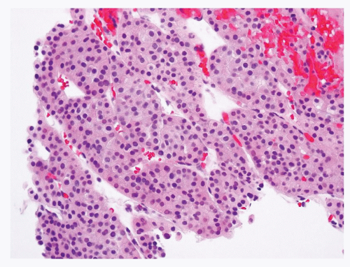 FIGURE 5.1 Renal oncocytoma typically is composed of solid nests of cells with oncocytic cytoplasm and uniform nuclei that lack nuclear membrane irregularity. |
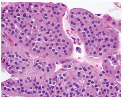 FIGURE 5.2 Centrally located small to conspicuous nucleoli in renal oncocytoma. |
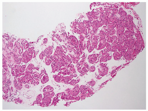 FIGURE 5.3 Tumor nests or tubules are often in a loose and hypocellular stroma that shows hyalinization or myxoid changes. |
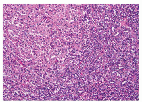 FIGURE 5.4 Renal oncocytomas occasionally contain oncoblasts, small cells with scant cytoplasm and hyperchromatic nuclei. |
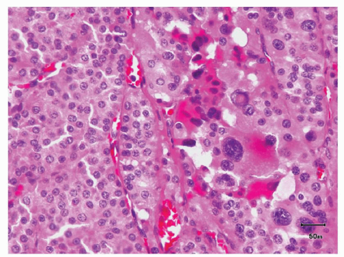 FIGURE 5.5 Scattered cells with large, pleomorphic, and hyperchromatic nuclei but dense, smudgy chromatin in renal oncocytomas. |
Although renal oncocytomas in surgical series are predominantly unilateral, they have been found to be bilateral and multifocal in approximately 5% to 10% of patients (6,7). Renal oncocytosis refers to a rare condition in which patients develop multiple oncocytic tumors in addition to numerous oncocytic nodules (Fig. 5.6) and other oncocytic changes in
the adjacent nonneoplastic renal parenchyma that can range from infiltrative growth of oncocytic renal tubules, oncocytic cortical cysts, to diffuse oncocytic changes in renal tubules (8). In a recently reported exceptional case, renal oncocytosis was diagnosed based on changes found in a renal core biopsy (9


the adjacent nonneoplastic renal parenchyma that can range from infiltrative growth of oncocytic renal tubules, oncocytic cortical cysts, to diffuse oncocytic changes in renal tubules (8). In a recently reported exceptional case, renal oncocytosis was diagnosed based on changes found in a renal core biopsy (9
Related posts:


Stay updated, free articles. Join our Telegram channel
Full access? Get Clinical Tree
