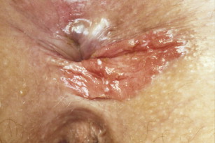
Pruritus ani is a common condition with multiple causes. Primary causes are thought to be fecal soiling or food irritants. Secondary causes include malignancy, infections including sexually transmitted diseases, benign anorectal diseases, systemic diseases, and inflammatory conditions. A broad differential diagnosis must be considered. A reassessment of the diagnosis is required if symptoms or findings are not responsive to therapy. The pathophysiology of itching, an overview of primary and secondary causes, and various treatment options are reviewed.
Key points
- •
Pruritus ani is a dermatologic condition characterized by itching or burning in the perianal area.
- •
Pruritus ani can be either primary (idiopathic) or secondary.
- •
There are a multitude of different causes of pruritus ani, making diagnosis and treatment options numerous.
Introduction
Pruritus ani is defined as a dermatologic condition characterized by itching and/or burning in the perianal region. Pruritus ani affects 1% to 5% of the population, is four times more prevalent in men than women, and is most commonly present in the fourth to sixth decades of life. Pruritus ani is categorized as either primary (idiopathic) or secondary. Although some papers state that 50% to 90% of pruritus ani cases are idiopathic, others claim that roughly 75% of cases have associated pathology. There are nearly 100 different causes for pruritus ani, making differential diagnoses and treatment options vast. This article provides a thorough review of secondary and idiopathic pruritus ani and outlines effective diagnostic and treatment options.
Historical perspective
The earliest known mention of pruritus ani is in the Chester Beatty Medical Papyrus. This ancient Egyptian papyrus was given to the British Museum by American industrialist Chester Beatty. Ten of its 41 remedies were devoted to management of anal itching and irritation. Because of a lack of understanding of the many conditions that may cause anal itching, pruritus ani was known as a “condition that eludes all attempts at cure.” Subsequently, numerous topical and injectable treatments for pruritus ani have been developed or reported. In 1966, Caplan demonstrated the role of soiling and fecal contamination of the skin as a cause of pruritus symptoms. This is generally considered to be the most common cause of symptoms when secondary causes have been ruled out. Anorectal physiology studies also support this theory. They demonstrated that patients with pruritus have a more pronounced accommodation of the internal anal sphincter with rectal distention compared with control subjects and leak sooner on a saline infusion test. Thus, most modern treatment protocols focus on eliminating irritants, skin protection, and proper anal hygiene.
Etiology of itch
Itch is an unpleasant sensation that leads to the desire to scratch and can be categorized into the following groups: cutaneous, neuropathic, neurogenic, and psychogenic. Cutaneous or pruritoceptive itch is caused by inflammation of the skin. Neuropathic itch is caused by damage to the peripheral nervous system and can be present anywhere along the afferent nerve pathway. Neurogenic itch is induced centrally. Lastly, psychogenic itch is caused by delusional states.
The sensation of itch can be brought on by various stimulus modalities including thermal; electrical; mechanical (heat, xerosis, and so forth); and chemical stimuli. Histamine has been studied extensively as a potential neuronal mechanism of itch; however, it is not the only substance that produces itching. Kallikrein, bradykinin, papain, and trypsin are all itch-mediating substances that are not responsive to blockades with classic histamine antagonists, such as diphenhydramine. As a consequence, antihistamines have proved ineffective in treating pruritus in many instances.
Itch is a surface phenomenon initiated by the stimulation of C-fibers in the epidermis and subepidermis. C-fibers are slow-conduction velocity unmylenated fibers with extensive terminal branches and transmit messages that the brain interprets as the sensation of itch. Itch receptors may be located more superficially than pain receptors and consequently, itch is believed to be a subthreshold of pain. The idea that itch receptors are located superficially is supported by the fact that minor mechanical stimuli can create the sensation of itch.
Scratching the affected skin provides inadequate feedback to inhibit itching and prolonged itching can cause damaging excoriations and infections, which provides additional itching stimuli. Thus, scratching results in a vicious cycle of itching and scratching that is difficult to break.
Secondary causes
Pruritus ani has been attributed to idiopathic and secondary causes. Secondary pruritus ani, which is pruritus induced by an underlying cause, can be divided into the following categories: inflammatory, nonsexual infectious, systemic, premalignant and malignant, and anorectal causes ( Table 1 ). Sexually transmitted causes of perianal itch and pathology is discussed separately elsewhere in this issue. In all of these cases pruritus ani is exacerbated by the scratch–itch cycle, which can lead to infections and further increase the itching frequency.
| Inflammatory Diseases | Nonsexual Infectious Diseases | Sexually Transmitted Diseases |
|---|---|---|
| Psoriasis | Pilonidal disease | Gonorrhea |
| Atopic dermatitis | Hidradentis suppurativa | Syphilis |
| Contact dermatitis | Crohn disease | Chancroid |
| Seborrheic dermatitis | Tinea cruris | Granuloma iguinale |
| Scleroderma | Herpes zoster | Molluscum contagiosum |
| Erythema multiforme | Trichomoniasis | Herpes simplex |
| Pemphigus vulgaris | Bilhartziasis | Condyloma acuminata |
| Dermatitis herpetiformis | Oxyurasis (pinworm) | Chlamydia |
| Lichem planus | Larva currens | |
| Lichen sclerosis et atrophicus | Cimicosis (bed bugs) | |
| Radiation dermatitis | Pediculosis (lice) | |
| Darier disease | Scabies |
| Premalignant and Malignant Diseases | Systemic Diseases | Anorectal Diseases |
|---|---|---|
| Acanthosis nigricans | Diabetes mellitus | Hemorrhoids |
| Leukoplakia | Leukemia and lymphoma | Anal creases |
| Mycosis fungoids | Hepatic diseases | Fistula in ano |
| Leukemia cutis | Thyroid disorders | Fissures |
| Squamous cell carcinoma | Renal failure | Rectal prolapse |
| Basal cell carcinoma | Iron deficiency anemia | |
| Bowen disease | Vitamin A and D deficiencies | |
| Melanoma | Aplastic anemia | |
| Dysplastic nevus | ||
| Paget disease |
Inflammatory Diseases
Numerous inflammatory diseases can manifest with perianal symptoms and puritus ani including psoriasis, atopic dermatitis, seborrheic dermatitis, lichen planus, and lichen sclerosis.
Psoriasis
Psoriasis has been shown in numerous studies to be a prevalent underlying cause of pruritus ani. Typical psoriasis presents on the trunk, knees, elbows, and scalp. Psoriasis present in the anus, groin, genitals, and axillae is referred to as “inverse psoriasis” because it presents as the inverse of the normal distribution. Although the exact incidence is unknown, one study found that a significant portion of patients (54%) with inverse psoriasis had involvement of the anus. Typical psoriasis presents as bright red plaquelike lesions, whereas inverse psoriasis is demarcated, paler in color, and has lesions without scales. Psoriasis cannot be cured but can be treated with short-term use of a low-to-mid potency steroid for up to 4 weeks. After the induction of remission the patient should switch to a nonsteroidal topical treatment, such as calcipotriene, for maintenance ( Fig. 1 ).
Atopic dermatitis
Atopic dermatitis is a chronic inflammatory, pruritic disease of the skin that is induced by an allergic response. People with atopic dermatitis are also likely to have asthma, eczema, and hay fever. Lesions are nonspecific diffuse erythema, dry and scaly, and often marked by evidence of excoriations. Biopsies are often inconclusive because of the mixed inflammatory infiltrate with eosinophils. Thus, a careful patient history provides the physician with the best chance of making a correct diagnosis. Atopic dermatitis has been shown to be associated with keratosis pilaris (rough, dry bumps) on the arms and thighs; Morgan folds (creases found beneath the eyes); “sniffers” (a crease found across the nose); urticaria; and white dermatographism. Treatment options include the use of strong moisturizing agents, anti-inflammatory agents, and antihistamines.
Seborrheic dermatitis
Seborrheic dermatitis is not a common cause of pruritus ani and is characterized by extensive, moist erythema in the perineum. Seborrheic dermatitis usually presents in the scalp, chest, ears, beard, and suprapubic area and is easily treated with 2% sulfur with 1% hydrocortisone lotion.
Lichen planus
Lichen planus is believed to be caused by an altered cell-mediated immune response to an unidentified source. Patients with lichen planus may also have another disease of altered immunity including myasthenia gravis, alopecia, vitiligo, ulcerative colitis, and lichen sclerosis. Lichen planus can also present in patients with chronic active hepatitis and primary biliary cirrhosis. Widespread lichen sclerosis should be looked for in these patients. Cutaneous lesions are shiny, flat-topped papules that are more darkly pigmented than the surrounding skin. Lesions typically develop on the flexural surfaces of the limbs with a generalized eruption developing after a week and a maximal spreading occurring between 2 and 16 weeks. Genital involvement is common and can be extremely pruritic. Direct immunofluorescence study reveals globular deposits of IgM. The condition usually resolves itself within 6 (>50%) to 18 months (85%). Treatment options include topical steroids or, for more severe cases, light therapy (narrow-band or broadband UVB therapy).
Lichen sclerosis
Lichen sclerosis, usually appearing as lichen sclerosus et atrophicus in dermatologic literature, is a chronic disease of unknown cause. Lichen sclerosis is five to six times more prevalent in women than men and involves the vulva and extends posteriorly to the perianal region. Typical lesions are porcelain-white papules and plaques. In females one should inspect the interlabial sulci, labia minora, clitoral hood, clitoris, and perineal body and in males the prepuce, coronal sulcus, and glans penis. The affected areas typically break down and reveal the underlying, raw tissue that can be extraordinarily painful and pruritic. As the underlying tissue heals, the area is replaced by chronic inflammation, sclerosis, and atrophy. During a physical examination one typically sees white patches in a pattern surrounding the vulva and anus. Squamous cell carcinoma is the most common malignancy described in association with anogenital lichen sclerosus. Women with lichen sclerosis have a 300-fold increased risk of developing cancer compared with those without the disease. Treatment of the condition does not reduce this risk. Short term (6–8 weeks) treatment with a potent topical steroid, such as clobetasol, is highly effective in reducing symptoms. Retinoids, testosterone creams, and tacrolimus ointment have also been described. Because of the risk of squamous cell carcinoma, nonresponding patients should have a skin biopsy to rule out malignancy.
Nonsexually Transmitted Infectious Diseases
Although bacterial, fungal, and parasitic infections make up the minority of pruritus ani causes, their contribution must not be underestimated.
Candida
Candida is responsible for 10% to 15% of pruritus ani. It is characterized by diffuse, marginated erythematous and many times macerated plaques. These lesions, localized to the inguinal and perianal region, are generally painful and very itchy. Such factors as old age, sweating, obesity, tight-fitting clothing, prolonged use of antibiotics, diabetes, and immunosuppressive therapy can predispose a person to Candida vulvitis or Candida balanitis . One review showed that surgical treatment of anal disorders (hemorrhoids, fissure, spasm, mucosal prolapse) eliminated Candida and dermatophyte infections in 20 out of 23 patients who cultured positive with symptomatic itching before the procedure. Diagnosis can be achieved using a culture or scraping of the lesion. Scrapings can be negative for hyphae if topical steroids were used; however, the use of steroids typically exacerbates dermatophyte growth. Topical and systemic antifungal agents have been used as treatment options.
Numerous nonsexually transmitted bacterial infections have been shown to lead to pruritus ani including Streptococcus , Staphylococcus aureus , and Corynebacterium minutissium . Weismann and colleagues found 19 patients with pruritus over a period of 1 to 20 years who had negative throat and nose cultures but were positive for either β hemolytic streptococci or S aureus in the perianal region. Erythrasma, a skin disease characterized by brown, scaly patches and caused by the bacterium C minutissium , typically presents with itching and can often be found at such sites as the toes and groin. It is best diagnosed with the Wood lamp, which shows a fluorescent coral pink or red color. False-negative results can be produced if the patient has recently showered. Erythromycin, tetracycline, and betamethasone lotion have been effective in the treatment of perianal C minutissium . Streptococcus is treated with amoxicillin, penicillin, and/or topical applications of mupirocin (bactroban). S aureus is treated with penicillin, doxycycline, or clindamycin.
Parasitic infections
Parasitic infections, specifically Enterobius vermicularis (pinworms), can be a common cause of pruritus ani in children. A cellophane tape test, applied in the early hours of the morning, can identify the adult worms and their eggs and confirm the diagnosis. Treatment is with albendazole or mebendazole. Other common parasites that induce pruritus ani include Sarcoptes scabei and Pediculosis pubis .
Herpes zoster
Herpes zoster, commonly referred to as shingles, is an infection of the varicella zoster virus. Herpes zoster can only occur in people who were previously infected with the virus and although the disease can occur at any age, it typically presents in patients older than the age of 50. The rash has a unique appearance and can usually be diagnosed visually. Herpes zoster causes a deep red rash with blisters that do not cross the midline of the body. Treatment options include antiviral medications including valacyclovir hydrochloride.
Systemic Diseases
Table 1 lists several systemic diseases associated with pruritus. These conditions are more often associated with generalized pruritus rather than pruritus ani specifically. In a prospective study of 55 patients presenting with generalized pruritus to a dermatology clinic, 12 were found to have a systemic cause of the pruritus. The underling conditions included iron deficiency anemia, hepatitis, uremia, diabetes mellitus, chronic lymphocytic leukemia, and lung cancer. Iron deficiency anemia was the most common diagnosis. Pruritus was the presenting symptom in 5 of the 12 patients.
Uremic pruritus
Uremic pruritus, or itching associated with end-stage renal disease, is a common complaint among patients on dialysis, affecting up to 90% of patients. Transplantation is the only known cure. Several biochemical mediators have been implicated in the pathogenesis this condition. Serotonin was initially thought to be a common cause of uremic and hepatic pruritus. However, several randomized controlled trials failed to find much clinical benefit with treatment of ondansetron, a selective serotonin antagonist. Gabapentin, omega-3 fatty acids, topical baby oil, sertraline, and pregabalin have all been used with some success to treated uremic pruritus.
Pruritus from cholestasis is a common condition in patients with liver disease. Cholestyramine, naltrexone, rifampin, and sertraline have all been used with variable success in treating this troublesome problem.
Premalignant and Malignant Diseases
There are numerous premalignant and malignant conditions that can lead to pruritus ani including intraepithelial neoplasia (AIN), or Bowen disease, and Paget disease. Half of patients with perianal Paget disease and perianal Bowen disease have associated itch.
Intraepithelial neoplasia
AIN is a condition associated with human papilloma virus and condylomata and can present inside or outside of the anal canal. AIN can range from low- to high-grade dysplasia, with high-grade dysplasia, also known as Bowen disease, being an intermediate stage toward malignant transformations into squamous cell carcinoma of the anus. Patients present with minor complaints and are usually diagnosed during evaluation of condylomata or pruritus. Patients with low-grade dysplasia can be treated topically. Alternatively, high-grade dysplasia is a premalignant condition. It is typically treated by mapping with punch biopsies and excision. Large amounts of uninvolved tissue may be excised to achieve clear margins. The recurrence rate with wide excision is 23.1% and the cancer rate is below 10% ( Fig. 2 ).


