Fig. 1.1
Primitive gastrointestinal tract and the various derivatives in the early weeks of embryo development. The liver is omitted in this and the following figures. CA celiac artery, CBD common bile duct, CHA common hepatic artery, IMA inferior mesenteric artery, IMV inferior mesenteric vein, LGA left gastric artery, LGEA left gastroepiploic artery, PHA proper hepatic artery, PV portal vein, RGA right gastric artery, RGEA right gastroepiploic artery, RGEV right gastroepiploic vein, SMA superior mesenteric artery, SMV superior mesenteric vein, SPA splenic artery, SPV splenic vein
1.3 Positional Changes of the Foregut
The foregut, midgut, and hindgut are supplied by the celiac artery (CA), superior mesenteric artery (SMA), and inferior mesenteric artery (IMA), respectively (Fig. 1.2a). These arteries independently originate from the aorta and run through the dorsal mesenteries. In contrast, the corresponding veins, that is, the splenic vein (SPV) from the foregut, superior mesenteric vein (SMV) from the midgut, and inferior mesenteric vein (IMV) from the hindgut, converge on the portal vein and drain into the liver (Fig. 1.2b). During the fifth week of development, the stomach appears as a fusiform dilatation of the foregut and rotates 90° clockwise around its longitudinal axis. The mesogastrium expands into the left upper abdomen to form the omental bursa and then becomes adherent to the dorsal parietal wall (Fig. 1.3). As this process continues, the spleen primordium appears as a mesodermal proliferation within the mesogastrium. The rotation of the stomach swings the duodenum from its initial midline position to the right side of the abdominal cavity. When the duodenum rotates to the right and becomes C-shaped, the ventral pancreatic bud moves dorsally and comes to lie below and behind the dorsal bud to form the uncinate process of the head of the pancreas. Later, the parenchyma and the duct system of both buds fuse (see Fig. 1.5). The duodenum and the head of the pancreas press against the dorsal body wall, and the posterior surface of the mesoduodenum fuses with the adjacent peritoneum via loose connective tissue. Thus, the pancreas is not a retroperitoneal organ but is a component of the mesoduodenum. This recognition is important for performing right hemicolectomy.

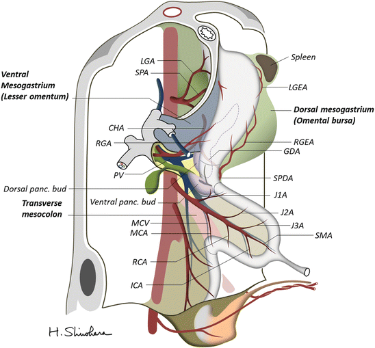
Fig. 1.2
Primary intestinal loop before rotation. The arteries originate from the aorta and run through the dorsal mesenteries (a). In contrast, the corresponding veins converge on the portal vein and drain into the liver (b). ARCV accessory right colic vein, ICV ileocolic vein, MCV middle colic vein. Refer to the above figure legend for other abbreviations
Fig. 1.3
Embryo during the fifth week of development. The stomach appears as a fusiform dilatation of the foregut and rotates 90° clockwise around its longitudinal axis. The ventral pancreatic bud moves dorsally and comes to lie below and behind the dorsal bud to form the uncinate process of the head of the pancreas. GDA gastroduodenal artery, ICA ileocolic artery, J1A first jejunal artery, J2A second jejunal artery, J3A third jejunal artery, RCA right colic artery. Refer to the above figure legend for other abbreviations
1.4 Rotation of the Midgut
Development of the midgut is initiated by elongation of the gut and its mesentery, resulting in formation of the primary midgut loop. It enters the extraembryonic cavity in the umbilical cord, the so-called physiological umbilical herniation, because the abdominal cavity temporarily becomes too small to contain the midgut. Occurring concurrently with elongation, the midgut loop rotates approximately 270° counterclockwise around an axis formed by the SMA. Figure 1.4 shows the intestinal loops after 90° counterclockwise rotation during herniation. The apex of the loop communicates with the yolk sac through the vitelline duct toward which the SMA runs through the proper mesentery of the midgut. After development, the cephalic limb of the midgut loop becomes the distal part of the duodenum, jejunum, and part of the ileum. The caudal limb becomes the terminal portion of the ileum, cecum, appendix, ascending colon, and proximal two-thirds of the transverse colon. Owing to the loop formation, the middle colic artery (MCA) that supplies the transverse colon branches from the SMA close to the origin of the first jejunal artery; the branching point is proximal to that of the ileocolic artery (ICA). During return of the intestinal loop into the abdominal cavity, the remaining 180° rotation occurs. Figure 1.5 shows the intestinal loops after the 180° rotation. The cephalic limb comes to lie on the caudal side, while the caudal limb lies on the cephalic side. The cecal bud appears as a small dilatation of the caudal limb of the midgut loop. The vitelline duct ordinarily ceases to exist during this process, but a small portion persists and forms Meckel’s diverticulum in some individuals. The SMV moves above the SMA. The right gastroepiploic vein (RGEV), MCV, and accessory right colic vein (ARCV) empty into the close part of the SMV. The jejunum and ileum form a number of coiled loops, but the large intestine does not. The dorsal pancreatic duct (duct of Santorini) enters the proximal part of the major papilla at the site of the minor papilla. The ventral pancreatic duct (duct of Wirsung) enters the common bile duct. The duct system of both buds eventually fuses to form the main pancreatic duct.
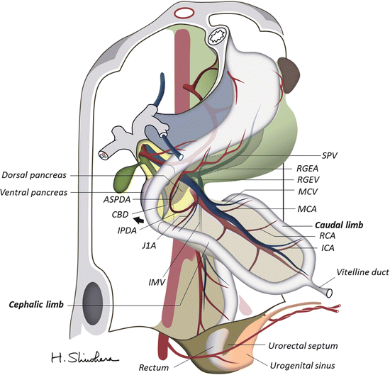
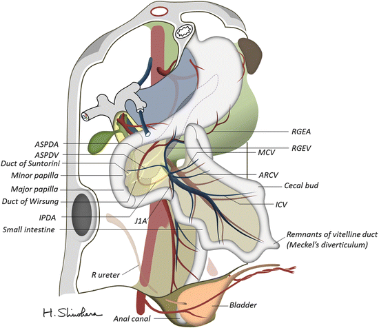
Fig. 1.4
The intestinal loops after a 90° counterclockwise rotation. The apex of the midgut loop communicates with the yolk sac through the vitelline duct toward which the superior mesenteric artery (SMA) runs through the proper mesentery. The cephalic limb of the midgut loop becomes the distal part of the duodenum, jejunum, and part of the ileum. The caudal limb becomes the terminal portion of the ileum, cecum, appendix, ascending colon, and the proximal two-thirds of the transverse colon. ASPDA anterior superior pancreatoduodenal artery, IPDA inferior pancreatoduodenal artery. Refer to the above figure legend for other abbreviations
Fig. 1.5
The intestinal loops after 180° midgut rotation. The cephalic limb comes to lie on the caudal side, while the caudal limb lies on the cephalic side. The superior mesenteric vein (SMV) moves above the superior mesenteric artery (SMA). The right gastroepiploic vein (RGEV), middle colic vein (MCV), and accessory right colic vein (ARCV) empty into the close part of the SMV. ASPDV anterior superior pancreatoduodenal vein. Refer to the above figure legend for other abbreviations
During the tenth week, the herniated midgut loop returns to the abdominal cavity. After completion of 270° rotation, the proximal portion of the jejunum comes to lie on the left side, and the transverse colon lies in front of the duodenum (Fig. 1.6). The SMV moves to the right side of the SMA: this is the definitive position of the two vessels. The cecum lies in the right iliac fossa, and the ascending colon and hepatic flexure are on the right side of the abdominal cavity. The distal end of the cecum forms a narrow diverticulum, the appendix. The rotation of the midgut loop swings the cephalic portion of the hindgut, placing the descending colon on the left side of the abdominal cavity.
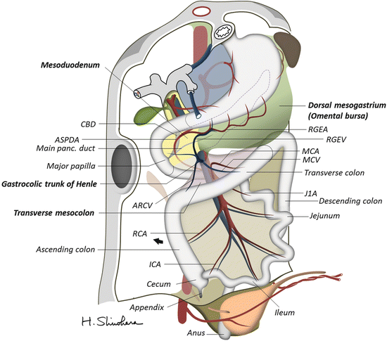
Fig. 1.6
 Evidence of Laparoscopic Surgery for Colorectal Cancer
 Laparoscopic Transverse Colectomy
 Robotic Total Mesorectal Excision
 Laparoscopic Right Lateral Pelvic Lymph Node Dissection (LPLND) with Pelvic Autonomic Nerve Preservation
 Laparoscopic Left-Sided Colectomy (Mobilization of Splenic Flexure and Sigmoidectomy)
 Transanal Minimally Invasive Surgery for Rectal Cancer


The intestinal loops after completion of 270° midgut rotation. The proximal portion of the jejunum comes to lie on the left side, and the transverse colon lies in front of the duodenum. Note that the right gastroepiploic vein (RGEV) and the accessory right colic vein (ARCV) form the gastrocolic trunk of Henle and flow into the superior mesenteric vein (SMV). Refer to the above figure legend for other abbreviations
Related posts:
Evidence of Laparoscopic Surgery for Colorectal Cancer
Laparoscopic Transverse Colectomy
Robotic Total Mesorectal Excision
Laparoscopic Right Lateral Pelvic Lymph Node Dissection (LPLND) with Pelvic Autonomic Nerve Preservation
Laparoscopic Left-Sided Colectomy (Mobilization of Splenic Flexure and Sigmoidectomy)
Transanal Minimally Invasive Surgery for Rectal Cancer
Stay updated, free articles. Join our Telegram channel
Full access? Get Clinical Tree
