Chapter 1.4
Physiology and function of the small intestine
Paul A. Blaker and Peter Irving
Guy’s and St Thomas’ NHS Foundation Trust, London, UK
The main functions of the small intestine are to complete the digestion of food through co-ordinated motility and secretion and to facilitate the absorption of water, electrolytes and nutrients. Approximately 9 L of fluid derived from oral intake (1.5 L) and exocrine secretions (7.5 L) enter the small intestine each day. Ninety per cent of this is reabsorbed in the small intestine with a further 8% absorbed in the colon. As such, only 100–150 mL of fluid is lost in faeces each day. The average length of the small intestine is 6.9 m but structural adaptations including mucosal folds, villi and microvilli mean that its surface area is 200–500 m2. The first 100 cm of the small intestine are highly adapted to the absorption of nutrients, whereas the more distal portions are involved in reclaiming fluid and electrolytes. The small intestine is able to absorb far in excess of the body’s requirements and as such, large portions of this organ can be removed without deleterious effects. However, changes in absorption and secretion homeostasis can rapidly lead to diarrhoea, dehydration, electrolyte disturbance and malnutrition.
1.4.1 Anatomy and histology
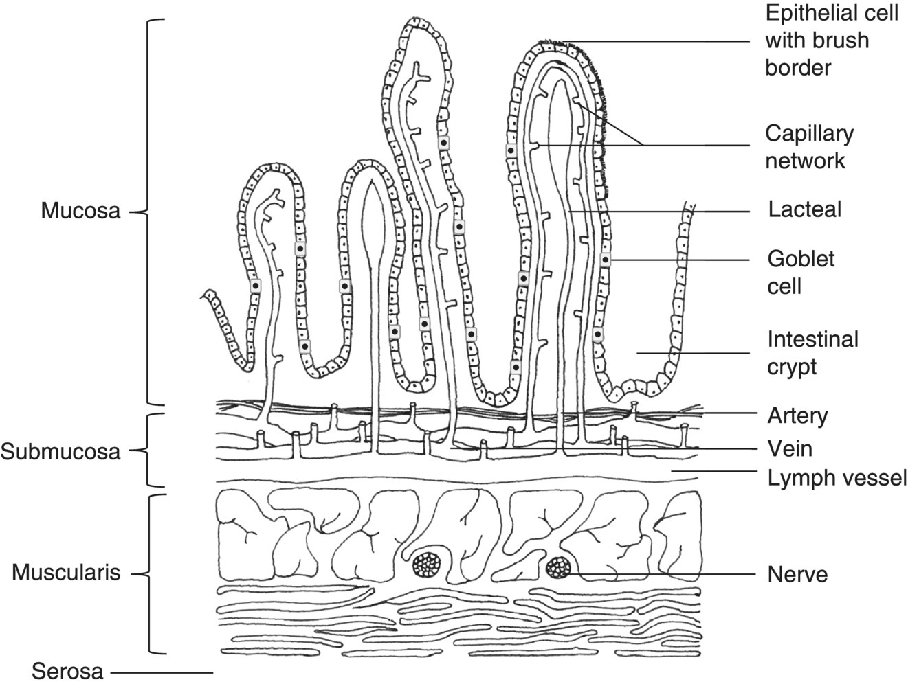
The small intestine includes three substructures termed the duodenum, jejunum and ileum, which extend sequentially from the gastric pylorus to the ileocaecal valve. The wall comprises an outer serous coat (tunica serosa), a layer of smooth muscle fibres (muscularis externa), submucosa consisting of dense connective tissue, a thin layer of smooth muscle (mucularis mucosa) and a mucosal layer (tunica mucosa) covered by epithelial cells (Figure 1.4.1). The tunica mucosa is thrown into numerous subfolds, creating the intestinal villi, which contain a dense blood capillary and lymphatic network that supplies the epithelial cells. Enterocytes are the most abundant epithelial cells (80%) and are characterised by the presence of enterocytic microvilli (brush border) that further increases the small intestinal surface area. Goblet cells are interspersed between enterocytes and secrete mucus that acts as a protective coat and lubricant. Tubular intestinal glands are found at the base of the villi (crypts of Lieberkuhn), which contain cells that differentiate into enterocytes, goblet cells, endocrine, paracrine and immune cells (Paneth cells). Changes in the cellular structure between sections of the small intestine allow for functional subspecialisation (Table 1.4.1).

Figure 1.4.1 Structure of the small intestine.
Table 1.4.1 Differences in the ultrastructure and function of the small intestine
| Layer | Duodenum | Jejunum | Ileum |
| Serosa | No change | No change | No change |
| Muscularis externa | Longitudinal and circular smooth muscle supplied by Auerbach’s plexus | Similar to duodenum | Similar to duodenum |
| Submucosa | Brunner’s glands +++ Meissner’s plexus | Brunner’s glands + | Brunner’s glands + |
| Muscularis mucosae | No change | No change | No change |
| Lamina propria | No Peyer’s patches | No Peyer’s patches | Peyer’s patches +++ |
| Intestinal epithelium | Simple columnar Goblet cells Endocrine cells Paracrine cells Paneth cells | Villi longer than duodenum | Villi shorter than duodenum |
| Sodium content | 145 mmol/L | 125 mmol/L | |
| Specialised functions | Iron and folate absorption | Iron and folate absorption in proximal jejunum Absorption of vitamin B1 and B2 | Vitamin B12 and bile salt absorption in terminal ileum Absorption of vitamin C |
Duodenum
The duodenum is approximately 25–35 cm in length and is split into four parts. It starts as the duodenal bulb, which arises from the gastric pylorus, and ends at the ligament of Treitz, where it joins the jejunum at the duodenojejunal flexure. The common bile duct enters the small intestine in the second part of the duodenum via the ampulla of Vater.
The duodenum is distinguished from other parts of the small intestine by the presence of numerous Brunner’s glands which secrete urogastrone (human epidermal growth factor), which is required for epithelial cell proliferation [1]. Consequently, the tips of the villi are continuously shed into the lumen and replaced by new cells from the crypts of Lieberkuhn. As such, the entire small intestine epithelium is renewed every 2–6 days.
Jejunum and ileum
The jejunum is approximately 2.5 m in length, whereas the length of the ileum is more variable (average 2–4 m). Both are contained within the peritoneum and are suspended by a mesentery. Most of the jejunum lies in the left upper quadrant of the abdomen, whereas the ileum mainly occupies the right lower quadrant. The jejunal folds are larger than those found in the duodenum or ileum.
1.4.2 Physiology and function
The gastric antrum sieves liquid chyme through the remaining solid matter in the stomach and delivers a continuous slow rate of gastric contents into the duodenum. The presence of chyme in the small intestine leads to the release of the hormones cholecystokinin (CCK) and secretin, which stimulate secretion of bicarbonate and pancreatic enzymes, and cause contraction of the gallbladder, which releases bile [2–4]. Proteins and peptides are degraded into amino acids through the action of pancreatic trypsin, chymotrypsin and elastase and subsequently by enzymes on the brush border. Lipids are degraded into fatty acids and glycerol and following emulsification by bile salts, triglycerides are split into free fatty acids and monoglycerides by pancreatic lipase. Carbohydrates may be broken down by pancreatic amylase into oligosaccharides or may pass into the colon where they are metabolised by GI microbiota. Brush border enzymes including dextrinase, glycoamylase, maltase, sucrase and lactase further break down oligosaccharides into monosaccharides prior to absorption. It is estimated that up to 65% of the adult population demonstrate a deficiency in lactase activity.
Reflex peristaltic waves mediated by musculomotor neurones propel the small intestinal contents at a rate of 1–2 cm/min, meaning that it takes an average of 2–6 h to reach the colon [5]. The intensity of the muscular contractions is influenced by the nature of the ingested food. Solid foods induce greater activity than liquid meals, and those that are high in glucose cause greater stimulation than ones high in fat.
Several mechanisms are involved in the absorption of nutrients by enterocytes, including passive diffusion, cytosis, active transfer and carrier-mediated transport [6]. Uptake of water is driven by the absorption of sodium (Na+), potassium (K+) and organic compounds and occurs through the formation of osmotic gradients. The absorption of Na+ is mediated by several different mechanisms including specific transmembrane carrier proteins.
1.4.3 Investigation of the small intestine
Correct diagnosis and management of small intestinal pathology are dependent on accurate history taking, clinical examination and specialist investigations. Non-bloody liquid stools greater than 1.5 L a day strongly suggest disease of the small intestine and weight loss may signify malabsorption. Here we summarise the key small intestinal investigations and describe their relevance to pathology.
Blood tests
Stay updated, free articles. Join our Telegram channel

Full access? Get Clinical Tree


