Chapter 1.2
Physiology and function of the oesophagus
Rami Sweis
University College London Hospital, London, UK
The oesophagus co-ordinates the transport of food and fluid from the mouth to the stomach. The oesophagogastric junction (OGJ) is a physiological barrier which reduces reflux of gastric contents. In harmony, these processes limit contact of the swallowed bolus, refluxed acid and other chemicals with oesophageal mucosa. Disruption of function can interrupt bolus delivery or induce gastro-oesophageal reflux. Symptoms produced may range in severity from heartburn and regurgitation to dysphagia and pain.
1.2.1 Anatomy
Oesophagus
The oesophagus is a muscular tube connecting the pharynx to the stomach. The cervical oesophagus extends distally from the cricopharyngeus and the thoracic oesophagus terminates at the hiatal canal before it flares into the gastric fundus. The muscularis propria consists of the outer longitudinal and inner circular muscle layers. The musculature is divided into the proximal striated and mid-distal smooth muscle. This proximal ‘transition zone’ is located one-third of the distance from the pharynx and is the site with the weakest force of peristaltic contractions [1].
Histologically, the oesophageal wall is composed of the mucosa, submucosa and muscularis mucosa. The oesophageal body is lined by non-keratinised stratified squamous epithelium which abruptly joins with the glandular gastric columnar epithelium at the squamocolumnar junction. This can be the site of mucosal change associated oesophagitis and Barrett’s oesophagus.
The antireflux barrier
The OGJ is not a clearly identifiable sphincter but its sphincter-like properties can be defined functionally as a high-pressure zone between the stomach and oesophagus. Sphincter competence is dependent on the integrity and overlap of the intrinsic lower oesophageal sphincter (LOS) and diaphragmatic crura. A separation, hiatus hernia, is associated with disruption of LOS integrity, loss of the intra-abdominal LOS segment and an increased susceptibility to gastro-oesophageal reflux.
1.2.2 Physiology and function
Voluntary swallowing initiates with ‘deglutitive inhibition’ of the smooth muscle oesophagus and LOS. This reflex relaxation is nitric oxide mediated and permits passage of the bolus with minimal resistance. The subsequent excitatory, predominantly cholinergic, activity produces a progressive wave of smooth muscle excitation. A co-ordinated peristalsis clears the bolus from the oesophagus.
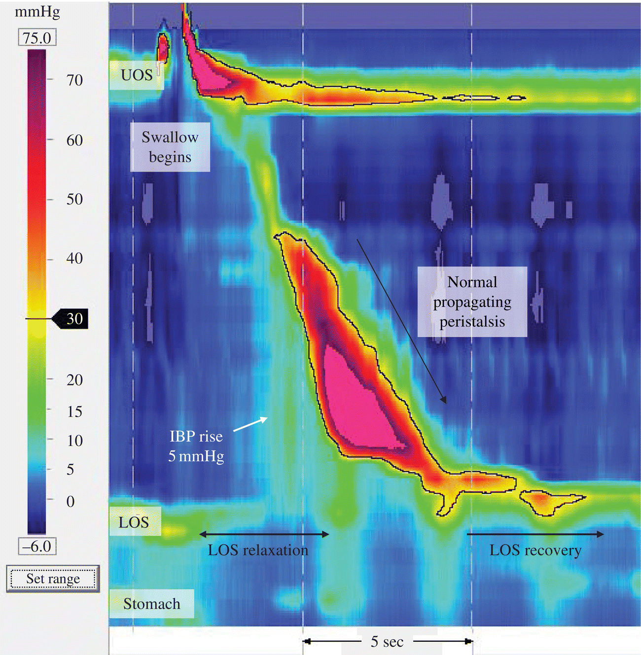
The LOS exhibits a continuous resting (basal) tone which relaxes on stimulation of the intramural nerves such as during deglutitive inhibition (swallowing). Disruption of this physiological process may impact on bolus transport and induce symptoms (Box 1.2.1). A representative normal swallow using high-resolution manometry is presented in Figure 1.2.1.

Figure 1.2.1 High-resolution manometry of a normal swallow, with pressure data presented as a spatiotemporal plot. Sensors are spaced at <2 cm intervals which provide a vivid depiction of oesophageal pressure activity from the pharynx to the stomach with changes in pressure represented as changes in colour (in clinical practice). Deglutitive inhibition is seen as the synchronous relaxation of the upper oesophageal sphincter (UOS) and lower oesophageal sphincter (LOS) followed by a co-ordinated peristalsis with increasing pressure duration as it progresses distally. Important landmarks are highlighted. Images acquired by 36-channel SSI Manoscan 360. IBP, intrabolus pressure.
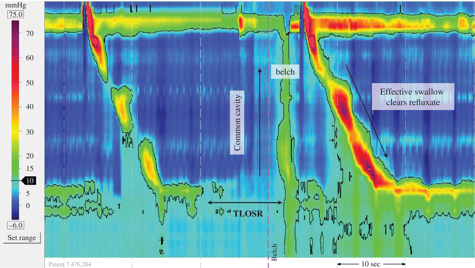
Spontaneous LOS relaxations normally occur as a response to gastric postprandial distension and bloating: ‘transient lower oesophageal sphincter relaxation’ (TLOSR). LOS relaxation can also follow peristaltic activity: ‘swallow-induced lower oesophageal sphincter relaxation’ (SLOSR). Gastro-oesophageal reflux and belch occur when there is equalisation of pressure between the stomach and oesophagus (common cavity) (Figure 1.2.2). Patients with gastro-oesophageal reflux disease (GORD) do not have an increased frequency of TLOSRs; rather, the tendency of reflux to occur during these events is greater [2]. The effectiveness of oesophageal clearance of refluxed material is an important contributor to the severity of GORD [3–5]. Other determinants of GORD include the presence and size of a hiatus hernia, increasing age and obesity as well as the calorie and fat content of the diet [6,7].

Figure 1.2.2 Transient lower oesophageal sphincter relaxation followed shortly afterwards by a common cavity during which there is equalisation of pressure between the stomach and oesophagus when reflux is most likely to occur. The event is terminated and the oesophagus is cleared of refluxed contents with the arrival of a well-co-ordinated primary peristalsis. Oesophageal and lower oesophageal sphincter pressures return to baseline levels following completion of peristalsis. TLOSR, transient lower oesophageal sphincter relaxation.
Measurement and assessment of function
In the absence of disease on endoscopy and failure to respond to empirical therapy, guidelines recommend manometry and ambulatory reflux testing [8,9]. Recent advances in technology provide better insight into the assessment of oesophageal function and disease.
Manometry
Peristalsis and OGJ activity can be measured with manometry. Conventional manometry (4–8 sensors) measures the circumferential contraction, pressure wave duration and peristaltic velocity of single water swallows. High-resolution manometry (HRM; 21–36 sensors) is an advance on conventional systems as it provides a compact, spatiotemporal representation of oesophageal pressure activity. In addition, it can measure the forces that drive movement of food and fluid through the oesophagus and OGJ [10]. An uninterrupted well-co-ordinated peristalsis defines oesophageal motility while the presence of a positive pressure gradient in the absence of obstruction describes whether this motility is effective and likely to clear the bolus [11] (see Figure 1.2.1). Thus HRM improves diagnostic sensitivity to peristaltic dysfunction as symptoms and mucosal damage are more likely to occur as a result of disturbed bolus transport and poor clearance [5]. Furthermore, recent advances in methodology have shown how HRM can also facilitate the assessment of swallowing behaviour (eating and drinking) when symptoms are more likely to be triggered [5,12,13] (Box 1.2.2).

Full access? Get Clinical Tree


