Reconstruction
Segmental resection
Proximal
Small bowel resection
Enteroenterostomy
Ileocecectomy
Ileo-ascending colostomy
Right colectomy
Ileo-transverse colostomy
Middle
Right extended colectomy
Ileo-transverse colostomy
Transverse colectomy
Colocolostomy
Left extended colectomy
Colocolostomy
Distal
Left colectomy
Colocolostomy
Sigmoidectomy
Colorectostomy
Low anterior resection
Colorectostomy
Proctectomy
Coloanal anastomosis
Nonsegmental resection
Subtotal colectomy
Ileo-sigmoid colostomy
Total abdominal colectomy
Ileorectostomy
Total proctocolectomy
Ileo-anal anastomosis
Perhaps the most critical point when dealing with the bowel that “does not reach” lies with preemptive planning. For any colorectal operation that will require reestablishment of gastrointestinal continuity, the surgeon should have a preoperative plan of what needs to be done for reconstruction after the specimen is resected. Patients should therefore be positioned to enable splenic flexure mobilization, for example, along with having the necessary equipment for mobilization maneuvers no matter the approach (open, hand-assisted laparoscopy, or purely laparoscopic). These strategies should be conveyed to both the patient and the surgical team so that any unexpected surprises can be mitigated.
Anatomic Constraints
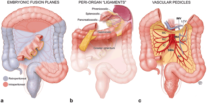
The primary concern in difficult bowel reconstruction is a tenuous and unsafe anastomosis. Multiple studies have demonstrated both “local” and “systemic” factors that contribute to poor anastomotic healing [1–4]. During an operation, the surgeon has immediate control of the local factors and a tension-free anastomosis with adequate blood supply is the most critical technical point that needs to be achieved to decrease the risk of anastomotic leak. Successful mobilization of the small bowel and colon to create tension-free anastomoses or stomas requires a clear understanding of their anatomic attachments. These attachments include (1) embryonic fusion planes, (2) peri-organ “ligaments,” and (3) vascular pedicles that can be ligated to maximize mobility while preserving necessary blood supply (Fig. 31.1).


Fig. 31.1
Anatomic constraints within the abdomen. Highlighted are the embryonic fusion planes (a), peri-organ ligaments (b), and vascular pedicles that are the targets of primary and secondary mobilization techniques (c). SMA superior mesenteric artery, IMA inferior mesenteric artery, IMV inferior mesenteric vein, IC ileocolic artery, RC right colic artery, MC middle colic artery, LC left colic artery, SA sigmoid arteries, LCV left colic vein. © Mayo Clinic
The small bowel is tethered to the posterior abdomen in an obliquely arranged mesentery that runs diagonally from the ligament of Treitz in the left upper quadrant to the right lower quadrant. The small bowel mesentery is usually very mobile with retroperitoneal fixation only at the ligament of Treitz and near the terminal ileum as it joins the retroperitoneal cecum and right colon. The right colon mesentery posteriorly abuts the right kidney, right ureter, and duodenum. After turning at the hepatic flexure, the transverse colon emerges from the retroperitoneum and its mesocolon is usually mobile before fixation into the splenic flexure. At this juncture, the left colon becomes retroperitoneal and its mesentery posteriorly abuts the left kidney. The splenic flexure is additionally fixated by the greater omentum and several peri-organ “ligaments” (splenocolic, renocolic, pancreatocolic, and phrenocolic ligaments). The sigmoid colon is nonperitonealized and usually held by a few lateral attachments as its mesentery courses over the left ureter and gonadal vessels. As the tenia disappears, the rectum begins intraperitoneally at the sacral promontory before traveling under the peritoneal reflection with its mesorectum to the pelvic floor and anorectal junction.
While mobilizing the small bowel and colon from the retroperitoneum and peri-organ attachments such as the spleen and omentum is often sufficient to provide needed reach, these “first-line” maneuvers simply free, but preserve embryonic planes. Secondary and more advanced maneuvers exploit the vascular tethers within the mesentery. These vessels include the superior mesenteric artery (SMA) and its branches (the ileocolic, right colic, and middle colic artery), the inferior mesenteric artery (IMA) and its branches (the left colic, sigmoid, and superior rectal artery), and the inferior mesenteric vein (IMV). Thoughtful and directed transection of these vessels while relying on collateral blood flow can provide significantly more reach while maintaining a tension-free anastomosis with adequate blood supply.
Diagnosing the Problem
Surgical trainees are taught that a successful anastomosis is one that is tension-free and well-vascularized. But is there a way to quantify how much tension is allowable for an anastomosis to be safe? Is there a way to quantify if adequate blood supply is reaching an anastomosis? These are critical questions that are always asked during mortality and morbidity conferences when presenting an anastomotic leak case, but unfortunately our ability to answer these questions with objective data is limited. On the contrary, we often rely on past experience and make clinical judgments when making these decisions.
Probably the simplest way to ask if an anastomosis is under tension is to lay the proximal and distal bowel ends in the field without any pulling or pushing. If the ends overlap each other by at least 5 cm, one can presume that there will be minimal to no tension on the anastomosis. When we need to pull inferiorly on the proximal end, or superiorly on the distal end, there will be problems and further mobilization needs to be performed. Similarly, during ileal-pouch anal anastomoses (IPAA), we use the inferior edge of the pubis symphysis as a rough estimate of adequate length if the apex of the pouch can reach it without tension.
Blood supply can be initially assessed with the gross appearance of the proximal and distal ends of the bowel. Completely ischemic tissue will have an obvious black-blue, discolored appearance, but this assessment is easiest at the extreme end of ischemia. In reality, bowel ends could be bruised, or “dusky,” and a clinical judgment needs to be made on its viability. In these cases, we observe whether there was bleeding at the anastomotic line during transection or use the Doppler to assess for blood flow. While somewhat rudimentary, we find these methods useful in those moments of doubt. Future diagnostic tests may include using intraoperative indocyanine green (ICG) angiography, which shows promise in distinguishing anastomotic ends with poor perfusion [5].
Specific Techniques: Making It Reach
When presented with the bowel that cannot reach, mobilization should begin in a sequential and logical fashion that uses defined technical principles to remove anatomic constraints (Table 31.2). Primary maneuvers include (1) mobilizing embryonic planes and (2) dividing peri-organ “ligaments” or attachments. Secondary maneuvers include directed ligation of vascular pedicles that restrict the mobility of the corresponding proximal bowel. Often, these vascular ligations are already part of the oncologic resection. Tertiary maneuvers include more extended bowel resections to reach a mobile proximal portion of bowel versus the construction of a stoma if no tension-free option is possible. To illustrate these principles, we describe several challenging operative situations in which multiple strategies may be necessary to achieve intestinal continuity.
Table 31.2
Mobilization techniques for difficult reconstructions
Maneuvers | Goals of maneuver | Examples |
|---|---|---|
Primary | Separation of embryonic fusion planes | Cattell and Mattox maneuvers |
Division of peri-organ “ligaments” | Splenic flexure mobilization | |
Secondary | Ligation of vascular pedicles | Ligating the ileocolic artery during IPAA |
Preservation of collateral blood supply | Preserving the middle colic artery to supply ileal pouch | |
Tertiary | Extended resection to mobile proximal bowel | Completion colectomy |
Stoma construction | End ileostomy or colostomy |
Colorectal and Coloanal Anastomosis
Primary reconstruction of the distal gastrointestinal tract after resection of the left colon, sigmoid, and/or rectum requires a colorectal or coloanal anastomosis. The construction of a tension-free anastomosis requires significant mobilization for the proximal bowel to reach into the pelvis and can be performed using open or minimally invasive techniques.
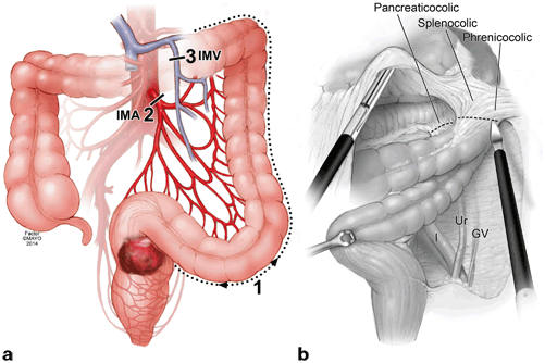
Primary maneuvers separate the left colon from its retroperitoneal and peri-organ attachments. This maneuver can be done using a lateral-to-medial or medial-to-lateral approach (Fig. 31.2). Either approach is effective and depends on the surgeon’s experience, training, and comfort level. The medial-to-lateral approach immediately identifies and ligates vascular pedicles such as the IMA and IMV before dissecting “underneath” the retromesenteric plane to the lateral line of Toldt and splenic flexure. The lateral-to-medial approach is more classically taught and equally effective, and both techniques have been thoroughly described before [6–8]. As such, we will go over general principles and add our specific commentary and operative pearls.


Fig. 31.2
Overview of primary and secondary maneuvers for colorectal and coloanal anastomoses. a Lateral-to-medial dissection proceeds with ( 1) mobilizing the line of Toldt and splenic flexure, ( 2) high ligation of the inferior mesenteric artery ( IMA), and ( 3) high ligation of the inferior mesenteric vein ( IMV) to provide maximum bowel length for a colorectal or coloanal anastomosis. A medial-to-lateral dissection proceeds in another order with ( 2) high ligation of the IMA, ( 3) high ligation of the IMV, and finally ( 1) mobilization of the retroperitoneal embryonic plane. b Critical retroperitoneal structures that can be identified during mobilization of the left colon are illustrated including the left iliac artery ( I), left ureter ( Ur), and left gonadal vessels ( GV). Splenic flexure mobilization involves ligating the splenocolic, phrenicocolic, and pancreaticolic ligaments. © Mayo Clinic
Lateral-to-Medial Approach
For a lateral-to-medial approach, we first open the line of Toldt at the pelvic brim to enter the retromesenteric space (Fig. 31.2). With firm counter-traction on the colon medially, the white, wispy, and avascular fibers marking the embryonic, retromesenteric fusion plane can be visualized and dissected bluntly, sharply, or with electrocautery. The retroperitoneum, gonadal vessels, and left ureter remain undisturbed posteriorly and the dissection is continued superiorly toward the splenic flexure. One of the teaching points during this maneuver is to keep closer to the colon edge and to avoid laterality once the line of Toldt is incised. If the latter is done, then the dissection will actually come around the retroperitoneum rather than the colon mesentery, and the left kidney will be elevated. The colon mesentery will often maintain a sheer glistening layer of parietal peritoneum that can be used to distinguish from the underlying fat of the retroperitoneum.
As the surgeon works superiorly, the left kidney will be encountered posteriorly with its overlying Gerota’s fascia. The kidney should remain undisturbed, and any bleeding suggests that the wrong plane has been entered. With firm medial and inferior traction on the colon, the splenic flexure can be approached laterally while staying close to the colon to avoid “wandering off” into the more lateral retroperitoneum and sometimes thick omental attachments. Tension lines should be demonstrated and sharply cut, cauterized, or divided with energy devices. The goal is to enter the lesser sac which would signify the surgeon coming “around the bend” of the splenic flexure. Often there is abundant omentum that will need to be dissected free from the distal transverse colon and its epiploica. If there is difficulty freeing the splenic flexure with a lateral, counterclockwise approach, then the surgeon should switch to a medial, clockwise approach by flipping the omentum superiorly and detaching the inferior omental leaflet from the mid-transverse colon to enter the lesser sac. Once the lesser sac is entered, then the surgeon can approach the splenic flexure medially to join the lateral dissection.
Mobilizing the splenic flexure is an important first step in distal reconstructions such as colorectal or coloanal anastomoses. Cadaveric studies have shown that an additional 10–28 cm of colonic length can be gained with mobilization of the splenic flexure and distal transverse colon [9, 10]. Some surgeons advocate splenic flexure mobilization at the very beginning of the operation to avoid any future debate at the end of a long resection, while others advocate selective use of the technique on an as-needed basis depending on colon redundancy [9]. It is our routine practice to mobilize the splenic flexure preceding an anticipated mid-rectal to coloanal anastomoses.
< div class='tao-gold-member'>
Only gold members can continue reading. Log In or Register to continue
Stay updated, free articles. Join our Telegram channel

Full access? Get Clinical Tree


