Fig. 5.1
The surgeon performs the procedure standing between the patient’s legs, with an assistant on the right side of the operating table and another one on the left side
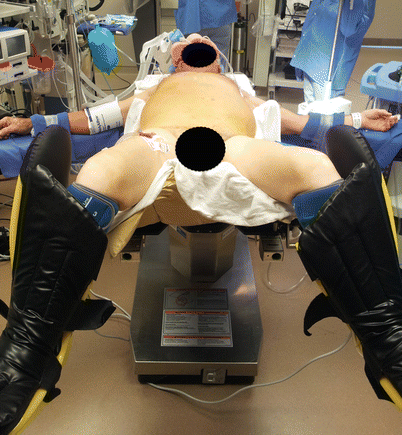
Fig. 5.2
The patient is positioned supine on the operating table. The legs are extended on stirrups, and the knees are flexed at angle of 20°–30°
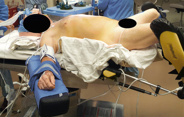
Fig. 5.3
The patient is positioned over a bean bag that is inflated to prevent sliding when a steep reverse Trendelenburg position is used
5.2 Trocar Positions for Laparoscopic Antireflux Surgery and Laparoscopic Heller Myotomy
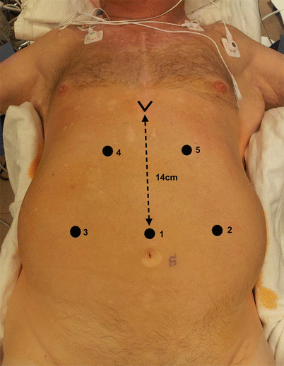
Using a Veress needle that is placed 14 cm below the xiphoid process, we initially inflate CO2 into the abdominal cavity to a pressure of 15 mmHg. Alternatively, a Hasson cannula can be used. We recommend using an optical trocar with a 0° scope to obtain access.
Figures 5.4 and 5.5 illustrate the positions of the five 11-mm trocars that are used for these operations. Trocar 1, which is used for the 30° camera, is placed in the same location as the Veress needle. Trocar 2 is placed in the left midclavicular line at the same level as trocar 1; it is used for insertion of a Babcock clamp, a grasper to hold the Penrose drain surrounding the esophagus, or a device to take down the short gastric vessels. Trocar 3 is placed in the right midclavicular line at the same level as the first two trocars; it is used for insertion of a retractor to lift the left lateral segment of the liver. Trocars 4 and 5 are placed under the right and left costal margins, so that their axes form an angle of about 120° with the camera. They are used for the dissecting and suturing instruments.