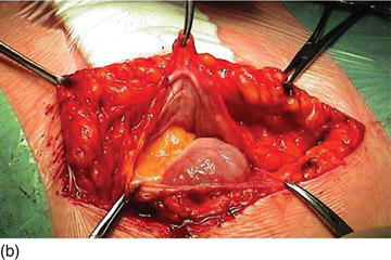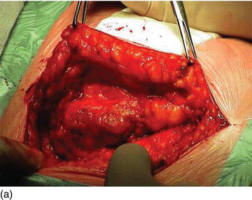
A lateral incision is made about 10 cm from the stoma through the skin into the subcutaneous tissue, well lateral to the stomahesive skin marks (Figure 10.1). The extent of the hernia defect is delineated and the hernia sac carefully preserved (Figure 10.2a
Stay updated, free articles. Join our Telegram channel

Full access? Get Clinical Tree


