Fig. 8.1
Hiatal hernia viewed on upper endoscopy
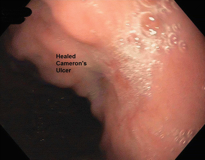
Fig. 8.2
Healed Cameron’s ulcer on upper endoscopy
8.1.2 Operation
8.1.2.1 Patient Positioning
Place the patient in a supine position with both arms tucked and padded. A Foley catheter is necessary for measurement of intraoperative urine output. A footboard and a waist belt strap are used to accommodate steep reverse Trendelenburg positioning. Prior to the beginning of the operation, antibiotics and chemical and mechanical venous thromboembolism prophylaxis are given, and an orogastric tube is inserted for stomach decompression.
8.1.2.2 Port Placement
The ports are placed with the purpose of having proper triangulation of the patient’s esophageal hiatus, keeping in mind that the hiatus is located slightly to the patient’s left and tends to be much more cephalad after pneumoperitoneum insufflation. We enter the abdomen with a Veress needle in the left upper quadrant (LUQ), and then proceed by placing an 11-mm visualization trocar between the xiphoid and the umbilicus. Three more working ports are placed as shown in Fig. 8.3 (two 11-mm trocars in the left upper quadrant and a 5-mm trocar in the right upper quadrant). A Nathanson liver retractor is inserted in the epigastrium.
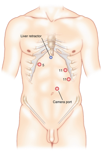

Fig. 8.3
Port placement for laparoscopic paraesophageal hernia repair
8.1.2.3 Operative Procedure
Step 1
Reduce the hernia sac
Place the patient in steep reverse Trendelenburg and right-side-down positioning and start pulling the contents of the hernia sac out of the mediastinum (Fig. 8.4a). We use the bipolar energy device to divide the short gastric vessels, just starting at the inferior margin of the spleen. Then continue to dissect out the left crus at the angle of His and enter the avascular plane in the mediastinum just anterior to the aorta (Fig. 8.4b). As the dissection is continued anteriorly, it is important to identify the left pleura and the anterior vagus nerve (Fig. 8.4c). Once the medial edge of the right crus is identified, we then move our dissection to that side in order to begin dividing the gastrohepatic ligament and further identify the right crus. We identify the posterior vagus nerve and then make our retroesophageal window in order to insert a Penrose drain for better retraction on the esophagus and stomach for the mediastinal dissection.
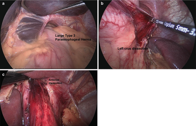

Fig. 8.4
(a) Pulling the contents of the hernia sac out of the mediastinum. (b) Dissection of the left crus. (c) Continuing anterior dissection, identifying the left pleura and the anterior vagus nerve
Step 2
Obtain 3 cm of intra-abdominal esophagus through extensive mediastinal dissection
Remove the orogastric tube and continue to retract the esophagus and the stomach with the Penrose drain at the gastroesophageal junction (GEJ). We use hook electrocautery to dissect higher in the mediastinum, to avoid bleeding from the vascular attachments to the esophagus (Fig. 8.5).
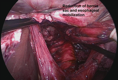

Fig. 8.5
Esophageal mobilization
Step 3
Create a tension-free posterior cruroplasty
Once we have achieved enough esophageal length to have at least 3 cm of intra-abdominal esophagus, we then proceed to place our interrupted sutures to plicate the crura posterior to the esophagus (Fig. 8.6a). We place these sutures approximately every 5–8 mm until we have closed the crura, leaving only enough space for an instrument. We then use an absorbable mesh to reinforce this cruroplasty, placing it securely to the crura as an overlay in a U-shaped fashion (Fig. 8.6b).
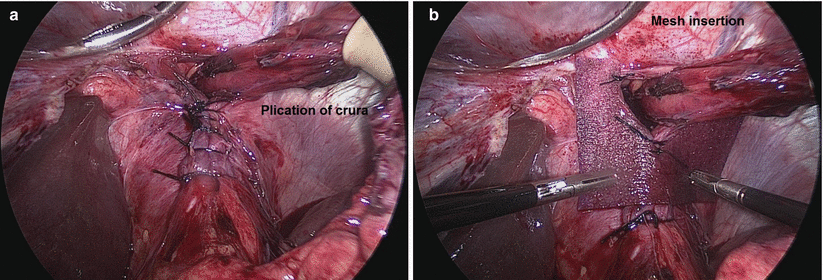

Fig. 8.6
(a) Plication of the crura posterior to the esophagus. (b) Insertion of absorbable mesh to reinforce cruroplasty
Step 4
Create an antireflux fundoplication
An antireflux fundoplication is then created, using either a complete or partial wrap. The choice between creating a Nissen or a Toupet fundoplication is made before going to the operating room, based on the results of the preoperative esophageal manometry study. Patients with poor motility are given a Toupet fundoplication in order to limit potential postoperative dysphagia (Fig. 8.7). In this patient, with preserved peristalsis, a Nissen fundoplication was performed. We located the GEJ and then found a location to grasp on the greater curvature of the stomach approximately 3 cm distal and posterior to the GEJ. This location was marked with a clip, which is then used to help guide the “shoeshine” maneuver and create a floppy, symmetrical wrap. The Nissen fundoplication is completed by connecting the opposing sides of the stomach with two interrupted sutures about 2 cm apart. (The sides of the stomach are connected to the esophagus in a Toupet fundoplication.) The wrap is then further secured to the hiatus at both crura on either side of the esophagus.
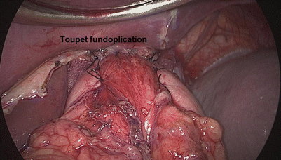

Fig. 8.7
Toupet fundoplication to limit postoperative dysphagia in patients with poor esophageal motility
Step 5
Perform completion endoscopy
We perform an endoscopy at the end of each operation in order to directly confirm that we have completed a symmetric wrap and have an easy entry at the GEJ (Fig. 8.8).
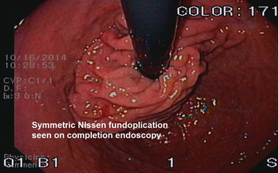

Fig. 8.8
Completion endoscopy confirming symmetric Nissen fundoplication
8.1.3 Postoperative Course
The patient is given a clear liquid diet after the operation and is advanced to a mechanical soft diet the next day. We do not feel that an upper GI series is indicated, and patients are usually discharged home on postoperative day 1. They maintain their soft diet for 2 weeks until their follow-up clinic visit, and their diet is advanced appropriately at that time. Patients are always seen again for follow-up at 6 months and 12 months after their operation.
8.2 Collis Gastroplasty
8.2.1 Case Presentation
The patient is a 70-year-old woman with a past medical history of worsening symptoms of heartburn, regurgitation, and dysphagia for the past month. She has a past surgical history of a laparoscopic hiatal hernia repair with nonabsorbable mesh and Nissen fundoplication. Given her prior surgical history with recurrent symptoms of gastroesophageal reflux disease, a workup of recurrent hiatal hernia was performed:
Barium swallow: large hiatal hernia with mild to moderate narrowing of the proximal stomach at the diaphragmatic hiatus and a dilated distal esophagus (Fig. 8.9)
Stay updated, free articles. Join our Telegram channel

Full access? Get Clinical Tree


